interaction of sensory and cognitive information underlying. Neuroscience and Behavioral Physiology, Vol. 36, No. 8, 2006. Disorders of Learning and Memory ...
Neuroscience and Behavioral Physiology, Vol. 36, No. 8, 2006
Disorders of Learning and Memory Processes in a Monkey Model of Alzheimer’s Disease: The Role of the Associative Area of the Cerebral Cortex K. N. Dudkin,1 I. V. Chueva,1 F. N. Makarov,1 T. G. Bich,2 and A. E. Roher2
Translated from Rossiiskii Fiziologicheskii Zhurnal imeni I. M. Sechenova, Vol. 91, No. 8, pp. 857–871, August, 2005. Original article submitted April 14, 2004, revised version received March 10, 2005. The processes of learning and storage of the results of learning were studied in a model of Alzheimer’s disease in two groups of rhesus macaques (three individuals in each group). Studies were performed after injection of neurotoxins (group I) and physiological saline (group II, controls). Two months after injections (stage C1), learning parameters were studied in monkeys of both groups using a new stimulus discrimination test (filled geometrical figures versus outline figures). There were significant differences between the animals of the two groups. Learning was hindered in monkeys of group I, with significant increases in the learning time (the time to achieve a stable probability of correct responding of 0.85) and in the probability of refusals. Monkeys of group II showed no learning impairment. Animals were trained to discriminate new stimuli (images of two monkeys) six months after injections (stage C3). Learning was impaired in animals of group I, such that learning measures had the same levels as previously; monkeys of group II showed no learning impairment. Analysis of the characteristics of working memory, which is involved in storing the results of new learning, was performed at stage C1; monkeys of group I showed significant degradation of these measures, with a significant decrease in the probability of correct solutions at stage C1 (to a level of 0.5), with some increase at stages C2 (at four months) and C3, along with a significant increase in the probability of refusals, values being similar at all time points. For monkeys of group II, these characteristics showed no degradation. Motor response times at stages C1, C2, and C3 were not different for the two groups of monkeys. The structural-functional organization of interactions between sensory and cognitive processes during learning and the storage of information in working memory are discussed, as is the role of the associative areas of the cortex in these interactions. KEY WORDS: rhesus macaque, learning processes, working memory, visual discrimination, model of Alzheimer’s disease, associative areas of the cortex.
Current concepts of the neurophysiological bases of consciousness include the position that there is an interaction between sensory and cognitive processes resulting in the production of behavior. We suggest that one of the principles underlying the organization of cognitive processes is the formation of functional systems defined by the behav-
ioral task, the motivational factor, the properties of objects, and the context obtaining at the time [1–6]. This leads to the formation of separate channels for the processing of sensory information, for example, information on the shapes and colors of objects and their spatial characteristics – position in the field of vision, orientation, size, and interrelationships in space. It follows from this hypothesis that there are individual, morphologically based processes for learning and working memory, these processes being components of cognitive processes. The identification of neuronal structures in the associative areas of the cortex determining the interaction of sensory and cognitive information underlying
1 Cognitive
Processes Modeling Group and Neuromorphology Laboratory, I. P. Pavlov Institute of Physiology, Russian Academy of Sciences, 6 Makarov Bank, 199034 St. Petersburg, Russia. 2 Health Research Institute, Sun City, 10515 Western Santa Fe, Arizona 85351, USA.
789 0097-0549/06/3608-0789 ©2006 Springer Science+Business Media, Inc.
790
Dudkin, Chueva, Makarov, et al.
ST3
OO
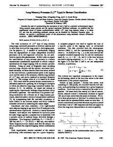
Fig. 1. Conditioned visual stimuli for new learning. ST3 were used for training two months after injections; OO were used at six months. The + sign identifies the reinforced stimuli and the – sign the negative (unreinforced) stimuli.
the ongoing recognition of objects in the visible world is an important task for understanding the neurophysiological bases of the organization of behavior. Despite extensive results, sufficient data for these hypothetical conceptualizations continue to accumulate. How do cognitive structures for object discrimination form? How does sensory information perceived from the external world interact with the “model world” already formed in the organism’s memory? What neurophysiological mechanisms are responsible for the segmentation of visual scenes, extraction of the features required for recognition, and storage of incoming information in working memory? What are the functional roles of the various areas of the cerebral cortex in these processes? Clear achievements have been made in recent years, though there are still no widely accepted answers to these questions. We suggest that a significant step in solving this problem would be provided by studies of impairments in the cognitive characteristics of learning and memory in a monkey model of Alzheimer’s disease. Alzheimer’s disease is known to be accompanied by neuropathological changes and neurotransmitter deficits, primarily with respect to acetylcholine, such that the cholinergic hypothesis of the origins of this disease is currently the most widely accepted [14–23]. The consequences of neurodegeneration – a set of cognitive functions – appear during the course of the disease [14, 15, 20]. The main sensory symptoms of Alzheimer’s disease in humans include impairment of working memory and the discrimination of the spatial relationships between objects, which accompany the deterioration of new learning [14, 15, 17]. These dysfunctions are subsequently supplemented by impairment of planning ability and the control of actions, speech disturbances, comprehension dis-
turbances, spatial disorientation, and other irreversible impairments of many other important behavioral functions [15, 17, 18]. These points illustrate the relevance and importance of modeling Alzheimer’s disease in animals. Studies of a monkey model of Alzheimer’s disease [9–11, 16] have provided new data related to the mechanisms of cognitive dysfunctions, furthering our understanding of the neurophysiological principles underlying the organization of cognitive processes. Our previous studies have demonstrated that injections into monkeys (rhesus macaques) of a neurotoxin selective for the cholinergic receptors (p75NTR) of neurons in Meynert’s basal nucleus and the enzyme DBH-saporin, which disturbs the function of noradrenergic neurons in the locus ceruleus, leads to significant deterioration of the discrimination of visual information even though it is stored in working (short-term) memory [9–11]. Injections of these neurotoxins led to creation of a monkey model of Alzheimer’s disease: data were obtained on impairments of the characteristics of working memory and their relationship with the type of visual information and the course of Alzheimer’s disease. The characteristics of impairments in the sensory and cognitive components of working memory were also identified. However, it remained unclear whether these characteristics are impaired in new learning and the storage of its results in working memory. The aim of the present work was to continue our studies of this model of Alzheimer’s disease. The present experiments were intended to produce complete or partial responses to the following questions. Is new learning of a visual discrimination task impaired during the development of Alzheimer’s disease in monkeys? Which characteristics
Disorders of Learning and Memory Processes in a Monkey Model of Alzheimer’s Disease of learning processes are most impaired? What are the characteristics of the storage of the results of this impaired learning in working memory? What is the role of the associative fields of the cortex in these impairments?
METHODS The methods used in the present studies have been described in detail elsewhere [11]. Alzheimer’s disease was modeled in monkeys of group I (identification Nos. 20, 21, and 22) by unilateral injection of the neurotoxin p75-saporin (the ribosomal toxin saporin linked to a monoclonal antibody to receptor p75NTR) into the lateral ventricles (A = 17, L = 1, D = 12) [13] at a dose of 118.5 µl (320 µg). This neurotoxin is known to induce irreversible degeneration of cholinergic neurons in the basal nucleus of Meynert [20]. In addition, the enzyme dopamine-beta-hydroxylase (DBHsaporin) was given at a dose of 47.6 µl (100 µg); this impairs the functions of noradrenergic neurons in the locus ceruleus [20]. Monkeys of group II (identification Nos. 23, 24, and 25) received injections of sterile physiological saline (0.9% NaCl) at a dose of 166.1 µl (420 µg). Studies were performed on the same six monkeys divided into two groups of three previously used for identification of the quantitative characteristics of working memory impairment [9–12]. Monkeys were trained to discriminate new stimuli (Fig. 1); training to discriminate stimuli ST3 was performed after the first two months post-injection, while training with stimuli OO was performed at six months. The ability to retain the results of learning the discrimination of stimuli ST3 in working memory was also studied at two, four, and six months (stages C1, C2, and C3, respectively) after neurotoxin administration. This was addressed by including delays of 1–4 sec in the animals’ motor responses into the experimental protocol, this delay recruiting the mechanisms of working memory for shortterm retention of visual information as the monkeys performed the behavior. The quantitative characteristics of new learning and the storage of its results in working memory were compared for the monkeys of the two groups: after injection of neurotoxins (group I) and after injection of physiological saline (group II). During experiments, animals were placed in a primate chair with a test panel with two suspended matte screens which also served as keys for making selections. Conditioned visual stimuli were projected onto the screens. A tuning stimulus (a sound signal of duration 1 sec) was presented at the beginning of the task. Conditioned stimuli were then presented in random order, simultaneously on both screens (left and right) for 2 sec: a positive stimulus (reinforced) and a negative stimulus (non-reinforced). Monkeys’ correct decisions were expressed by pressing the screen on which the positive stimulus appeared, and this was accompanied by automatic delivery of food reinforcement via a
791
nozzle attached to the mouth [2]. Incorrect selections were not reinforced. Monkeys were regarded as trained when the level of correct decisions reached 85%. Each of 20 sequential (training) series consisted of 20 presentations of pairs of stimuli; series were separated by intervals of 5–10 min. The reinforcement used in these experiments consisted mainly of various fruit juices and sweetened water. Motivation was maintained at a sufficiently high level on the basis of the animals’ individual preferences. After experiments, monkeys received normal food and drink rations, including vitamins and trace elements. Monkey keeping and all experiments were in accord with the requirements of the “Regulations for Scientific Experiments Using Experimental Animals,” Presidium of the Academy of Sciences of the USSR, 2 April 1980. An automatic experiment control program [2] was used to form the animals’ behavior and to record the numbers of stimuli presented (n), the total number of all (positive and negative) correct responses (m), the number of correct responses (l), the number of refusals (n – m), and the motor reaction time (T). The probabilities of correct responses (PR(C) = 1/m) and refusals (P(RF) = n – m/n) were calculated. The effects of experimental factors on the characteristics of new learning were studied in both groups (two and six months after injections); effects on the characteristics of storage in working memory of the results of new learning (discrimination of stimulus ST3) were studied at two, four, and six months (stages C1, C2, and C3) using multifactorial dispersion analysis (MANOVA). Quantitative criteria for behavioral responses were provided by the probability of correct responses, the probability of refusals, and the motor reaction time. Significant factors and interactions between them were identified using Fisher’s F test, where P is the statistical significance at a level of 95%.
RESULTS Monkeys of both groups – those given neurotoxin injections (group I) and physiological saline (group II) – were used for studies of the characteristics of the learning of new stimuli and the storage of this learning in working memory during the development of Alzheimer’s disease. At two months post-injection, monkeys were trained to discriminate new stimuli: filled geometrical figures versus outline figures (Fig. 1, ST3). The characteristics of the storage of the results of learning in the monkeys’ working memory were studied by testing in a delayed discrimination task at two, four, and six months (sages C1, C2, and C3). At six months after neurotoxin injection, new learning was studied using an additional stimulus – images consisted of two monkeys (Fig. 1, stimulus OO). The results of training the animals are shown in Fig. 2, I, II (discrimination of ST3) and III, IV (discrimination of OO). This shows that despite individual differences, the
792
Dudkin, Chueva, Makarov, et al.
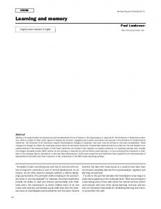
Fig. 2. Learning curves for monkeys trained to visual discrimination of conditioned stimuli two (I, II) and six (III, IV) months after injections. I, III) Monkeys after neurotoxin injections; A, B, and C are monkeys Nos. 20, 21, and 22, respectively; II, IV) monkeys after injection of physiological saline; A, B, and C are monkeys Nos. 23, 24, and 25, respectively. The abscissas show training session sequence numbers; the ordinates on the left show the probability of correct responses (continuous line, white circles); the ordinates on the right show the probability of refusals to take decisions (dotted line, black diamonds). The horizontal thick lines show the learning criterion (a level of correct responses of 85%) and the vertical thick lines show the threshold series at which the learning criterion was stably achieved. For explanation see text.
monkeys within each group showed common features in their learning curves, while there were significant differences between groups (Fig. 2, I, III and II, IV). Each plot shows two learning curves demonstrating the relationship between time and the probability of correct responses (light circles) and the probability of refusals (dark diamonds),
which characterize the learning process. We note that the learning criterion used here consisted of stable correct responding at a level of 85% (shown by the straight horizontal lines on plots). In monkeys of group I (Fig. 2, I, III), learning was significantly hindered both two and six months after neurotoxin administration. Learning became
Disorders of Learning and Memory Processes in a Monkey Model of Alzheimer’s Disease
793
Fig. 2. Continued.
unstable and the time required to achieve the 85% level of correct responses increased in comparison with that in monkeys of group II (Fig. 2, II, IV). Monkey No. 20 had not reached this learning level at two months. It is evident from Fig. 2 that the probability of refusals for monkeys of group I increased from that in monkeys of group II, such that the refusal probability curves were more unstable and oscillatory than those for monkeys of group II. This applied to both learning situations. Additional quantitative information for comparing the monkeys’ learning results at two and six months was obtained by dispersion analysis (Fig. 3).
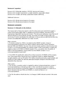
The probability of correct responses for each animal was averaged for all training series (Fig. 3, A). Monkeys of group I (Nos. 20, 21, and 22) showed significant decreases in both cases of new learning (at two and six months postinjection) such that values were significantly different (F = 18.4, p = 0.0000) from values in monkeys of group II (Nos. 23, 24, and 25), not reaching a level of 80% for both stimuli (ST3, OO). There were no significant differences between the results obtained for one stimulus compared with the other (F = 0.06, p = 0.8110). This result provides evidence that learning for new stimuli is hindered regardless of the time since neurotoxin administration.
794
Dudkin, Chueva, Makarov, et al.
Fig. 3. Mean probabilities of correct responses, probabilities of refusals, and motor reaction times for correct responses calculated for all training series for each monkey. On the abscissa, the numbers 20, 21, and 22 show monkeys after neurotoxin injections; 23, 24, and 25 show monkeys injected with physiological saline. Ordinates: A) mean probability of correct responses; B) mean probability of refusals; C) mean motor response time. Stimulus ST3 shows training two months after injections; stimulus OO shows training at six months. Points show arithmetic means and bars show the interval corresponding to the Fisher least significant difference (LSD). The differences in each pair of means with non-overlapping LSD values are statistically significant. For further details see text.
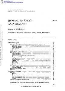
The probability of refusals averaged for all training series for each animal (Fig. 3, B) increased significantly for monkeys of group I for both stimuli (ST3, OO; F = 24.10, p = 0.0000) as compared with monkeys of group II, which showed no deterioration in these characteristics. We note that for monkeys of group I, the increases in refusals also showed no differences for the two stimuli (F = 2.36, p = 0.1258), as was also the case for the decreases in the probability of correct responses. Motor response times averaged for all series for each animal (Fig. 3, C) demonstrated significant individual differences between monkeys of groups I and II (F = 53.61; p = 0.0000); monkeys Nos. 20, 21, and 22 showed no significant differences associated with the stimuli (F = 2.10, p = 0.1491). However, among the monkeys of group II, Nos. 23 and 24 showed significant differences in motor response times associated with the type of visual information (F = 4.02, p = 0.0016). Which characteristics of working memory are associated with the storage of the results of new learning (for stimulus ST3)? We will consider data for monkeys of the two groups averaged for the behavioral characteristics of all animals of each group and all delays (Fig. 4, I). The probability of correct responses for monkeys of group I (Fig. 4, I, A, 1) decreased significantly (F = 356.5, p = 0.0000) as compared with monkeys of group II (Fig. 4, I, A, 2). This decrease was more significant (to a level of 0.52) at stage C1 (two months post-injection), after which there was a significant increase at stages C2 and C3 (four and six months respectively, F = 6.04, p = 0.0043), which demonstrates some compensation of the impairment. The probability of refusals for monkeys of group I (Fig. 4, I, B, 1) increased significantly (F = 238.45; p = = 0.0000) compared with monkeys of group II (Fig. 4, I, B, 2), such that the values for monkeys of the two groups showed no significant difference at all time stages (F = 0.52, p = 0.5989).
These differences, as compared with the probabilities of correct responses, demonstrate significant features of the neuronal structures responsible for these behavioral characteristics. Motor reaction times for correct responses at stages C1, C2, and C3 were not different in animals of the two groups (Fig. 4, I, C, F = 0.45, p = 0.5057); all mean values showed large spreads. The relationships between values of all behavioral characteristics for monkeys of each group averaged for all animals at all stages (C2, C2, and C3) and the duration of the delay are presented in Fig. 4, II. The probability of correct responses in animals of group I (Fig. 4, II, A, 1) was significantly smaller (F = = 176.42, p = 0.0000) than in monkeys of group II (Fig. 4, II, A, 2), for all delays, with significant differences for all delay durations (F = 17.54, p = 0.0000). We emphasize the point that values associated with no delay (delay duration 0) also differed significantly, demonstrating a change in the mechanisms of long-term memory after neurotoxin injection. The probability of refusals in monkeys of group I (Fig. 4, II, B, 1) was significantly greater (F = 238.56, p = 0.0000) than in monkeys of group II (Fig. 4, II, B, 2); unlike the probability of correct responses, the probability of refusals was not significantly related to the delay duration (F = 1.64, p = 0.1908). This provides a further indication of differences in the neurophysiological structures responsible for the behavioral characteristics studied here, i.e., monkeys’ correct responses and refusals. Motor reaction times for correct responses for monkeys of both groups (Fig. 4, II, C, 1 and 2) were not significantly different (F = 0.83, p = 0.4822). The values of these measures were not significantly dependent on the delay duration (F = 0.44, p = 0.8507) and were characterized by large dispersion.
Disorders of Learning and Memory Processes in a Monkey Model of Alzheimer’s Disease
795
Fig. 4. Relationships between the mean probability of correct responses (A), the probability of refusals (B), and motor reaction times for correct responses (C) and the post-injection period (I) and the delay duration (II) in which working memory stored the results of new learning for stimulus ST3. On abscissa I: C1, C2, and C3 show post-injection time points (two, four, and six months); on abscissa II: 0, 1, 2, and 4 show delay durations, sec. Mean data for each group of monkeys: 1) after neurotoxin injections; 2) after physiological saline injections. For further details see caption to Fig. 3. For further explanation see text.
DISCUSSION The results obtained here provide evidence that rhesus macaques given neurotoxin injections to model Alzheimer’s disease [9–12], as compared with control monkeys given injections of physiological saline, showed impairments of the mechanisms of new learning. We note that the animals learned different new stimuli at two and six months after neurotoxin injections. In both cases, the monkeys showed significant impairments of two components of the learning process: visual discrimination of new stimuli, leading to a
significant increase in the learning time (to achieve a stable level of 85% correct responses), and a significant increase in the probability of refusals (Fig. 2). These data show that learning to discriminate new stimuli is hindered by two months after neurotoxin administration, though impairments of the mechanisms of learning over the six months after injections were not seen (Fig. 2, I–IV, Fig. 3). Experiments on marmosets given injections of the selective cholinergic immunotoxin ME20.4 IgG-saporin into Meynert’s nucleus [16] showed that high neurotoxin doses produced significant impairment of the processes of
796
Dudkin, Chueva, Makarov, et al. Decision-taking, executive control, awareness
Spatial discriminatory features
Prefrontal cortex Parietal cortex
Lower temporal cortex Visual cortex
Dicriminatory features for non-spatial objects
First-level discriminatory features Ongoing sensory description
Fig. 5. Functional scheme showing the organization of the associative areas of the cortex on interaction of sensory and cognitive processes. For description see text.
learning visual discrimination. The authors of [16] suggested that the deterioration of learning was not associated with visual, motor, or motivational impairments, but was due to degeneration of neuronal structures in the frontal and temporal areas of the cortex responsible for perceptual discrimination. It should be noted that the only behavioral criterion used in this study was correct responses. However, we believe that a more complete description of the animal’s behavior requires use not only of a behavioral criterion based on correct responses, but also use of refusals [10–12]. Our data on impairments of two components of the learning process widen existing concepts. We suggest that one of these (the “sensory” component) is associated with the processing of sensory information (segmentation, sign extraction). The second component (the “cognitive”) reflects the processes involved in decision-taking, which involve the mechanisms of purpose, motivation, need, and attention. These mechanisms are responsible for the formation of control processes immediately involved in decision-taking. The increase in refusals to take decisions in monkeys given neurotoxin injections compared with control monkeys therefore points to impairments of these
mechanisms. First, it would appear that in our type of experiment, impairments are reflected as degradation of the neuronal structures responsible for motivation and selective attention, which support decision-taking during the discrimination of conditioned visual stimuli. In addition, the authors of the study cited above [16] found no significant impairments in the storage of the results of learning. However, our data also provide evidence of a deficit of working memory associated with the storage of the results of new learning during the development of Alzheimer’s disease (Fig. 4). Studies of the characteristics of working memory demonstrated that monkeys of group I (Fig. 4, I, A, 1) showed significant deterioration in these characteristics after neurotoxin injections as compared with group II monkeys (Fig. 4, I, A, 2) given physiological saline. This deterioration depended on the stage of disease. At two months after neurotoxin injections (stage C1), the probability of correct responses in group I monkeys was significantly smaller than at subsequent stages (C2 and C3), indicating some compensation for the impairments over time. The probability of refusals, however, increased significantly two months after injections (C1) and was not signif-
Disorders of Learning and Memory Processes in a Monkey Model of Alzheimer’s Disease icantly different at the subsequent stages. These results again demonstrate a difference between the “cognitive” and “sensory” components of working memory [10–12]. The reaction time for correct motor responses at all stages postinjection showed no significant difference from pre-injection values, which appears to be evidence for the absence of impairments in structures responsible for motor responses. Group II monkeys (Fig. 4, I, 2) showed no significant changes in behavioral characteristics with time from injection of physiological saline, either in the probability of correct responses (Fig. 4, I, A), the probability of refusals (Fig. 4, I, B), or the motor reaction time (Fig. 4, I, A). Our previous studies [3–5, 7] showed that the cholinergic structures of the visual and associative (prefrontal and lower temporal) areas of the cortex in rhesus macaques play a significant role in various components of behavior and are involved in the processes of visual recognition and working memory. For example, in experiments with delayed visual discrimination in conditions of reversible modification of cholinergic structures produced by the M-cholinoreceptor blocker amizil [3–5, 7], the characteristics of working memory depended on the properties of the stimuli. The processes of working memory for different types of information were impaired at different doses of amizil, which is evidence for at least some difference in the neurotransmitter mechanisms of short-term information storage. In other words, the mechanisms of working memory include a set of cholinergic structures differing in terms of their organization and responsible for the characteristics of the short-term storage of different types of visual information [7]. It should be emphasized that reversible modification of cholinergic structures had no effect on visual discrimination in the absence of a delay, demonstrating a fundamental difference between the mechanisms of long-term and short-term memory. The data obtained in the present studies showed that cholinergic neurotoxins, conversely, impaired visual discrimination involving storage of the results of learning not only in working memory, but also in long-term memory (in the absence of a delay). We emphasize that both components of the processes of storage of the results of learning were impaired (Fig. 4, II, A, B). We have previously demonstrated that the deficit in working memory in the model of Alzheimer’s disease in rhesus macaques results from significant features in the characteristics of two of its components – the “sensory” and the “cognitive” [10–12]. These features determined the nature of the increase in the corresponding indeterminacy (entropy) of visual discrimination. For entropy due to impairments in the “sensory” component, the relationship with the type of visual information identified before neurotoxin injection persisted after disease onset. Such relationships were insignificant for entropy due to impairments in the “cognitive” component. This result may mean that the cholinergic mechanisms mediating sensory processing differ from those responsible for decision-taking. They may
797
differ not only in terms of their structural-functional organization, but also in terms of their localization in the cortex. The mechanisms of decision-taking may evidently have a complex structural organization. Their functions include comparison of the results of processing ongoing sensory information with the contents of long-term memory and extracted to working memory by control processes responsible for motives (motivation), purpose, and attention. Results obtained from studies of monkeys with irreversible modification of neuronal structures in the parietal and prefrontal areas of the cortex [8, 12] provide a deeper understanding of this structural organization. They provide evidence that individual removal of these structures from these areas and simultaneous removal are both followed by significant deterioration in the characteristics of the two components of working memory. Extirpation has also been shown to lead to deterioration in the characteristics of longterm memory. The results provide evidence that field 7 and the sulcus principalis are involved in the system assessing spatial relationships and the mechanisms of long- and shortterm memory. As compared with data obtained in a model of Alzheimer’s disease, there were only moderate differences in the impairments of the two components of working memory after lesioning of structures in the prefrontal and parietal cortex. The entropy of visual discrimination associated with impairments in the “sensory” component did not retain its relationship with the type of information characteristic of animals of the control group. At the same time, the entropy of visual discrimination due to impairments of the “cognitive” component was significantly different for some stimuli as compared with others and was dependent on the location of the extirpation [12]. Comparison of these data with those obtained from studies of a model of Alzheimer’s disease show that neurotoxins apparently induced the development of impairments not only in the prefrontal and parietal areas of the cortex. In fact, Meynert’s nucleus is known to project to such areas of the neocortex as the visual prefrontal, parietal, and lower temporal, and also to the amygdala and hippocampus; all these areas show neurodegenerative changes affecting cholinergic structures [16–19]. This means that the prefrontal cortex plays a larger role in the performance of higher visual functions than previously recognized [21–23]. The principal characteristic of this area of the cortex – its structural and functional heterogeneity – is due to both structural and functional differences in its individual fields. In rhesus macaques, it is known that such cytoarchitectonic fields as fields 8–13, 45, and 46 of the prefrontal cortex have extensive connections with other areas of the cortex and subcortex [24], so the origin of the functional heterogeneity of this part of the cortex is understandable, as is its important role in cognitive processes. In relation to the sulcus principalis, this structure is known to receive information from field 7 of the lower parietal cortex, along with information from areas TEO and TE of the lower temporal cortex [24].
798 The sulcus principalis of the prefrontal cortex integrates object and spatial information. It is clear that lesioning of the neuronal structures of the sulcus principalis leads to impairment of the projections of field 7 of the lower parietal cortex and areas TEO and TE of the lower temporal cortex to the prefrontal cortex [24]. However, structures of the parietal, lower temporal, and visual areas of the cortex, which must support the processing and discrimination of spatial relationships (field 7), images of animals (areas TEO and TE), and spatial frequency spectra (visual cortex) are also impaired by cholinergic neurotoxins. The significant increase in the probability of refusals seen in the present studies reflects deterioration of control processes, particularly the processes of motivation and selective attention, which are involved in decision-taking and the performance of executive control or executive function. Many authors have suggested that the prefrontal cortex makes the most significant contributions to these higher cognitive processes – decision-taking, awareness, planning, prediction, and control of executive responses [15, 21, 22]. Our results [8, 12] widen our knowledge of the functional organization of these areas of the cortex, providing evidence that the parietal cortex (field 7) may also be involved in performing these functions. This conclusion is reinforced by published data showing that fields 9 and 46 of the prefrontal cortex receive information from field 7 of the parietal area. How are cognitive structures formed in long-term memory? How does sensory information perceived from the external world interact with the “world model” already present in the organism’s memory? What neurophysiological mechanisms support the processing of visual information – segmentation of visual scenes, formation of features during learning, and storage of the resulting information? Our previous data [8, 12] show that the result of sensory processing is the formation of several (at least three) functional visual informational streams, which are used by different areas of the cortex. These data, along with results obtained by modeling Alzheimer’s disease [9–11], allow us to propose a scheme for the interaction of the sensory and cognitive components in the associative areas of the cortex, as shown in Fig. 5. Ongoing sensory information arrives in the visual cortex from a number of underlying neuronal levels: the retina and subcortical structures. On learning to discriminate visual objects, visual discriminatory features of the first level are formed by functional systems including both the visual areas of the cortex and the lower temporal and prefrontal areas [1, 2]. The learning of spatial discriminatory features involves the creation of a functional system consisting of the visual, parietal, and prefrontal areas of the cortex [8, 12]. All these features, acquired as a result of learning, are stored in long-term memory and are extracted during the process of decisiontaking into working memory, whose functional mechanism also result from cooperative interactions between neuronal structures in the associative areas of the cortex.
Dudkin, Chueva, Makarov, et al. It can be suggested that our data demonstrating significant decreases in the probability of correct responses in new learning and the storage of their results after injection of neurotoxins provide evidence of impairments of the mechanisms responsible for processing sensory information and forming and storing discriminatory features. Decisiontaking processes in our suggested scheme are supported by the mechanisms of the prefrontal cortex in interaction with other cortical and subcortical structures, particularly the parietal and lower temporal areas (Fig. 5). We suggest that the significant increase in the probability of refusals seen in the present studies, independent of the delay duration, may be evidence for impairment of such cooperative cortical neuronal mechanisms. This study was supported by the Russian Foundation for Basic Research (Grant No. 01-04-49502). REFERENCES 1. 2.
3.
4.
5.
6.
7.
8.
9.
10.
11.
12.
K. N. Dudkin, Visual Perception and Memory [in Russian], Nauka, Leningrad (1985). K. N. Dudkin, V. K. Kruchinin, Yu. V. Skryminskii, and I. V. Chueva, Methods of Automated Studies of the Neuronal Mechanisms of Behavior [in Russian], Nauka, Leningrad (1989). K. N. Dudkin, V. K. Kruchinin, and I. V. Chueva, “Processes of visual recognition in monkeys and their neuronal correlates in the visual cortex: the effects of an M-cholinoreceptor blocker,” Ros. Fiziol. Zh. im. I. M. Sechenova, 78, No. 10, 36–43 (1992). K. N. Dudkin, V. K. Kruchinin, and I. V. Chueva, “Involvement of the cholinergic structures of the prefrontal and lower temporal cortex in the processes of visual recognition in monkeys,” Ros. Fiziol. Zh. im. I. M. Sechenova, 79, No. 2, 31–42 (1993). K. N. Dudkin, V. K. Kruchinin, and I. V. Chueva, “Synchronization processes in the mechanisms of short-term memory in monkeys: involvement of cholinergic and glutamatergic cortical structures,” Ros. Fiziol. Zh. im. I. M. Sechenova, 81, No. 8, 128–1234 (1995). K. N. Dudkin and I. V. Chueva, “Relationship between measures of learning in rhesus macaques and the properties of visual objects,” Ros. Fiziol. Zh. im. I. M. Sechenova, 81, No. 9, 25–34 (1995). K. N. Dudkin and I. V. Chueva, “Specific characteristics of cholinergic mechanisms of short-term memory for different types of visual information in monkeys: the effects of amizil,” Ros. Fiziol. Zh. im. I. M. Sechenova, 83, No. 10, 16–23, (1997). K. N. Dudkin, I. V. Chueva, and F. N. Makarov, “The roles of the prefrontal and parietal areas of the cortex in learning and memory processes in monkeys,” Ros. Fiziol. Zh. im. I. M. Sechenova, 86, No. 11, 1458–1471 (2000). K. N. Dudkin, I. V. Chueva, F. N. Makarov, T. G. Beach, and A. R. Roher, “Impairment of working memory processes in monkeys in a model of Alzheimer’s disease,” 383, No. 4, 562–564 (2002). K. N. Dudkin, I. V. Chueva, F. N. Makarov, T. G. Beach, and A. E. Roher, “Impairments of the sensory and cognitive components in working memory processes in monkeys in a model of Alzheimer’s disease,” 389, No. 4, 1–3 (2003). K. N. Dudkin, I. V. Chueva, F. N. Makarov, T. G. Beach, and A. E. Roher, “Impairments of working memory processes and decision-taking in monkeys with modeled Alzheimer’s disease,” Ros. Fiziol. Zh. im. I. M. Sechenova, 89, No. 9, 1033–1045 (2003). K. N. Dudkin, I. V. Chueva, and F. N. Makarov, “Interaction of sensory and cognitive processes in visual recognition: the role of the associative areas of the cerebral cortex,” Ros. Fiziol. Zh. im. I. M. Sechenova, 89, No. 10, 1226–1239 (2003).
Disorders of Learning and Memory Processes in a Monkey Model of Alzheimer’s Disease 13. 14.
15.
16.
17.
18.
P. Bailey and C. von Bonin, The Neocortex of Macaca mulatta, Urbana (1947). R. T. Bartus, “On neurodegenerative diseases, models, and treatment strategies: lessons learned and lessons forgotten a generation following the cholinergic hypothesis,” Exptl. Neurol., 163, 495–529 (2000). A. Collie and P. Maruff, “The neuropsychology of preclinical Alzheimer’s disease and mild cognitive impairment,” Neurosci. Biobehav. Rev., 24, 365–374 (2000). A. Fine, C. Hoyle, C. J. Maclean, T. L. Levatte, H. F. Baker, and R. M. Ridley, “Learning impairments following injection of a selective cholinergic immunotoxin, ME20.4 IgG-saporin, into the basal nucleus of Meynert in monkeys,” Neurosci., 81, 331–343 (1997). P. R. Hof, C. Bourgas, J. Constantinidis, and J. H. Morrison, “Selective disconnection of specific visual association pathways in cases of Alzheimer’s disease presenting with Balint’s syndrome,” J. Neuropathol. Exptl. Neurol., 49, No. 2, 168–184 (1990). A. D. Lawrence and B. J. Sahakian, “The cognitive psychopharmacology of Alzheimer’s disease: focus on cholinergic systems,” Neurochem. Res., 23, No. 5, 787–794 (1998).
19.
20.
21.
22.
23.
24.
799
J. McGaughy, B. J. Everitt, T. W. Robbins, and M. Sarter, “The role of cortical cholinergic afferent projections in cognition: impact of new selective immunotoxins,” Behav. Brain Res., 115, 251–263 (2000). A. E. Roher, Y. M. Kuo, P. E. Potter, M. R. Emmerling, R. A. Durham, D. G. Walker, L. I. Sue, W. G. Honer, and T. G. Beach, “Cortical cholinergic denervation elicits vascular Aβ deposition,” Ann. N.Y. Acad. Sci., 829, 172–181 (2000). D. H. Salat, J. A. Kaye, and J. A. Janowski, “Selective preservation and degeneration within the prefrontal cortex in aging and Alzheimer disease,” Arch. Neurol., 58, No. 9, 1403–1408 (2001). M. Sarter, and J. P. Bruno, “Cognitive functions of cortical acetylcholine: toward a unifying hypothesis,” Brain Res. Rev., 23, No. 1–2, 28–46 (1997). J. Schroder, M. S. Buchsbaum, L. Shihabuddin, et al., “Patterns of cortical activity and memory performance in Alzheimer’s disease,” Biol. Psychiatry, 49, No. 5, 426–436 (2001). M. J. Webster, J. Bachevalier, and L. G. Ungerleider, “Connections of inferior temporal areas TEO and TE with parietal and frontal cortex in macaque monkeys,” Cerebral Cortex, 4, No. 5, 470–483 (1994).