Behavioral Neuroscience 1997, Vol. Ill, No. 6,1184-1196
Copyright 1997 by the American Psychological Association, Inc. 0735-7044/97/S3.00
Disruption of Spatial but Not Object-Recognition Memory by Neurotoxic Lesions of the Dorsal Hippocampus in Rats Christopher A. Duva, Stan B. Floresco, Glen R. Wunderlich, Terrance L. Lao, John P. J. Pinel, and Anthony G. Phillips
This document is copyrighted by the American Psychological Association or one of its allied publishers. This article is intended solely for the personal use of the individual user and is not to be disseminated broadly.
University of British Columbia
Ischemia-induced cell loss in the CA1 region of the dorsal hippocampus results in severe deficits on delayed non-matching-to-sample (DNMS), whereas hippocampectomy produces little or no impairment, suggesting that partial hippocampal damage is more detrimental to DNMS performance than total ablation. To test this hypothesis, rats with or without preoperative DNMS training were given partial cytotoxic lesions of the dorsal hippocampus. When tested, neither group displayed any DNMS deficits despite widespread cell loss in the CA1 and other regions of the dorsal hippocampus. In the final experiments, rats tested previously on DNMS were found to be impaired on the Morris water maze. The finding that partial hippocampal lesions disrupt spatial memory while leaving object-recognition memory intact indicates a specialized role for the hippocampus in mnemonic processes.
Considerable effort has been directed at understanding the medial temporal lobe (MTL) amnesic syndrome. First described as a consequence of bilateral medial temporal lobectomy performed to alleviate intractable neurological disorders (Scoville & Milner, 1957), MTL amnesia is characterized by profound anterograde and less severe retrograde amnesia against a background of relatively normal cognitive function (Squire & Cohen, 1984). The development of monkey (Mishkin, 1978) and rat (Aggleton, 1985; Mumby, Pinel, & Wood, 1990); Rothblat & Hayes, 1987) models of the human disorder has allowed the relationship between locus of brain damage within the MTL and memory deficits to be investigated systematically. To assess the behavioral effects of damage to specific MTL structures, animal models have relied heavily on tests of object-recognition memory. One such task, the nonrecurring-items delayed non-matching-to-sample (DNMS) task, has become a central component of both the monkey and rat models (Mishkin & Murray, 1994; Mumby, Pinel, Kornecook, Shen, & Redilla, 1995; Mumby, Wood, & Pinel, 1992; Squire, 1992; Zola-Morgan & Squire, 1985b), and it is also sensitive to human amnesia (Squire, Zola-Morgan, & Chen, 1988). In the DNMS task, subjects are required to hold a representation of a stimulus object in memory (for Christopher A. Duva, Stan B. Floresco, Glen R. Wunderlich, Terrance L. Lao, John P. J. Pinel, and Anthony G. Phillips, Department of Psychology, University of British Columbia, Vancouver, British Columbia, Canada. Glen R. Wunderlich is now at the University of Toronto, Toronto, Ontario, Canada. This research was supported by Grant 7808 from the Natural Sciences and Engineering Research Council of Canada and by a university graduate fellowship from the University of British Columbia. Correspondence concerning this article should be addressed to Anthony G. Phillips, Department of Psychology, University of British Columbia, 2136 West Mall, Vancouver, British Columbia, Canada V6T 1Z4. Electronic mail may be sent via Internet to
[email protected].
intervals from a few seconds up to many minutes), so that when they are presented with that object and a new object they can chose the new object to be rewarded. As work with the animal models has progressed, views on the relative importance of the various MTL structures to objectrecognition memory have undergone several revisions (Mishkin & Murray, 1994; Murray, 1992,1996). Initially, it was believed that combined bilateral hippocampal-amygdala lesions were necessary to produce DNMS impairments in monkeys and, by extension, MTL amnesia in humans (Mishkin, 1978; Murray & Mishkin, 1984; Saunders, Murray, & Miskhin, 1984). When it was later demonstrated that bilateral hippocampal ablation alone could impair DNMS performance (Mahut, Zola-Morgan, & Moss, 1982; Zola-Morgan & Squire, 1986), this view was supplanted by the hypothesis that the hippocampus itself is critical for object-recognition memory. However, the anterior-ventral surgical approach used to aspirate the hippocampus, amygdala, or both in monkeys necessarily damages the adjacent, underlying entorhinal and perirhinal cortices (referred to here collectively as the rhinal cortex), and early researchers paid little attention to the contribution of rhinal cortex damage to the amnesic effects of large MTL lesions (Mishkin & Murray, 1994; Murray, 1992, 1996). Results of recent experiments have demonstrated that discrete rhinal cortex lesions in monkeys can produce severe DNMS deficits (Meunier, Bachevalier, Mishkin, & Murray, 1993; Zola-Morgan, Squire, Amaral, & Suzuki, 1989; ZolaMorgan, Squire, Glower, & Rempel, 1993) and, conversely, that circumscribed neurotoxic (Murray & Mishkin, 1996) or radio frequency (Alvarez, Zola-Morgan, & Squire, 1995) lesions of the monkey hippocampus (made without damaging the rhinal cortex) produces little if any impairment on DNMS. An identical pattern of results has emerged from the study of the rat model. Bilateral hippocampectomy or amygdalectomy, performed separately or in combination produced little or no DNMS impairments (Aggleton, Hunt, & Rawl-
1184
This document is copyrighted by the American Psychological Association or one of its allied publishers. This article is intended solely for the personal use of the individual user and is not to be disseminated broadly.
DISRUPTION OF SPATIAL BUT NOT OBJECT MEMORY
ins, 1986; Glenn & Mumby, 1996; Mumby et al., 1995, 1996, 1992; Rothblat & Kromer, 1991), whereas bilateral damage restricted to the rhinal cortex produced a severe deficit (Mumby & Pinel, 1994; Kornecook, Lui, Duva, Anzarut, & Pinel, 1995). Accordingly, the rhinal cortex, not the hippocampus, amygdala, or both, is now thought to be the critical MTL structure subserving object-recognition memory (Mishkin & Murray, 1994; Mumby & Pinel, 1994; Murray, 1992, 1996). This conclusion, based on the results of the surgical lesions studies, is difficult to reconcile with the amnesic effects of cerebral ischemia. Cerebral ischemia, which is the interruption of the oxygen supply to the brain (SchmidtKastner & Freund, 1991), can produce amnesia in humans (Kartsounis, Rudge, & Stevens, 1995; Volpe & Hirst, 1983; Zola-Morgan, Squire, & Amaral, 1986) and lead to DNMS impairments in both monkeys (Bachevalier & Mishkin, 1989; Zola-Morgan, Squire, Rempel, Glower, & Amaral, 1992) and rats (Mumby et al., 1996; Wood, Mumby, Pinel, & Phillips, 1993). Ischemia-induced neuropathology is most severe in the dorsal CA1 region of the hippocampus, and memory impairments following ischemia are typically attributed to this lesion (Zola-Morgan et al., 1986). Paradoxically, ischemia-induced DNMS impairments can be considerably more severe than the mild deficits sometimes observed after bilateral hippocampectomy: Some ischemic animals are never able to learn the DNMS task, and those that do are typically impaired even at short retention intervals (Bachevalier & Mishkin, 1989; Mumby et al., 1996; Wood et al., 1993). How can an ischemia-induced lesion to one subfield of the hippocampus result in a profound DNMS impairment when complete hippocampal ablations have little if any effect on DNMS performance? Bachevalier and Mishkin (1989) proposed two possible explanations to account for this discrepancy: First, they suggested that partial hippocampal lesions may produce a functional disorganization of the remaining hippocampal circuitry. Although the hippocampus itself is not critical for DNMS performance, a partially damaged, malfunctioning hippocampus may interfere with afferent brain structures that actually do participate in objectrecognition memory (i.e., the hippocampal-interference hypothesis). Second, Bachevalier and Mishkin suggested that ischemia-induced DNMS deficits may be the product of extrahippocampal neuropathology that cannot be detected with commonly used histological techniques such as Nissl staining (i.e., the extrahippocampal-damage hypothesis). CA1 lesions may accompany only the neuropathology elsewhere in the brain that is actually responsible for ischemia-induced memory impairments. The primary purpose of the current research was to determine whether partial lesions of the dorsal hippocampus would be sufficient to produce deficits on DNMS. The lesions in this research damaged much of the same area as ischemia (the dorsal CA1) but were made with a neurotoxin and thereby did not subject the entire forebrain to the sequalea of pathophysiological events induced by ischemia. By substantially reducing the possibility of extrahippocampal neuropathology, this approach permitted a test of Bach-
1185
evalier and Mishkin's (1989) hippocampal-interference and extrahippocampal-damage hypotheses. In Experiment 1, rats were given extensive preoperative DNMS training before receiving neurotoxic lesions of the dorsal hippocampus. In Experiment 2, the rats were naive to DNMS at the time of receiving the neurotoxic lesions and received all training and testing postsurgically. In neither condition did neurotoxic lesions of the dorsal hippocampus significantly affect DNMS performance. Another purpose of this research was to characterize further the mnemonic functions of the hippocampus. The hippocampus is widely believed to be involved in spatial memory (Jarrard, 1993; Nadel, 1991; O'Keefe & Nadel, 1978), and complete hippocampal ablations (Morris, Garrud, Rawlins, & O'Keefe, 1982; Olton, Becker & Handelmann, 1979; Sutherland, Kolb, & Wishaw, 1982) and circumscribed lesions to the various subfields (Gayoso et al., 1994; Handelmann & Olton, 1981; Nunn & Hodges, 1994; Ylinen et al., 1991) have been found to disrupt performance on spatial memory tasks. In a third experiment, tests of spatial memory were administered to a single group of rats with neurotoxic lesions of the dorsal hippocampus. By comparing the results of partial hippocampal damage on spatial versus nonspatial memory tasks, we could determine whether the hippocampus acts as general "declarative"-type memory structure (Squire, 1992; Squire & Zola-Morgan, 1991; Zola-Morgan, Squire, & Ramus, 1994), which would be suggested by a deficit in both spatial and nonspatial tasks, or whether it has a more specialized function (Nadel, 1991; O'Keefe & Nadel, 1978), as would be suggested by deficits only on specific tasks.
Experiment 1: Effects of Neurotoxic Lesions of the Dorsal Hippocampus on DNMS Performance in Rats With Preoperative Training Ischemic monkeys and rats that received preoperative training were found to be severely impaired on the DNMS task. Monkeys displayed significant postoperative reacquisition and performance impairments (Bachevalier & Mishkin, 1989). In rats, ischemia produced a severe reacquisition deficit and performance impairments at all delays tested: 4, 15, 60, 120, and 300 s (Mumby et al., 1996; Wood et al., 1993). In both cases, these deficits were much more severe than those produced by bilateral hippocampal ablation (Mishkin, 1978; Mumby et al., 1996,1992). The purpose of Experiment 1 was to assess the effects of partial neurotoxic lesions of the dorsal hippocampus on object-recognition memory. If partial ischemia-induced hippocampal lesions are sufficient to produce DNMS impairments, then partial neurotoxic lesions to the same area should have a similar effect. However, if ischemia-induced DNMS impairments are the result of extrahippocampal neuropathology, then neurotoxic lesions of the dorsal hippocampus should have no effect on DNMS performance. Method Subjects. The subjects were 11 experimentally naive male Wistar rats (Charles River Laboratories, Quebec, Ontario, Canada)
This document is copyrighted by the American Psychological Association or one of its allied publishers. This article is intended solely for the personal use of the individual user and is not to be disseminated broadly.
1186
DUVA ET AL.
that weighed 300-350 g at the beginning of the experiment. The rats were housed individually and maintained on a 12-hr dark-light cycle (light from 8 a.m. to 8 p.m.). Rats were allowed free access to water when in their home cages. Before behavioral testing, their body weights were reduced to 85% of ad-lib levels. This weight was maintained throughout the experiments by providing each rat with a daily ration of 15-25 g Rat Chow (Ralston-Purina, St. Louis, MO). Behavioral training began after the rats were on the restricted feeding regimen for 14 days. Apparatus. The DNMS testing apparatus has been described in detail elsewhere (Mumby et al., 1990). It consisted of an elevated runway that was separated from identical goal areas at each end by opaque guillotine doors. Each goal area contained two food wells in which 45-mg food pellets (Bio-Serv Inc., Frenchtown, NJ) could be delivered by hand through plastic tubes. Test stimuli consisted of more than 400 objects of various sizes, shapes, colors, and textures. Each object was large enough to cover the food well but light enough to be displaced easily by the rats. No objects with obvious odors were used, and the objects were washed every few days in a solution of water and chlorine bleach. Preoperative training. All training and testing took place during the light phase of the dark-light cycle 14-21 hr after the rats' last meal. Animals were tested no more than once per day and no fewer than five times per week. Training consisted of three phases: habituation, simple object discrimination, and DNMS. The habituation and simple object-discrimination procedures have been described previously (Mumby et al., 1990). Briefly, rats were habituated to the apparatus and shaped to alternate between opposite ends of the box to receive reinforcement. Once they were retrieving food pellets consistently, the operation of the guillotine doors was introduced. After habituation, the rats were trained on a simple object-discrimination task. Two dissimilar objects were chosen as stimuli and were randomly assigned as S+ (reward) and S- (no reward). At the start of each session, one door was closed and the S+ and S— were positioned over the food wells behind the door. The rat was placed in the center of the apparatus, and, when it approached the closed door, the door was opened, thereby allowing access to the test objects. If the rat displaced the S+ from over its food well, then a food pellet was delivered; if the rat displaced the S—, no reward was delivered. This procedure was then repeated at the opposite end of the apparatus. Rats were allowed to correct themselves only during the first session. All rats were trained until a criterion of at least 22 of 25 correct choices on two consecutive sessions was reached. Once a rat reached criterion on simple object discrimination, DNMS training began. For each trial a different pair of objects was used. A trial began with the rat in the center of the box and both doors closed. Two objects were chosen, one of which was designated the sample object and the other the unfamiliar object. The objects were placed over randomly designated food wells, one at each end. One of the doors was then opened to allow the rat access to the sample object. The rat displaced the sample object and received a food reward that was concealed beneath it. The sample object was removed immediately and placed at the other end of the apparatus over the vacant food well. This process took approximately 4 s, after which time the other door was opened and the rat was allowed to displace one of the two objects. If the rat chose the novel object, it was rewarded and both objects were removed. If it chose the sample object, both objects were removed and no reinforcement was delivered. Only when a rat actually displaced an object was it considered to have made a choice; merely touching an object was not considered a choice. As with object discrimination, correction was allowed only for the first session. Rats were then allowed to return to the center of the box, at which time both doors were closed and a new trial was begun. Rats were not handled
during the DNMS session. Each rat received 20 trials per session until it reached a criterion of at least 17 of 20 (85% correct) on 2 consecutive days. After reaching criterion at the 4-s retention interval, each rat received training at delays of 60,120, and 300 s. Training at each of these delays continued until a rat reached criterion or completed five sessions, whichever came first. Once the training at the various delays was complete, rats then received eight sessions of mixeddelay testing. During these sessions, rats were tested in an ascending delay manner, starting with the shortest delay and finishing with the longest. This pattern was then reversed and repeated until a total of 20 trials was run. The mean score for each retention interval obtained during mixed-delay testing served as the rat's baseline measure of performance to which postsurgery scores would be compared. On the basis of their behavioral performance, the rats were divided into two matched groups that were given different surgical manipulations. Surgical procedure. The procedure for producing the neurotoxic lesions was adapted from Volpe, Davis, Towle, and Dunlap (1992) and was designed to mimic the distribution of cell loss observed in the hippocampus of rats subjected to cerebral ischemia induced by the two-vessel occlusion (2VO) method (Smith et al., 1984), defined as a greater cell loss in the septal pole of the CA1 with less pronounced loss in the temporal pole (Auer, Jensen, & Wishaw, 1989; Wood et al., 1993). Six rats received bilateral neurotoxic lesions of the dorsal hippocampus made with Af-methyl, D-aspartate (NMDA), and 5 rats received a sham-surgical procedure. Rats were anesthetized with sodium pentobarbitol (65 mg/kg ip) and placed in a stereotaxic apparatus. The scalp was incised to reveal the skull, and holes were drilled over the dorsal hippocampus with a dental drill. For the NMDA-lesioned group, bilateral injections of NMDA (0.7 ul, 40 nmol/ul; Sugaimachi et al., 1992) were made at three sites along the septotemporal extent of the dorsal hippocampus at the following coordinates: bregma = — 3.3 mm, midline = ±2.0 mm, and depth = —2.6 mm; bregma = —4.0 mm, midline = ± 2.5 mm, and depth = —2.7 mm; and bregma = —4.5 mm, midline = ±2.9 mm, and depth = —2.9 mm. Delivery of the neurotoxin was accomplished via a 10-ul Hamilton syringe and a Harvard Apparatus (South Natick, MA) pump set at an infusion rate of 0.35 ul/min. After infusion, the injection cannulas were left in place for 5 min to allow the neurotoxin to diffuse into the surrounding tissue. The cannulas were then removed, and the wound was closed with stainless steel auto clips. Sham-lesioned rats received exactly the same treatment, but, to reduce the possibility of inadvertent hippocampal damage, an infusion of vehicle was not made. Immediately after surgery, the rats were housed in individual plastic cages and their condition was closely monitored. Postsurgical survival was 100%. The rats were allowed 3 weeks to recover before postoperative behavioral testing was initiated. Postoperative testing. Postoperative testing began with the reacquisition of the nonmatching-to-sample rule. Postsurgery criterion was the same as the presurgery criterion: at least 17 of 20 correct on 2 consecutive days. Once a rat had reached criterion, it received eight mixed-delay sessions that were identical to those that it received preoperatively. Histological procedure. After the completion of behavioral testing, rats were anesthetized with sodium pentobarbital (100 mg/kg ip) and transcardially perfused with 10% formalin in 0.05% phosphate buffer (pH = 7.4). Their brains were immediately removed and stored in phosphate-buffered formalin for at least 24 hr. The brains were then dehydrated in graded ethanols and xylene and embedded in paraffin. Coronal sections, 10 um thick, were cut throughout the septotemporal extent of the hippocampus. Every
1187
DISRUPTION OF SPATIAL BUT NOT OBJECT MEMORY 10th section was mounted on a gelatin-coated slide and stained with 0.1% cresyl violet. The extent of each lesion was determined by projecting the slide onto a schematic of the corresponding hippocampal level from the atlas of Paxinos and Watson (1986) and tracing the damage by hand. Five septotemporal levels distributed approximately evenly across the long axis of the hippocampus (Auer et al., 1989; Wood et al., 1993) were analyzed.
300
T
o
200
This document is copyrighted by the American Psychological Association or one of its allied publishers. This article is intended solely for the personal use of the individual user and is not to be disseminated broadly.
Results Behavior. The NMDA-lesioned group required a mean of 66.6 trials to learn the simple object discrimination, and the sham-lesioned group required a mean of 60 trials. This difference was not significant, t(9) = 0.83, p = .22, and demonstrated that both groups of rats could perform a simple discrimination equivalently at the beginning of the experiment. The mean number of DNMS trials to reach criterion both before and after surgery for both groups is shown in Figure 1A. A two-variable repeated measures analysis of variance (ANOVA) demonstrated that there was no significant difference between the mean number of trials for the NMDAlesioned (M = 220) and the sham-lesioned (M = 264) groups to reach criterion before surgery, F(l, 20) = 0.273, p = .60. Similarly, there was no significant difference in the mean number trials (lesioned = 85, sham lesioned = 65) taken by both groups to reattain criterion postsurgically (F < 1); both groups of animals reacquired the DNMS task significantly faster postsurgically than they had initially learned it, F(l, 20) = 36.37, p = . 0001. Figure IB shows the mean percentage of correct responses, averaged across each delay in the eight mixeddelay sessions, for both groups of rats before and after surgery. Both groups exhibited a similar pattern of results. Pre- and postsurgery scores were at or above 85% at the 4-s interval and declined monotonically as the delay was increased to 300 s. A three-variable ANOVA (Group X Time X Delay) revealed no significant main effect for group, F(l, 9) = 1.60, p = .24, or time (pre- vs. postsurgery; F < 1). There was a significant main effect for delay, F(3, 27) = 31.00, p < .0001, indicating that the scores at the long delays were significantly lower than scores at the short delays. There was no significant Delay X Group interaction, F(3, 27) = 2.32, p = .10, or Delay X Time interaction, F(3, 27) = 1.16,;? = .34, indicating that neither the lesion nor time of testing (i.e., pre- or postsurgery) contributed to the declining scores across delays. Histology. Figure 2 shows a schematic representation of the extent of the neurotoxic lesions at five septotemporal levels of the hippocampus. All 6 lesioned rats had partial damage to the dorsal hippocampus. The lesions included most of the CA1 across the transverse and long axes of the dorsal hippocampus and were most extensive in the septal pole, the area that also receives the greatest amount of damage after 2VO ischemia (Auer et al., 1989; Wood et al., 1993). Additional but inconsistent damage to the CA2, CA3, CA4, and dentate gyrus also occurred, predominantly at the more septal levels. Some incidental damage to the anterior parietal cortex also occurred as a result of the injection cannulas and residual NMDA. Sham-lesioned rats had only
6 « 100 _co K
Lesion
Sham
100
90
o O 80 4-*
70
I 0> Q.
60 50 4
60
120
300
Delay (s) Figure 1. A: Mean number of trials before (pre) and after (post) surgery to reach criterion on delayed non-matching-to-sample for Af-methyl-D-aspartate-lesioned rats (n = 6) and for the shamlesioned rats (n = 5). B: Mean percentage of correct responses at each delay in the mixed-delay sessions for the two groups before and after surgery. Error bars represent standard errors of the mean.
minor hippocampal and cortical damage as a result of cannula implantation.
Discussion The results of Experiment 1 demonstrate that partial damage to the dorsal hippocampus as a result of neurotoxic lesions is not sufficient to produce DNMS deficits in rats. Preoperatively trained rats that received NMDA lesions reattained criterion on DNMS at a rate equivalent to the sham-lesioned rats. Postsurgery scores at delays of 4, 60, 120, and 300 s also were equivalent between groups. Additionally, there was no within-groups difference between pre- and postsurgery scores for either the NMDA-lesioned or the sham-lesioned groups. Although the neurotoxic lesions in this experiment were not restricted to the CA1, they did destroy a large portion of this region along most of the septotemporal and transverse axes of the dorsal hippocampus. On the basis of the known
1188
DUVA ET AL.
damage to extrahippocampal brain regions, not CA1 neuropathology (Bachevalier & Mishkin, 1989; Mumby et al., 1996).
This document is copyrighted by the American Psychological Association or one of its allied publishers. This article is intended solely for the personal use of the individual user and is not to be disseminated broadly.
-2.56
-3.14
-3.80
-4.52
-5.20
Figure 2. Extent of the neurotoxic lesions at five septotemporal levels of the hippocampus for rats in Experiment 1. Hatched areas indicate the size of the smallest lesion on the specific section indicated, and the solid black areas represent the largest lesions that extend beyond the shaded regions.
diversity and extent of intrahippocampal connections (Amaral & Witter, 1995), these lesions should have been sufficient to produce some degree of functional disorganization in the remaining hippocampus, similar to what may putatively be produced by ischemia (Bachevalier & Mishkin, 1989). Our behavioral results contrast sharply with those obtained from rats that received partial damage to the hippocampus as a result of cerebral ischemia (Mumby et al., 1996; Wood et al., 1993): Postoperatively, ischemic rats required significantly more trials than sham-ischemic rats to reattain criterion on DNMS; they were impaired at all delays relative to sham-ischemic animals, and they performed significantly worse on the DNMS task than they did preoperatively. The discrepancy between these findings and our results suggests that partial hippocampal lesions are not responsible for the DNMS deficits observed in ischemic rats. The lack of support for Bachevalier and Mishkin's (1989) hippocampalinterference hypothesis lends support to the idea that ischemia-induced DNMS deficits occur as a consequence of
Experiment 2: Effects of Partial Neurotoxic Lesions of the Dorsal Hippocampus on DNMS Performance in Naive Rats The amount of preoperative DNMS experience subjects receive has been shown to affect the magnitude of a DNMS impairment produced by hippocampal aspiration lesions (Zola-Morgan & Squire, 1986). In monkeys, DNMS impairments were greatest when there was no preoperative training (Mahut et al., 1982; Zola-Morgan & Squire, 1986), mild when some preoperative training was given (Mishkin, 1978), and absent when there was extensive preoperative training (Murray & Mishkin, 1984). Similarly, in rats, hippocampal ablations after extensive preoperative training result in only slight DNMS impairments at long delays (i.e., 10 min; Mumby et al., 1992), whereas without such training the DNMS deficit is somewhat greater (Mumby et al., 1995). The reason for this effect is not entirely clear. It may be that the greater deficits in naive animals are not true memory impairments but are attributable to some other nonmnemonic cause. Alternatively, the DNMS task may be more sensitive in detecting an anterograde memory impairment in naive animals, impairments that are somehow masked by extensive preoperative training. Cerebral ischemia can also impair DNMS performance in animals with no preoperative DNMS training. Untrained monkeys are able to learn the task to criterion at a normal rate but are impaired when longer delays are introduced (Zola-Morgan et al., 1992). In rats, however, there are both severe acquisition and performance deficits. Some rats are unable to learn the task to criterion even after 1,000 trials, and rats that do manage to learn the task are typically impaired at short retention intervals (i.e., 15 s) when tested during the mixed-delay sessions (Wood et al., 1993). The purpose of Experiment 2 was to determine whether partial neurotoxic lesions of the dorsal hippocampus would impair DNMS performance in rats with no preoperative DNMS experience. Failure to observe a DNMS deficit in naive rats with NMDA lesions of the dorsal hippocampus would provide further evidence that ischemia-induced DNMS impairments are not the result of partial hippocampal damage but the result of extrahippocampal neuropathology.
Method Subjects and apparatus. The subjects were 13 experimentally naive male Wistar rats (Charles River, Quebec, Ontario, Canada). Housing and food deprivation procedures were identical to those used in Experiment 1, as was the testing apparatus. Surgical procedure. Seven of the rats received bilateral NMDA lesions to the dorsal hippocampus and 6 received a sham-surgical procedure. The lesioning method was identical to that used in Experiment 1. However, to produce less damage to the dentate gyrus, CA2, CA3, CA4, and overlying parietal cortex, we used a smaller volume (0.4 ul) of the same concentration of NMDA.
DISRUPTION OF SPATIAL BUT NOT OBJECT MEMORY Behavioral procedure. Behavioral training and testing were the same as those used in Experiment 1, except that all training and testing were conducted postsurgically. Rats were first habituated to the apparatus and then taught a simple object discrimination. They then were trained to criterion on DNMS at the 4-s delay and given training at delays of 60,120, and 300 s. Finally, they received eight sessions of mixed-delay testing.
This document is copyrighted by the American Psychological Association or one of its allied publishers. This article is intended solely for the personal use of the individual user and is not to be disseminated broadly.
Results Behavior. The NMDA-lesioned rats required a mean of 67 trials to learn the object discrimination, whereas the sham-lesioned rats required a mean of 75 trials. This difference was not statistically significant, f(ll) = 0.63, p = .54, indicating that both groups of rats performed the discrimination equivalently at the start of the experiment. The mean number of trials required by the NMDAlesioned rats to reach criterion on the DNMS task was 330. Sham-lesioned rats required a mean of 334 trials. This difference was not statistically significant, r(ll) = 0.09, p = .93. The mean percentage of correct scores at each delay (averaged across the eight mixed-delay sessions) for the NMDA-lesioned and sham-lesioned rats are presented in Figure 3. Both groups scored approximately 90% correct at the 4-s delay, and their performance declined monotonically as the delay was increased to 300 s. A two-variable repeated measures ANOVA. (Group X Delay) revealed that there was no significant main effect of group, F(3,11) = 0.54, p = .48, indicating that, overall, the lesioned and the sham-lesioned groups did not differ significantly. There was a main effect for delay, F(3, 11) = 39.98, p = .0001, indicating that the decline in performance with increasing delays was statistically significant. The Group X Delay interaction was not significant, F(3, 11) = 0.89, p = .46, demonstrating that the decline in performance with increasing delays was comparable between the groups.
1189
Histology. Rats from this experiment also were used in Experiment 3, and detailed histological results for these rats are discussed in the neuropathology section of that experiment. Briefly, however, the NMDA lesions damaged most of the CA1 subfield of the hippocampus, with the damage being most consistent in the medial and septal aspects. Damage to other hippocampal fields and the parietal cortex was less than in Experiment 1.
Discussion NMDA lesions of the dorsal hippocampus did not impair the learning or performance of the DNMS task even in rats that had no preoperative training. NMDA-lesioned rats were able to acquire the nonmatching rule in approximately the same number of trials as sham-lesioned rats; the acquisition scores for both groups (approximately 330 trials) fell within the range typical for intact rats (Mumby et al., 1990, 1992; Wood et al., 1993). Furthermore, once trained, the lesioned and sham-lesioned rats performed the DNMS task comparably at all delays: 4,60,120, and 300 s. These findings are in sharp contrast to those obtained in ischemic rats naive to DNMS. Such rats are impaired in both the acquisition and performance of the DNMS task (Wood et al., 1993). These results indicate that NMDA and ischemia must produce changes in neural function that differ with respect to the locus of damage and possibly the type of cellular response (i.e., receptor regulation). The rats in our study sustained damage to much of the same hippocampal area that was damaged by ischemia in the Wood et al. (1993) study, namely, the dorsal-septal CA1. Similar to Experiment 1, this damage could be reasonably expected to produce some type of functional disorganization in the remaining hippocampus. The fact that these rats were not even slightly impaired on the DNMS task suggests that ischemia-induced memory impairments are not likely the result of a functional disorganization of the hippocampus produced by partial hippocampal damage.
100
tso
Experiment 3: Effects of Neurotoxic Lesions of the Dorsal Hippocampus on Spatially Guided Behavior
90
o 80 O c 70 CD
o3
Q_
60 50
60
120
300
Delay (s) Figure 3. Mean percentage of correct responses at each delay in the mixed-delay sessions for the NMDA-lesioned (n = 7) and the sham-lesioned (n = 6) rats after surgery. Error bars represent standard errors of the mean. NMDA = W-methyl-D-aspartate.
The hippocampus is widely believed to be involved in spatial memory (Jarrard, 1993; Nadel, 1991; Okeefe & Nadel, 1978). Among the strongest support for this view is that bilateral hippocampectomy produces deficits in the performance of spatial memory tasks (Morris et al., 1982; Olton et al., 1979; Sutherland et al., 1982). Even partial hippocampal lesions produce spatial memory impairments. For example, CA1 cell loss induced by high-frequency stimulation of the perforant path has been linked to disruptions in water-maze performance (Ylinen et al., 1991), as has adrenalectomy-induced cell loss in the dentate gyrus (Conrad & Roy, 1993). Neurotoxic lesions of the CA1 (Gayoso et al., 1994; Nunn & Hodges, 1994) and the CA3 (Handelmann & Olton, 1981) also have been found to impair spatial memory. Damage to the dorsal aspects of these hippocampal subfields may be the critical factor in producing these deficits because partial aspiration lesions of the dorsal
This document is copyrighted by the American Psychological Association or one of its allied publishers. This article is intended solely for the personal use of the individual user and is not to be disseminated broadly.
1190
DUVA ET AL.
hippocampus impair water-maze performance and ventral lesions have little effect (Moser, Moser, & Andersen, 1993). Ischemia-induced hippocampal CA1 damage also has been linked to spatial memory impairments (Auer et al., 1989; Hagan & Beaughard, 1990; Kiyota, Miyamato, & Nagaoka, 1991; Volpe, Pulsinelli, Tribuna, & Davis, 1984; Whishaw, Rod, & Auer, 1994). However, attempts to correlate CA1 cell loss with these deficits have produced mixed results. Some researchers have reported a significant correlation (Rod, Whishaw & Auer, 1990; Volpe et al., 1992), whereas others have found no (Nunn et al., 1994) or only a weak (Olsen, Scheel-Kruger, Moller, & Jensen, 1994) correlation, suggesting that CA1 cell loss may not be the only determinant of impaired spatial performance by ischemic rats (Nunn & Hodges, 1994). From a neuroanatomical perspective, the disruptive effect of partial lesions seems reasonable: The widespread intrahippocampal connections, the topographically organized extrahippocampal projections, and the systematic circuitlike nature of the hippocampal formation (Amaral & Witter, 1995) suggest that partial hippocampal lesions would produce a major disruption of information flow and concomitant behavioral impairments. The purpose of this experiment was twofold: (a) to replicate previous findings that partial hippocampal lesions can produce deficits on the Morris water maze and (b) to compare the effects partial lesions of the dorsal hippocampus on the performance of spatial versus nonpatial memory tasks in the same group of rats. We hypothesized that the NMDA-lesioned rats from Experiment 2, which showed no deficits on a nonspatial object-recognition task, would display deficits in spatial learning on the water maze. This finding would indicate that partial hippocampal lesions are sufficient to impair some, but not all, types of memory and would suggest a selective role for the hippocampus in mnemonic functions.
Method Subjects. The subjects were the same rats that were used in Experiment 2. For this experiment, the rats were given free access to food and water when in their home cages. Apparatus. The testing room measured 3 X 7 X 3.5 m and was illuminated by a 120-W incandescent light bulb in the ceiling near the center of the room. The walls of the room were painted beige, and the room contained numerous extramaze cues that the rats could use as landmarks. The experimenter was separated from the water maze by a large brown room divider. Between trials, the rats were kept in a plastic bin that was lined with paper towels and bedding. The Morris water maze (Morris et al., 1982) consisted of a large white plastic pool 180 cm wide and 54 cm high. The pool was filled with 20 cm of water, which was rendered opaque by adding water-soluble, nontoxic, white powdered paint. The water temperature was approximately 22 °C. The submerged escape platform was 18 cm high and 9 cm wide with a piece of wire mesh 1 3 X 1 3 cm fixed to the top. Animal movement in the pool was recorded by an overhead tracking system consisting of a videocamera linked to a computer equipped with tracking software (HVS Image, Hampton, England). Before each trial the rats were marked with a black felt marker along the head and back to provide contrast with the water
required by the video tracking system. The computer system tracked the swim paths, recorded the latency to find the submerged platform, and measured the path length for each rat as it searched for the platform. Procedure. Training on the Morris water maze began approximately 2 weeks after the completion of Experiment 2. For a single training trial, rats were placed tail first into the water facing toward the pool wall. They were then allowed 90 s to search for the submerged platform. If a rat failed to find the platform in the allotted time, the experimenter manually guided it toward the platform. Once on the platform, the rat was allowed 15 s to observe the extramaze cues. The rat was then removed from the platform and placed in the holding bin until the next trial. The intertrial interval was approximately 4 min. The escape platform was always located in the NW quadrant of the pool. The release points (N, S, E, or W) were varied randomly from trial to trial. Animals were given eight trials per day for 4 days. The data from every four trials were averaged to form eight trial blocks, which were used in the final analysis.
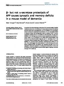
Results Behavior. Figure 4B shows the mean latency to find the hidden platform for the NMDA-lesioned and sham-lesioned groups over blocks of eight trials. Both groups improved in their ability to find the submerged platform with daily practice sessions. A two-variable repeated measures ANOVA (Trial Block X Group) revealed a significant main effect for group, F(l, 11) = 7.48, p = .02, indicating that the rats with lesions to the dorsal hippocampus took significantly longer than the sham-lesioned rats to find the submerged platform. The effect of trial block and the Trial Block X Group interaction were both nonsignificant (F < 1). Figure 4A shows the mean path length to reach the submerged platform for both groups over blocks of eight trials. The rats in the NMDA-lesioned group had longer path lengths than the rats in the sham-lesioned group. A two-variable repeated measures ANOVA (Trial Block X Group) revealed a significant main effect for group, F(l, 11) = 17.32,p = .002, indicating that the lesioned rats traveled significantly farther in the pool to find the submerged platform than did the sham-lesioned rats. The main effect for trial block and the Group X Trial Block interaction were both nonsignificant (F < 1). Histology. Histology preparation and estimation of the neurotoxic lesions were identical to that of Experiment 1. Figure 5 shows the extent of the neurotoxic lesions at five septotemporal levels of the hippocampus. All 7 of the NMDA-lesioned rats sustained partial lesions to the dorsal hippocampus. The lesion included most of the CA1 across the dorsal hippocampus and was most severe in the septal pole. Figure 6 shows a photomicrograph of an NMDA lesion restricted to the CA1 subfield. All lesions, however, included some additional but inconsistent damage to the CA2, CAS, CA4, and dentate gyrus, predominantly at the more septal levels. Some incidental unilateral damage to the anterior parietal cortex also occurred as a result of the injection cannulas and residual NMDA. Damage to the CA2, CA3, CA4, and the anterior parietal cortex was less than in Experiment 1. Sham-lesioned rats had minor damage to the hippocampus and parietal cortex as a result of cannula implantation.
DISRUPTION OF SPATIAL BUT NOT OBJECT MEMORY
1191
1200
fpOOO£ 800
-2.56
CO
g 600 £ 400This document is copyrighted by the American Psychological Association or one of its allied publishers. This article is intended solely for the personal use of the individual user and is not to be disseminated broadly.
S. 200
-3.14
0 0
2
B
4 6 Trial Block -3.80
80 70-
o c CD
-4.52
50 40 30
-5.20
20 10 0
0
2
4 6 Trial Block
8
Figure 4. A: Mean path length for the same rats to find the submerged platform over blocks of eight trials. B: Mean latency for Af-methyl-D-aspartate-lesioned rats (n = 7) and sham-lesioned rats (n = 6) to find a submerged platform over blocks of eight trials. Error bars represent standard errors of the mean.
Figure 5. The extent of the neurotoxic lesions at five septotemporal levels of the hippocampus for rats in Experiments 2 and 3. Hatched areas indicate the size of the smallest lesion on the specific section indicated, and the solid black areas represent the largest lesions that extend beyond the shaded regions.
Discussion Partial NMDA-induced lesions of the dorsal hippocampus produced an acquisition deficit on a spatial version of the Morris water maze. Lesioned rats took longer to find the submerged platform and exhibited more random searching than did the sham-lesioned animals. These findings are in accordance with other reports of spatial memory deficits in rats after partial hippocampal lesions (Gayoso et al., 1994; Handlemann & Olton, 1981; Moser et al., 1993; Nunn & Hodges, 1994; Ylinen et al., 1991). Our findings are particularly relevant to the spatial memory impairments that have been attributed to ischemiainduced CA1 lesions. The NMDA lesions in this experiment produced a water-maze impairment and widespread cell loss in the dorsal-septal CA1, similar to that found after cerebral ischemia (Kiyota et al., 1991; Nunn et al., 1994; Olsen et al., 1994). Additional but inconsistent damage also was present
Figure 6. Photomicrograph of a cresyl-violet-stained section of the dorsal hippocampus in a rat with an W-methyl-D-aspartate lesion restricted primarily to the CA1.
This document is copyrighted by the American Psychological Association or one of its allied publishers. This article is intended solely for the personal use of the individual user and is not to be disseminated broadly.
1192
DUVA ET AL.
in the CA2, CAS, CA4, and dentate gyms, also at the more septal levels. The possibility that some of this damage contributed to the water-maze impairments cannot be ruled out. However, cell loss in some these regions also has been reported after cerebral ischemia in both rats (SchmidtKastner & Freund, 1991; Wood et al, 1993) and primates (Bachevalier & Mishkin, 1989), but their relationship to memory impairments has received little attention. Damage to the posterior partietal cortex has also been shown to effect water-maze performance (Kolb, Buhrman, MacDonald, & Sutherland, 1994). However, in this study, parietal cortex damage occurred primarily in the anterior parietal cortex and was minor compared with lesions that result in spatial memory deficits. Partial hippocampal lesions may impair spatial memory by disrupting the functional integrity of the hippocampus, thereby simulating a complete lesion. According to this view, damage to any of the different hippocampal subfields would be expected to produce a comparable memory impairment. Conversely, each subfield may make a unique contribution to spatial memory such that lesions of the various subfields, separately or in combination, may produce qualitatively different behavioral effects (Nunn & Hodges, 1994), effects that may be revealed only through the use of sophisticated test batteries. The impairments on the Morris water maze resulting from partial hippocampal lesions contrast with the lack of an impairment on DNMS in the same group of animals. This finding is consistent with other recent reports demonstrating a dissociation between the neural substrates underlying spatial and object-recognition memory (Aggleton, Warburton, & Nakamura, 1996; Glenn & Mumby, 1996). The implications of these findings for the role of the hippocampus in memory are addressed next.
General Discussion Compared with the effects of hippocampectomy, the effects of cerebral ischemia are paradoxical: Bilateral hippocampal ablation produced little or no deficits on DNMS performance (Alvarez et al., 1995; Mumby et al., 1995, 1996, 1992; Murray & Mishkin, 1996), whereas cerebral ischemia, which purportedly produces selective CA1 damage, produced a severe impairment ('Bachevalier & Mishkin, 1989; Mumby et al., 1996; Wood et al., 1993). Bachevalier and Mishkin (1989) proposed two hypotheses to account for this paradox: the hippocampal-interference hypothesis and the extrahippocampal-damage hypothesis. According to the hippocampal-interference hypothesis, cerebral ischemia produces a greater disruption of DNMS than bilateral ablation, not because the hippocampus is critical for DNMS performance but because partial hippocampal lesions cause a functional disorganization of the remaining hippocampus, which in turn interferes with afferent structures that subserve object-recognition memory. By contrast, the extrahippocampal-damage hypothesis attributes the greater memory impairments found in ischemic animals to extrahippocampal neuropathology that is not detectable with conventional histological procedures.
The main purpose of this research was to determine whether partial neurotoxic lesions of the dorsal hippocampus can produce deficits on DNMS. The use of a neurotoxin to produce dosal hippocampal lesions reduced or eliminated the possibility of damaging extrahippocampal structures involved in object-recognition memory. Rats in Experiment 1 that received preoperative training and were then given NMDA lesions were not impaired on the reacquisition of the nonmatching rule or at the performance of the DNMS task at delays of 4, 60, 120, and 300 s. Similarly, in Experiment 2, naive rats with NMDA lesions learned the nonmatching rule at a normal rate and were unimpaired on DNMS performance at any retention interval. These results are consistent with previous studies in rats in which bilateral hippocampectomies were found to have little or no effect on DNMS performance (Aggleton et al., 1986; Mumby et al., 1995, 1996,1992; Rothblat & Kromer, 1991). These findings, however, are in marked contrast to the severe DNMS impairments that have been attributed to partial hippocampal damage induced by ischemia, despite the fact that our NMDA lesions produced widespread cell loss in much of the same area, the dorsal CA1, that is damaged as a result of ischemia. The diverse and widespread connections along both the septotemporal and transverse axes of the hippocampus (Amaral & Witter, 1995) suggest that the extent of the lesions could reasonably be considered to produce some type of functional disorganization of the remaining hippocampus. Therefore, these data do not support the hippocampal-interference hypothesis (Bachevalier & Mishkin, 1989) and suggest that partial damage to the dorsal hippocampus in ischemic animals is not critical to their DNMS impairment. Mumby et al. (1996) reached a similar conclusion when it was found that removal of the functionally compromised hippocampus did not attenuate existing DNMS impairments in ischemic rats. It is possible that the NMDA lesions in Experiments 1 and 2 failed to produce a DNMS deficit because the pattern of brain damage produced by the neurotoxin was not identical to that produced by ischemia. Three aspects of this research address this concern. First, there was some variance in the size of the NMDA lesions: Some lesions were primarily restricted to the CA1, whereas others included portions of the CA2, CA3, CA4, and the dentate gyrus. The important point is that despite this variance, which may be thought to produce different patterns of functional disorganization, the variance in the behavioral data was negligible: No subject displayed any deficits in DNMS. Second, lesioned rats were tested both with and without preoperative training: In neither case did the NMDA lesion produce a detectable deficit. Third, although the NMDA lesions of the dorsal hippocampus had no effect on DNMS performance in Experiment 2, in Experiment 3 they did significantly disrupt performance on the water maze. This demonstrates that the lesions were extensive enough to produce an impairment in a spatial task known to be sensitive to partial hippocampal damage (Moser et al., 1993; Nunn & Hodges, 1994) while leaving nonspatial object-recognition memory intact. Our results also are similar to those found after partial, nonischemic hippocampal lesions in monkeys. Ibotenic acid
This document is copyrighted by the American Psychological Association or one of its allied publishers. This article is intended solely for the personal use of the individual user and is not to be disseminated broadly.
DISRUPTION OF SPATIAL BUT NOT OBJECT MEMORY
lesions that destroyed 85% of the monkey hippocampus and amygdala produced no impairments in postsurgical DNMS reacquisition or performance (O'Boyle, Murray, & Mishkin, 1993). Monkeys with electrolytic lesions that damaged 38-75% of the hippocampus were able to learn the DNMS task at a normal rate and performed normally at delays up to 120 s (Alvarez et al., 1995). They displayed a deficit at only the longest delays (10 and 40 min), and their removal from the testing environment during these delays may have produced possible confounds. Murray and Mishkin (1996) found no deficits even at delays of 40 min after excitotoxic hippocampal lesions in monkeys if they were left in the testing apparatus. Taken in combination, there are now considerable data indicating that partial hippocampal lesions in monkeys and rats, when produced by methods other than ischemia, have little effect on DNMS performance. This is indirect but strong evidence that the disruptive effects of cerebral ischemia on DNMS is a consequence of extrahippocampal neuropathology. Four pieces of behavioral evidence suggest that extrahippocampal neuropathology plays a significant role in ischemiainduced memory impairments: (a) the findings of our research, which demonstrate that partial hippocampal lesions designed to mimic the damage produced by ischemia in amnesic rats did not disrupt DNMS performance; (b) the demonstration by Mumby et al. (1996) that ablating a hippocampus suspected of producing interference and concomitant DNMS impairments did not attenuate these impairments; (c) the lack of a strong correlation between CA1 cell loss and spatial memory deficits (Nunn & Hodges, 1994; Olsen et al., 1994); and (d) the observation that animals with neurotoxic lesions of the dorsal hippocampus and CA1 had a different profile of memory impairments in the Morris water maze than did rats with ischemia-induced hippocampal damage (Nunn & Hodges, 1994). Because a growing body of evidence now indicates that ischemia is causing amnesia due to pathological changes outside the hippocampus, it is critical to determine where these changes are and by what mechanisms they might be produced. Because the prefrontal cortex (Bachevalier & Mishkin, 1986; Otto & Eichenbaum, 1992), the basal forebrain (Aigner et al., 1986; Kornecook, Kippin, Pinel, & Wood, 1993), the rhinal cortex (Meunier et al., 1993; Mumby & Pinel, 1994), and the medial thalamus (Mumby, Pinel, & Dastur, 1993; Zola-Morgan & Squire, 1985a) have all been implicated in DNMS, ischemia-produced neuropathology in any of these areas could be mediating the amnesic effects of cerebral ischemia. Indeed, recent neuroanatomical studies using the sensitive Fink and Heimer (1967) silver impregnation technique have documented the existence of ischemia-produced cell loss in many of these areas (Grain, Westerkam, Harrison, & Nadler, 1988; Freund, Buzsaki, Leon, & Somogyi, 1990; Nunn & Jarrard, 1994). Of course, the object-recognition deficits produced by cerebral ischemia may be the consequence of pathological functional changes rather than cell loss. Cerebral ischemia can cause widespread functional changes in the hippocampus (Schmidt-Kastner & Freund, 1991), but few researchers have examined extrahippocampal functional changes after
1193
ischemia. Those who have found that ischemia can result in long-lasting functional alterations throughout the brain. For example, Dietrich, Ginsberg, and Busto (1986) found changes in metabolic responsiveness in a somatosensory circuit of rats that were subjected to 30 min of ischemia. Additionally, Peruche, Klaassens, and Kreiglstein (1995) found a decrease in the amplitude of electrocorticograms of rats that had been subjected to 10 min of ischemia. One study (Ishimaru, Takahashi, Ikarashi, & Maruyama, 1995), which examined only the hippocampus, may be relevant when evaluating the possibility of functional changes in the absence of observable neuropathology. Gerbils were given pentobarbital as a neuroprotective agent prior to ischemia. Although this treatment prevented hippocampal CA1 cell loss completely, additional assays showed that hippocampal acetylcholine levels were reduced, as was the release of acetylcholine. Thus, there were ischemia-induced changes in neurotransmitter levels in the hippocampus with no observable neuropathology. In accordance with this, water-maze deficits after 2VO ischemia have been reported in rats with no detectable CA1 cell loss (Jaspers, Block, Heim, & Sontag, 1990). Wherever the putative extrahippocampal neuropathology hypothesized to produce DNMS deficit may be occurring, it appears to be mediated by the postischemic hippocampus, as hippocampectomy before or immediately after ischemia blocks the development of DNMS impairments (Mumby et al., 1996). One process occurring in the postischemic brain that may produce widespread neuropathology is seizure activity. The temporal lobe, particularly the amygdala and hippocampus, can become focal points for seizures after a variety of neurological insults (Gale, 1992; McEwen, 1994). Prolonged seizure activity after ischemia may occur in the hippocampus and produce damage in afferent structures that receive excitatory projections. Postischemic seizure activity may offer an explanation as to why complete hippocampal ablations prevent ischemia-induced DNMS deficits (Mumby et al., 1996). Hippocampectomy before or immediately after ischemia may significantly reduce postischemic seizures and thereby prevent extrahippocampal neuropathology and concomitant memory deficits. Importantly, ischemic rats that do not display overt seizures do not develop significant DNMS deficits (Yates, Schattman, & Mumby, 1996). Although there are numerous detailed theories about the mnemonic functions of the hippocampus (Eichenbaum, Otto, & Cohen, 1994; Hirsch, 1974; Mishkin, Malamut, & Bachevalier, 1984; O'Keefe, & Nadel, 1978; Olton et al., 1979; Rawlins, 1985; Sutherland & Rudy, 1989; Worden, 1992), the broader question of whether the hippocampus is a general memory structure or one with a more specialized function will be addressed here. Onfe theory (Squire, 1992; Squire & Zola-Morgan, 1991; Zola-Morgan et al., 1994) posits that the hippocampus (and related structures) is the critical brain region mediating declarative memory (i.e., consciously accessible memory for information such as facts, events, and relations among stimuli). In animals, object-recognition memory tasks are believed to tap into mnemonic processes analogous to human declarative memory, an idea supported by the fact that amnesic humans
This document is copyrighted by the American Psychological Association or one of its allied publishers. This article is intended solely for the personal use of the individual user and is not to be disseminated broadly.
1194
DUVA ET AL.
perform poorly on DNMS (Squire et al., 1988). Because the hippocampus no longer appears to be involved critically object-recognition memory, recent studies with animals, including our experiments, suggest that it can no longer be considered a general "declarative memory structure," such that damage to it would produce a universal impairment in all types of declarative memory (Squire, 1992; Squire & Zola-Morgan, 1991; Zola-Morgan etal., 1994). Recent evidence is consistent with a more specialized role for the hippocampus in memory. One aspect of hippocampal function that has received the greatest amount of support is its role in spatial memory. Hippocampal involvement in spatial memory is well documented in rats (Jarrard, 1993; O'Keefe & Nadel, 1978), but similar results have also been found in monkeys (Parkinson, Murray, & Mishkin, 1988) and humans (Cave & Squire, 1991). Nadel (1991) suggested that the hippocampus may act as a "spatial module" in terms of memory processing. Our findings are in agreement with a specialized role for the hippocampus in memory. Partial hippocampal lesions produced a deficit in a spatial memory task (i.e., the Morris water maze) while leaving other forms of declarative memory unaffected (i.e., performance on DNMS). The hippocampus also may be involved in the long-term retention of information. Rawlins (1985) suggested that the hippocampus acts as a temporary memory store or buffer. This idea is consistent with the DNMS impairments seen at long delays in monkeys (Alvarez et al., 1995) and rats (Mumby et al., 1992) and with the retrograde amnesia attributed to hippocampal damage (Squire, 1992). Moreover, Vnek and Rothblat (1996) found long-term deficits in the retention of three concurrent object discriminations in rats with lesions to the dorsal hippocampus. Initial levels of performance between lesioned and control groups were equal; however, when tested 3 weeks later, the rats with hippocampal lesions were impaired on the retention of the object-discrimination problems. The normal initial performance in combination with the impairments after a delay of 3 weeks led us to conclude that the hippocampus may be involved in the retention of information for weeks to possibly years. Murray (1996) stated that the MTL can no longer be viewed as a single functional unit because damage to discrete regions leads to qualitatively different types of mnemonic impairments. Thus, the hippocampus can be seen as being one component of a larger MTL memory system, a component that most likely performs a limited subset of "declarative type" functions (Nadel, 1992). Structures in the MTL themselves, however, appear to be part of a larger memory system (Mishkin & Murray, 1994) comprising diverse brain regions such as the diencephalon, prefrontal cortex, and basal forebrain. Identification of the critical brain structures responsible for ischemia-induced object-recognition deficits may provide important clues about the true nature of this extensive memory system.
References Aggleton, J. P. (1985). One-trial object recognition by rats. Quarterly Journal of Experimental Psychology, 37, 279-294.
Aggleton, J. P., Hunt, P. R., & Rawlins, J. N. P. (1986). The effects of hippocampal lesions upon spatial and non-spatial tests of working memory. Behavioural Brain Research, 19, 133-146. Aggleton, J. P., Warburton, E. C., & Nakamura, R. K. (1996). Further evidence of a dissociation between the neural systems underlying object recognition and spatial memory in the rat. Society for Neuroscience Abstracts, 22, 1119. Aigner, T. G., Mitchell, S. J., Aggleton, J. P., Belong, M. R., Struble, R. G., Price, D. L., Wenk, G. L., Pettigrew, K. D., & Mishkin, M. (1991). Transient impairment of recognition memory following ibotenic-acid lesions of the basal forebrain in macaques. Experimental Brain Research, 86, 18-26. Alvarez, P., Zola-Morgan, S., & Squire, L. R. (1995). Damage limited to the hippocampal region produces long-lasting memory impairment in monkeys. Journal of Neuroscience, 15, 37963807. Amaral, D. G., & Witter, M. P. (1995). Hippocampal formation. In G. Paxinos (Ed.), The rat nervous system (pp. 443—493). San Diego, CA: Academic Press. Auer, R. N., Jensen, M. L., & Whishaw, I. Q. (1989). Neurobehavioral deficit due to ischemic brain damage limited to half of the CA1 sector of the hippocampus. Journal of Neuroscience, 9, 1641-1647. Bachevalier, J., & Mishkin, M. (1986). Visual recognition impairment follows ventromedial but not dorsolateral prefrontal lesions in monkeys. Behavioural Brain Research, 20, 249-261. Bachevalier, J., & Mishkin, M. (1989). Mnemonic and neuropathological effects of occluding the posterior cerebral artery in Macaca mullata. Neuropsychologia, 27, 83-105. Cave, C. B., & Squire, L. R. (1991). Equivalent impairment of spatial and nonspatial memory following damage to the human hippocampus. Hippocampus, 1, 329-340. Conrad, C. D., & Roy, D. J. (1993). Selective loss of hippocampal granule cells following adrenelectomy: Implications for spatial memory. Journal of Neuroscience, 13, 2582-2590. Crain, B. J., Westerkam, W. D., Harrison, A. H., & Nadler, J. V. (1988). Selective neuronal death after transient forebrain ischemia in the Mongolian gerbil: A silver impregnation study. Neuroscience, 27, 387-402. Dietrich, W. D., Ginsberg, M. D., & Busto, R. (1986). Effect of transient cerebral ischemia on metabolic activation of a somatosensory circuit. Journal of Cerebral Blood Flow and Metabolism, 6, 405-413. Eichenbaum, H., Otto, T., & Cohen, N. J. (1994). Two functional components of the hippocampal memory system. Behavioral and Brain Sciences, 17, 449-518. Fink, R. P., & Heimer, L. (1967). Two methods for selective silver impregnation of degenerating axons and their synaptic endings in the central nervous system. Brain Research, 4, 369-374. Freund, T. F., Buzsaki, G., Leon, A., & Somogyi, P. (1990). Hippocampal cell death following ischemia: Effects of brain temperature and anesthesia. Experimental Neurology, 108, 251260. Gale, K. (1992). Subcortical structures and pathways involved in convulsive seizure generation. Journal of Clinical Neurophysiology, 9, 264-277. Gayoso, M. J., Primo, C., Al-Majdalawi, A., Fernandez, J. M., Garrosa, M., & Iniguez, C. (1994). Brain lesions and water-maze learning deficits after systemic administration of kainic acid to adult rats. Brain Research, 653, 92-100. Glenn, M. J., & Mumby, D. G. (1996). Place- and objectrecognition deficits following lesions of the hippocampus or perirhinal cortex in rats: A double dissociation. Society for Neuroscience Abstracts, 22, 1120. Hagan, J. J., & Beaughard, M. (1990). The effects of forebrain
This document is copyrighted by the American Psychological Association or one of its allied publishers. This article is intended solely for the personal use of the individual user and is not to be disseminated broadly.
DISRUPTION OF SPATIAL BUT NOT OBJECT MEMORY ischemia on spatial learning. Behavioural Brain Research, 41, 151-160. Handelmann, G. E., & Olton, D. S. (1981). Spatial memory following damage to hippocampal CAS pyramidal cells with kainic acid: Impairment and recovery with preoperative training. Brain Research, 217, 41-58. Hirsch, R. (1974). The hippocampus and contextual retrieval of information from memory: A theory. Behavioral Biology, 12, 421-444. Ishimaru, H., Takahashi, A., Dcarashi, Y., & Maruyuma, Y. (1995). Pentobarbitol protects against CA1 pyramidal cell death but not dysfunction of hippocampal cholinergic neurons following transient ischemia. Brain Research, 673,112-118. Jarrard, L. E. (1993). On the role of the hippocampus in learning and memory in the rat. Behavioral and Neural Biology, 60, 9-26. Jaspers, R. M. A., Block, R, Heim, C., & Sontag, K.-H. (1990). Spatial learning is affected by transient occlusion of common carotid arteries (2VO): Comparison of behavioural and histopathological changes after "2VO" and "four-vessel" occlusion in rats. Neuroscience Letters, 117, 149-153. Kartsounis, L. D., Rudge, P., & Stevens, J. M. (1995). Bilateral lesions of the CA1 and CA2 fields of the hippocampus are sufficient to cause a severe amnesic syndrome in humans. Journal of Neurology, Neurosurgery, and Psychiatry, 59, 95-98. Kiyota, Y, Miyamato, M., & Nagaoka, A. (1991). Relationship between brain damage and memory impairments in rats exposed to transient forebrain ischemia. Brain Research, 528, 295-302. Kolb, B., Buhrman, K., MacDonald R., & Sutherland, R. J. (1994). Dissociation of the medial prefrontal, posterior parietal, and posterior temporal cortex for spatial navigation and recognition memory in the rat. Cerebral Cortex, 4, 664-680. Kornecook, T. J., Kippin, T. E., Pinel, J. P. J., & Wood, E. R. (1993). Impairment of reacquisition and performance of delayed nonmatching-to-sample following lesions to the medial septum and diagonal band in rats. Society for Neuroscience Abstracts, 19, 1233. Kornecook, T. J., Lui, M., Duva, C. A., Anzarut, A., & Pinel, J. P. J. (1995). Effects of perirhinal or rhinal cortex lesions on objectmemory tasks in the rat. Society for Neuroscience Abstracts, 21, 1935. Mahut, H., Zola-Morgan, S., & Moss, M. (1982). Hippocampal resections impair associative learning and memory in the monkey. Journal of Neuroscience, 2, 1214-1229. McEwen, B. S. (1994). The plasticity of the hippocampus is the reason for its vulnerability. Seminars in the Neurosciences, 6, 239-246. Meunier, M., Bachevalier, J., Mishkin, M., & Murray, E. A. (1993). Effects on visual recognition of combined and separate ablations of the entorhinal and perirhinal cortex in rhesus monkeys. Journal of Neuroscience, 13, 5418-5432. Mishkin, M. (1978). Memory in monkeys severely impaired by combined but not by separate removals of amygdala and hippocampus. Nature, 273, 297-298. Mishkin, M., Malamut, B., & Bachevalier, J. (1984). Memories and habits: Two neural systems. In G. Lynch, J. L. McGaugh, & N. M. Weinberger (Eds.), Neurobiology of learning and memory (pp. 65-77). New York: Guilford Press. Mishkin, M., & Murray, E. A. (1994). Stimulus recognition. Current Opinions in Neurobiology, 4, 200-206. Morris, R. G. M., Garrud, P., Rawlins, J. N. P., & O'Keefe, J. (1982). Place navigation impaired in rats with hippocampal lesions. Nature, 297, 681-683. Moser, E., Moser, M., & Andersen, P. (1993). Spatial learning impairment parallels the magnitude of dorsal hippocampal
1195
lesions, but is hardly present following ventral lesions. Journal of Neuroscience, 13, 3916-3925. Mumby, D. G., & Pinel, J. P. J. (1994). Rhinal cortex lesions and object recognition in rats. Behavioral Neuroscience, 108, 11-18. Mumby, D. G., Pinel, J. P. J., & Dastur, F. N. (1993). Mediodorsal thalamic lesions and object recognition in rats. Psychobiology, 21, 27-36. Mumby, D. G., Pinel, J. P. J., Kornecook, T. J., Shen, M. J., & Redila, V. A. (1995). Memory deficits following lesions of the hippocampus or amygdala in rat: Assessment by an objectmemory test battery. Psychobiology, 23, 26-36. Mumby, D. G., Pinel, J. P. J., & Wood, E. R. (1990). Nonrecurringitems delayed nonmatching-to-sample in rats: A new paradigm for testing nonspatial working memory. Psychobiology, 18, 321-326. Mumby, D. G., Wood, E. R., Duva, C. A. Kornecook, T. J., Pinel, J. P. J., & Phillips, A. G. (1996). Ischemia-induced objectrecognition deficts in rats are attenuated by hippocampal ablation before or soon after ischemia. Behavioral Neuroscience, 110, 266-281. Mumby, D. G., Wood, E. R., & Pinel, J. P. J. (1992). Objectrecognition memory is only mildly impaired in rats with lesions of the hippocampus and amygdala. Psychobiology, 20, 18-27. Murray, E. A. (1992). Medial temporal lobe structures contributing to recognition memory: The amygdaloid complex versus the rhinal cortex. In J. P. Aggleton (Ed.), The amygdala: Neurobiological aspects of emotion, memory, and mental dysfunction (pp. 453^70). London: Wiley-Liss. Murray, E. A. (1996). What have ablation studies told us about the neural substrates of stimulus memory? Seminars in the Neurosciences, 8, 13-22. Murray, E. A., & Mishkin, M. (1984). Severe tactual as well as visual memory deficits follow combined removal of the amygdala and hippocampus in monkeys. Journal of Neuroscience, 4, 2565-2580. Murray, E. A., & Mishkin, M. (1996). 40-minute visual recognition memory in rhesus monkeys with hippocampal lesions. Society for Neuroscience Abstracts, 22, 281. Nadel, L. (1991). The hippocampus and space revisited. Hippocampus, 1, 221-229. Nadel, L. (1992). Multiple memory systems: What and why. Journal of Cognitive Neuroscience, 4, 179-188. Nunn, J., & Hodges, H. (1994). Cognitive deficits induced by global cerebral ischemia: Relationship to brain damage and reversal by transplants. Behavioural Brain Research, 65, 1-31. Nunn, J. A., & Jarrard, L. E. (1994). Silver impregnation reveals neuronal damage in cingulate cortex following 4 VO ischemia in the rat. NeuroReport, 5, 2363-2366. Nunn, J. A., LePeillet, E., Netto, C. A., Hodges, H., Gray, J. A., & Meldrum, B. S. (1994). Global ischemia: Hippocampal pathology and spatial deficits in the water maze. Behavioural Brain Research, 62, 41-54. O'Boyle, V. J., Murray, E. A., & Mishkin, M. (1993). Effects of excitotoxic amygdalo-hippocampal lesions on visual recognition in rhesus monkeys. Society for Neuroscience Abstracts, 19, 438. O'Keefe, J., & Nadel, L. (1978). The hippocampus as a cognitive map. London: Oxford University Press. Olsen, G. M., Scheel-Kruger, J., Moller, A., & Jensen, L. H. (1994). Relation of spatial learning of rats in the Morris water maze task to the number of viable CA1 neurons following four-vessel occlusion. Behavioral Neuroscience, 108, 681-690. Olton, D. S., Becker, J. T., & Handelmann, G. E. (1979). Hippocampus, space, and memory. Behavioral and Brain Sciences, 2, 313-365. Otto, T., & Eichenbaum, H. (1992). Complimentary roles of the
This document is copyrighted by the American Psychological Association or one of its allied publishers. This article is intended solely for the personal use of the individual user and is not to be disseminated broadly.
1196
DUVA ET AL.
orbital prefrontal cortex and the perirhinal-entorhinal cortices in an odor-guided delayed-nonmatching-to-sample task. Behavioral Neuroscience, 106, 762-775. Parkinson, J. K., Murray, E. A., & Mishkin, M. (1988). A selective mnemonic role for the hippocampus in monkeys: Memory for the location of objects. Journal of Neuroscience, 8, 4159^167. Paxinos, G., & Watson, C. (1986). The rat brain in stereotaxic coordinates (2nd ed.). San Diego, CA: Academic Press. Peruche, B., Klaassens, H., & Krieglstein, J. (1995). Quantitative analysis of the electrocorticogram after forebrain ischemia in the rat. Pharmacology, 50, 229-237. Rawlins, J. N. P. (1985). Associations across time: The hippocampus as a temporary memory store. Behavioral and Brain Sciences, 8, 479^96. Rod, M. R., Whishaw, I. Q., & Auer, R. N. (1990). The relationship of structural ischernic brain damage to neurobehavioural deficit: The effect of postischemic MK-801. Canadian Journal of Psychology, 44, 196-209. Rothblat, L. A., & Hayes, L. L. (1987). Short-term object recognition memory in the rat: Nonmatching with trial-unique stimuli. Behavioral Neuroscience, 101, 587-590. Rothblat, L. A., & Kromer, L. F. (1991). Object-recognition memory in the rat: The role of the hippocampus. Behavioural Brain Research, 42, 25-32. Saunders, R. C., Murray, E. A., & Mishkin, M. (1984). Further evidence that amygdala and hippocampus contribute equally to recognition memory. Neuropsychologia, 22, 785-796. Schmidt-Kastner, R., & Freund, T. F. (1991). Selective vulnerability of the hippocampus in brain ischemia. Neuroscience, 40, 599-636. Scoville, W. B., & Milner, B. (1957). Loss of recent memory after bilateral hippocampal lesions. Journal of Neurology, Neurosurgery, and Psychiatry, 20, 11-21. Smith, M. L., Bendek, G., Dahlgren, N., Rosen, L, Wieloch, T, & Seisjo, B. K. (1984). Models for studying long term recovery following forebrain ischemia in the rat: 2. A 2-vessel occlusion model. Acta Neurologica Scandinavica, 69, 385-401. Squire, L. R. (1992). Memory and the hippocampus: A synthesis from findings with rats, monkeys, and humans. Psychological Review, 99, 195-231. Squire, L. R., & Cohen, N. J. (1984). Human memory and amnesia. In G. L. Lynch, J. L. McGaugh, & N. M. Weinberger (Eds.), Neurobiology of learning and memory (pp. 3-64). New York: Guilford Press. Squire, L. R., & Zola-Morgan, S. (1991). The medial temporal lobe memory system. Science, 253, 1380-1386. Squire, L. R., Zola-Morgan, S., & Chen, K. (1988). Human amnesia and animal models of amnesia: Performance of amnesic patients on tests designed for the monkey. Behavioral Neuroscience, 11, 210-221. Sugaimachi, K., Izawa, K., Nakamura, K., Kimura-Iwasaki, K., Yamaguchi, M., Nakagawa, H., & Oshino, N. (1992). Impairment of working memory by neuronal degeneration with NMDA in rat hippocampal CA-1. Behavioral Pharmacology, 3, 379385. Sutherland, R. W, Kolb, B. E., & Wishaw, I. Q. (1982). Spatial mapping: Definitive disruption by hippocampal or medial frontal cortical damage. Neuroscience Letters, 31, 271-276. Sutherland, R. W, & Rudy, J. W. (1989). Configural association theory: The role of the hippocampal formation in learning, memory, and amnesia. Psychobiology, 17, 129-144. Vnek, N., & Rothblat, L. A. (1996). The hippocampus and long-term object memory in the rat. Journal of Neuroscience, 16, 2780-2787. Volpe, B. T, Davis, H. P., Towle, A., & Dunlap, W. P. (1992). Loss
of hippocampal CA1 pyramidal neurons correlates with memory impairment in rats with ischernic or neurotoxin lesions. Behavioral Neuroscience, 106, 457-464. Volpe, B. T, & Hirst, W. (1983). The characterization of the amnesic syndrome after hypoxic ischemic injury. Archives of Neurology, 40, 436-440. Volpe, B. T., Pulsinelli, W. A., Tribuna, J., & Davis, H. P. (1984). Behavioral performance of rats following transient forebrain ischemia. Stroke, 15, 558-562. Whishaw, I. Q., Rod, M. R., & Auer, R. N. (1994). Behavioral deficits revealed by multiple tests in rats with ischemic damage limited to half of the CA1 sector of the hippocampus. Brain Research Bulletin, 34, 283-289. Wood, E. R., Mumby, D. G., Pinel, J. P. J., & Phillips, A. G. (1993). Impaired object-recognition memory in rats following ischemiainduced damage in the hippocampus. Behavioral Neuroscience, 107, 51-62. Worden, R. P. (1992). Navigation by fragment fitting: A theory of hippocampal function. Hippocampus, 2, 165-187. Yates, J., Schattman, L., & Mumby, D. G. (1996). Hippocampal damage does not impair object recognition in rats with no presurgery training. Society for Neuroscience Abstracts, 22, 1119. Ylinen, A., Lahtinen, H., Sirvio, J., Partanen, J., Asikainen, A., Gulyas, A., Freund, T. F, & Riekkinen, P. (1991). Behavioural, electrophysiological and histopathological changes following sustained stimulation of the perforant pathway input to the hippocampus: Effect of the NMDA receptor antagonist, CGP 39551. Brain Research, 553, 195-200. Zola-Morgan, S., & Squire, L. R. (1985a). Amnesia in monkeys following lesions of the mediodorsal nucleus of the thalamus. Annals of Neurology, 17, 558-564. Zola-Morgan, S., & Squire, L. R. (1985b). Medial temporal lesions in monkeys impair memory on a variety of tasks sensitive to human amnesia. Behavioral Neuroscience, 99, 22-34. Zola-Morgan, S., & Squire, L. R. (1986). Memory impairment in monkeys following lesions limited to the hippocampus. Behavioral Neuroscience, 100, 155-160. Zola-Morgan, S., Squire, L. R., & Amaral, D. G. (1986). Human amnesia and the medial temporal region: Enduring memory impairment following a bilateral lesion limited to the field CA1 of the hippocampus. Journal of Neuroscience, 6, 2950-2967. Zola-Morgan, S., Squire, L. R., Amaral, D. G., & Suzuki, W. A. (1989). Lesions of the perirhinal and parahippocampal cortex that spare the amygdala and hippocampal formation produce severe memory impairment. Journal of Neuroscience, 9, 43554370. Zola-Morgan, S., Squire, L. R., Clower, R. P., & Rempel, N. L. (1993). Damage to the perirhinal cortex exacerbates memory impairment following lesions to the hippocampal formation. Journal of Neuroscience, 13, 251-265. Zola-Morgan, S., Squire, L. R., & Ramus, S. J. (1994). Severity of memory impairment in monkeys as a function of locus and extent of damage within the medial temporal lobe memory system. Hippocampus, 4, 483^1-95. Zola-Morgan, S., Squire, L. R., Rempel, N. L., Clower, R. P., & Amaral, D. G. (1992). Enduring memory impairment in monkeys after ischemic damage to the hippocampus. Journal of Neuroscience, 12, 2582-2596.
Received April 14,1997 Revision received May 14, 1997 Accepted June 3,1997