Cell Host & Microbe
Primer Dissecting Regulatory Networks in Host-Pathogen Interaction Using ChIP-on-chip Technology Claudia Sala,1 David C. Grainger,2 and Stewart T. Cole1,* 1Global
Health Institute, Ecole Polytechnique Fe´de´rale de Lausanne, Station 15, CH-1015 Lausanne, Switzerland of Biological Sciences, University of Warwick, Coventry CV4 7AL, UK *Correspondence:
[email protected] DOI 10.1016/j.chom.2009.04.007 2Department
Understanding host-microbe interactions has been greatly enhanced by our broadening knowledge of the regulatory mechanisms at the heart of pathogenesis. The ‘‘transcriptomics’’ approach of measuring global gene expression has identified genes involved in bacterial pathogenesis. More recently, chromatin immunoprecipitation (ChIP) and hybridization to microarrays (chIP-on-chip) has emerged as a complementary tool that permits protein-DNA interactions to be studied in vivo. Thus, chIP-on-chip can be used to map the binding sites of transcription factors, thereby teasing apart gene regulatory networks. In this Review, we discuss the ChIP-on-chip technique and focus on its application to the study of host-pathogen interactions. The availability of bacterial genome sequences—including those of all the major pathogens—ushered in the postgenomic era. Today, a major goal in bacterial genomics is to understand the gene regulatory networks of bacterial cells and the response of these networks to environmental signals. Thus, there has been an explosion in high-throughput techniques designed to measure the interplay between transcription factors, DNA- and RNA-associated proteins and RNA polymerase on a global scale. Here we discuss recent technical advances in the development of chromatin immunoprecipitation (ChIP) as a tool for unraveling regulatory networks in bacteria and in the hostpathogen interplay. The development of the DNA microarray was a significant breakthrough in the study of microbial gene regulation; for the first time, researchers were able to quantify each individual transcript in a cell’s total pool of RNA. This ‘‘transcriptomics’’ approach has been widely used to identify genes expressed differentially in response to environmental signals and in mutant strains. Unsurprisingly, this technique found many applications in the study of host-pathogen interactions, and there are several excellent examples from in vitro, ex vivo, and in vivo studies (Grifantini et al., 2002, 2003; Schnappinger et al., 2003; Talaat et al., 2004). However, while transcriptome analysis provides information about the RNA content of the cell, it is unable to reveal the actual regulatory processes that shape gene expression: the precise binding targets of transcription factors. ChIP represents a powerful and complementary tool, since it identifies protein-DNA interactions in vivo (see Table 1 for global comparison of RNA profiling and chIP approaches). Briefly, ChIP involves fixing cells using formaldehyde, thereby crosslinking DNA-binding proteins to the chromosome, followed by cell lysis and shearing of DNA by sonication. The protein of interest is then immunoprecipitated with specific antibodies together with any crosslinked DNA fragments. After reversal of the crosslinks and purification, DNA can be analyzed in order to detect enrichment of the sequences bound by the protein of interest. Thus, using ChIP in conjunction with DNA microarray analysis 430 Cell Host & Microbe 5, May 21, 2009 ª2009 Elsevier Inc.
(ChIP-on-chip) permits DNA binding to be measured on a chromosome-wide scale (Figure 1). ChIP and chIP-on-chip were first developed for eukaryotic cells and have since found application in organisms as diverse as bacteriophages (Gonzalez-Huici et al., 2004), yeast (Harbison et al., 2004; Lee et al., 2002) and mammals (Boyer et al., 2005; Bulyk, 2006; Cawley et al., 2004), where the binding of several transcription factors as well as of chromatin-associated proteins was investigated. Among bacteria, ChIP-on-chip has been applied most to Escherichia coli (Grainger et al., 2004, 2005, 2006, 2007), with a smaller number of studies focused on other bacteria, such as Bacillus subtilis (Ben-Yehuda et al., 2005; Eichenberger et al., 2004; Molle et al., 2003a, 2003b), Helicobacter pylori (Danielli et al., 2006), Caulobacter crescentus (Laub et al., 2002), Mycobacterium bovis BCG (Rodrigue et al., 2007), and Mycobacterium tuberculosis (Sala et al., 2009). Despite already being used widely, the range of ChIP applications continues to expand (see Table 2 for a list of potential uses). For example, recent work has shown that ChIP can be used to study RNA-binding proteins by coupling immunoprecipitation with reverse-transcription PCR analysis. Using this approach, it was shown that the Drosophila melanogaster Ash1 transcription factor binds to three noncoding chromatin-associated regulatory RNAs (Sanchez-Elsner et al., 2006). Advantages of ChIP-on-chip over Traditional Techniques ChIP has many advantages over traditional genetic and biochemical approaches for studying interactions made by gene regulatory proteins. For example, there is no need to construct mutant strains, meaning that essential proteins can be studied and that potential polar effects on gene expression are avoided. Additionally, ChIP experiments are performed under physiological conditions, unlike in vitro biochemical assays of DNA binding. Most importantly, ChIP can be applied in vivo, on a chromosomewide scale. This allows the regulons of transcription factors to be determined directly, avoiding indirect effects on transcription
Cell Host & Microbe
Primer Table 1. RNA Profiling and ChIP-on-chip: A Comparison Transcriptomics
ChIP-on-chip
In vivo technology
In vivo technology
Genome-wide approach
Genome-wide approach
Unable to reveal the precise binding sites for the protein of interest
Identification of the targets for the protein under study
Unable to reveal regulatory cascades
Regulatory cascades can be defined as well as multiprotein complexes
mRNA stability problems
No stability issues
Mutant strains required as comparison
No need to make mutants
No antibody requirement
Need to make antibodies or to construct strains expressing tagged proteins
Various tools available for analysis
Analysis complicated by the paucity of bioinformatic devices, especially for prokaryotes
Set up, well established and widely used
Complex and long; several steps involved
that hinder transcriptomic analyses, such as regulatory cascades, where one regulator controls the expression of many others (see Table 1). Moreover, since ChIP-on-chip can be used to quantify the occupancy of RNA polymerase across the chromosome, the technique represents an alternative to transcriptomics that does not require RNA isolation, thus avoiding potential biases due to differential RNA stability. Designing a ChIP-on-chip Experiment Planning a ChIP-on-chip experiment involves collecting information, when possible, on the protein of interest with respect to its expression profile (i.e., exponential or stationary phase), requirement of cofactors, and need for specific conditions for activity (e.g., low oxygen tension). Then, the appropriate reference sample for normalization should be chosen; this is usually represented by total genomic DNA. Moreover, a number of technical and biological replicates should be planned in order to overcome the background noise that comes within these genome-wide approaches. Finally, there are some technical issues to be addressed, starting from the crosslinking protocol to the choice of the antibody to the appropriate controls to perform in order to estimate the success of the experiment; most of these points are discussed in the following sections. Technical Considerations for ChIP-on-chip Experiments Formaldehyde crosslinking is supposed to proceed through the formation of a Schiff base intermediate between the DNA backbone and formaldehyde, followed by reaction with lysine or arginine side chains (Fujita and Wade, 2004). This first step requires some optimization in terms of time and incubation temperature. In fact, insufficient fixation results in inability to identify protein-DNA interaction and extended crosslinking times may cause overaggregation and unnatural interactions. Sometimes, different crosslinkers have been used: for example,
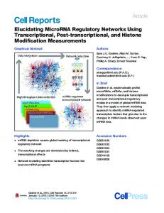
Figure 1. Schematic of the ChIP-on-chip Technology Cells are crosslinked with formaldehyde in order to introduce reversible covalent bonds between proteins and DNA. After cell lysis and shearing of DNA, the protein of interest is immunoprecipitated together with bound DNA fragments, which are then decrosslinked, purified, labeled, and hybridized to microarrays. Finally, after scanning, ChIP-on-chip signals are plotted against genome coordinates to visualize the protein binding sites.
bifunctional imidoester reagents were a good choice in mapping the distribution of chromatin-remodeling complexes (Fujita and Wade, 2004). Fortunately, the requirement of antibodies for performing chIP-on-chip does not necessarily involve expression and purification of the protein under study. Tagged proteins can also be used in ChIP-on-chip assays, thus simplifying the logistics, provided that the tag does not affect protein folding and function, and does not alter the cellular phenotype (i.e., growth rate, colony morphology). Once the protein of interest is tagged and the expressing strain made, immunoprecipitation can be performed with commercial antibodies. For example, a recent study in M. bovis BCG exploited the Myc tag: it was fused to the genes coding for all 13 sigma factors, and ChIP-on-chip was carried out using an anti-tag antibody (Rodrigue et al., 2007). Mooney and colleagues reported the use of the hemagglutinin epitope (HA) as a tag for the NusG regulator in E. coli (Mooney et al., 2009). HA was also used to tag H-NS in Salmonella (Navarre et al., 2006), obtaining results similar to those achieved when using native antibodies (Lucchini et al., 2006). ChIP assays in mammalian and plant cells took advantage of the biotin, FLAG, HA, and Cell Host & Microbe 5, May 21, 2009 ª2009 Elsevier Inc. 431
Cell Host & Microbe
Primer Table 2. Applications of ChIP and ChIP-on-chip Applications
Method
Examples
Identification of the DNA-binding sites for transcription factors
ChIP-on-chip
(Shimada et al., 2008; Wade et al., 2007)
Study chromatin-associated proteins
ChIP-on-chip
(Ben-Yehuda et al., 2005; Breier and Grossman, 2007; Navarre et al., 2006)
Study RNA-binding proteins
ChIP associated with RT-PCR
(Sanchez-Elsner et al., 2006)
Define the role of unknown proteins
ChIP-on-chip
(Sala et al., 2009)
Dissecting dynamics of macromolecular complexes
ChIP-on-chip
(Mooney et al., 2009)
Study response to environmental signals
ChIP-on-chip
(Danielli et al., 2006; Grainger et al., 2004, 2007)
Study response to drug treatment
ChIP-on-chip
(Grainger et al., 2005; Sala et al., 2009)
Analysis of host-pathogen interaction
ChIP associated with PCR
(Chang et al., 2006; Kay et al., 2007; Venza et al., 2008)
V5 (short peptide from simian virus 5) tags (de Folter et al., 2007; Kolodziej et al., 2009). However, whatever the method of immunoprecipitation, complex topologies in protein-DNA or protein-protein interactions may mask the epitope(s), and cross-reactivity with nonspecific targets increases background noise, therefore making the quality of the antibody one of the primary limitations. Thus, some ChIP-on-chip experiments are prone to false negative and false positive measurements (Lee et al., 2006). Controls can be performed before hybridizing immunoprecipitated DNA to microarrays. When possible, quantitative PCR on known binding sites may help in order to evaluate the success of the ChIP protocol. Finally, results obtained by chIP-on-chip are usually supported by traditional techniques. For example, the binding profiles of E. coli RutR and of M. tuberculosis BlaI were validated by electrophoretic mobility shift assays (EMSA) and DNase footprinting, and localization of the operator sequence was further corroborated by 50 -ends mapping (Sala et al., 2009; Shimada et al., 2008). Analyzing ChIP-on-chip Results Analysis of chIP-on-chip data can be challenging, particularly when global DNA-binding proteins are being studied or multiple datasets are compared. The problem is compounded for microbiologists because few custom-designed software packages are available. Currently, ChIP-on-chip data are most frequently visualized by uploading binding profiles into genome browsers, such as Artemis (Rutherford et al., 2000) or the UCSC Genome Browser (http://genome.ucsc.edu), where peaks correspond to binding of the factor under study. However, substantial technical expertise is required to perform screens of binding data that automatically extract protein-binding sites and their genomic context. Statistical analysis may be necessary when large data sets with technical or biological replicates are being examined. For instance, the genome-wide distribution of SpoIIID in B. subtilis was analyzed using the Rosetta Resolver statistical package (Eichenberger et al., 2004). When performing ChIP associated with high-throughput sequencing technologies (ChIPSeq), all the sequence reads have to be mapped to a reference genome. This step can be carried out using algorithms such as BLAT (Kent, 2002) or SSAHA (Ning et al., 2001), specifically designed to hold thousands of short tags. Additional software solutions are represented by MAQ (Li et al., 2008a) and SOAP (Li et al., 2008b), 432 Cell Host & Microbe 5, May 21, 2009 ª2009 Elsevier Inc.
which can improve accuracy in the alignment process. Finally, downstream analysis involves mapping reads to genome browsers and peak identification: FindPeaks (Fejes et al., 2008) and QuEST (Valouev et al., 2008) may be helpful in this last step of the process. Data obtained from either ChIP-on-chip or ChIPSeq experiments may require auxiliary software in order to analyze the immunoprecipitated DNA sequences. This can reveal simple trends, like any bias in GC content of the precipitated DNA, as in the case of H-NS binding to DNA (Grainger et al., 2006; Navarre et al., 2006), or more complicated patterns (for example consensus binding sites for transcription factors can be determined). At present, some algorithms are available for the identification of binding motifs: BioProspector, MDscan, and BioOptimizer (Jensen and Liu, 2004; Liu et al., 2004) have been successfully exploited in several works (Ben-Yehuda et al., 2005; Eichenberger et al., 2004; Wade et al., 2006). ChIP-on-chip in Axenic Cultures Studies performed in E. coli represent the paradigm for ChIP-onchip applications in axenic bacterial cultures (Grainger et al., 2005). The distribution of several transcription factors (MelR, CRP, FNR, RutR, LexA) has been determined, as has the chromosome-wide binding profile of RNA polymerase with various sigma factors (Grainger et al., 2004, 2005, 2007; Shimada et al., 2008; Wade et al., 2005, 2006). Thus, ChIP-on-chip has revealed that some bacterial transcription factors recognize single binding sites (as in the case of MelR) while others have more complex distributions (for example CRP and LexA). These studies have also provided new and deeper insights into gene regulation. For instance, most binding sites for transcription factor RutR were mapped within coding regions, suggesting that it may have some as yet undiscovered function (Shimada et al., 2008). Similarly, 25% of the binding sites for the RNA polymerase s32 subunit were located in genes (Wade et al., 2006). Furthermore, extensive overlap between s32 and s70 regulons was described, accounting for the ability of s32 mutants to transcribe heat-shock genes (Wade et al., 2006). Mooney and coworkers recently highlighted the use of ChIPon-chip to dissect multiprotein transcription complexes in E. coli (Mooney et al., 2009). The distribution of RNA polymerase, s70, NusA, NusG and r throughout the genome was analyzed and revealed close association of the sigma factor with RNA polymerase at promoter regions, whereas r, NusA, and NusG
Cell Host & Microbe
Primer subsequently join the enzyme during transcript elongation (Mooney et al., 2009). The paper constitutes an example of how in vivo crosslinking and hybridization to arrays could be used to explain the intricate dynamics of formation and remodeling of macromolecular complexes. The availability of high-density DNA microarrays for bacteria other than E. coli has facilitated the application of ChIP-on-chip to other bacterial systems, including several pathogens. For instance, the H. pylori Fur protein has been studied and found to bind to about 200 genomic loci in an iron-dependent manner, supporting the idea that this protein acts as a pleiotropic regulator (Danielli et al., 2006). Eichenberger and colleagues provided an extensive analysis of cell differentiation during the sporulation process in B. subtilis (Eichenberger et al., 2004). Their work represents an excellent example of integration of transcriptomics and ChIP-on-chip data as it reports gene expression studies corroborated by the SpoIIID genome-wide regulatory circuit. Surprisingly, SpoIIID was found to bind to some targets where it presumably acts as an architectural protein, in addition to its role as transcriptional regulator (Eichenberger et al., 2004). Therefore, chIP-on-chip assays revealed a new unexpected role for a previously described transcription factor. Another example of combination of expression profiling with ChIP-on-chip data resulted in global analysis of the CtrA regulon in C. crescentus (Laub et al., 2002). This protein is one of the four master regulators of cell-cycle progression (Shen et al., 2008) and was found to directly control at least 55 genes, some of them involved in polar morphogenesis (Laub et al., 2002). The BlaI regulon from M. tuberculosis was recently analyzed (Sala et al., 2009). Studies with this protein were of particular interest because, prior to ChIP-on-chip analysis, the biological role of this protein was unknown. ChIP-on-chip revealed that the M. tuberculosis BlaI regulon comprises five DNA loci including the blaI gene itself and others involved in resistance to b-lactam antibiotics (Sala et al., 2009). Thus, when applied to nonmodel organisms, ChIP-on-chip is an extremely powerful approach to define the role of unknown proteins. Additionally, ChIP-on-chip can reveal unexpected pathways that might shape the drug discovery process. For example, BlaI binds upstream of the operon coding for ATP synthase (Sala et al., 2009), suggesting possible links between cell-wall damage and ATP production. Targeting an important function such as the b-lactaminduced molecular mechanism that controls ATP synthesis opens a new avenue to rational drug design. Response to drug treatment represents an important topic when studying pathogenic bacteria. Different drugs determine different responses in terms of activation or repression of genes. This was demonstrated in M. tuberculosis using the transcriptomics approach (Boshoff et al., 2004): each drug generates a typical transcriptional signature, and related drugs share most of their expression profile. Consequently, knowledge of transcriptional signatures can be used to predict the mechanism of action of new compounds. Importantly, ChIP-on-chip permits changes in DNA binding by transcription factors, induced by environmental stimuli, to be measured. For instance, MelR binding to its target region was shown to occur both in the presence and in the absence of melibiose (Grainger et al., 2004), FNR was found to bind to the operator sites only upon anaerobic conditions (Grainger et al., 2007),
and BlaI was released from its binding sites upon b-lactam treatment (Sala et al., 2009). Furthermore, studies performed using antibodies against RNA polymerase confirmed that ChIP-onchip can be exploited to study the global distribution of the transcriptional machinery in different environmental conditions, including drug treatment (Grainger et al., 2005). For example, the response of E. coli to rifampicin was described in terms of relocalization of RNA polymerase as compared to the mocktreated sample: consistent with the predicted mechanism of action of this drug (Campbell et al., 2001), the enzyme was mainly associated with promoter sequences (Grainger et al., 2005). Response to salicylic acid was also investigated in E. coli by means of ChIP-on-chip: genes encoding stable RNAs and proteins were switched off whereas those required to survive the stress were transcribed (Grainger et al., 2005). As a consequence, ChIP-on-chip may become a new useful tool for predicting the mode of action of new molecules by identifying the different molecular ‘‘signatures’’ generated in terms of binding profiles of a given protein or enzyme. RacA and Spo0J: ChIP-on-chip of Proteins Involved in Chromosome Segregation As outlined in the examples described so far, ChIP-on-chip has been mainly applied to transcription factors and to proteins playing relevant roles in mRNA synthesis. However, the technique can also be useful to study proteins responsible for chromosome partitioning, sporulation, and segregation, as in the case of B. subtilis RacA and Spo0J (Ben-Yehuda et al., 2005; Breier and Grossman, 2007). In particular, Ben-Yehuda and coworkers demonstrated that RacA binds to at least 25 regions spread across the chromosomal origin of replication, thus defining a centromere-like element (Ben-Yehuda et al., 2005). On the other hand, Spo0J binds to 10 parS sites throughout the B. subtilis chromosome and spreads around each one, contributing to chromosome partitioning (Breier and Grossman, 2007). Therefore, ChIP-on-chip can be exploited for examining biological phenomena other than transcription. H-NS Binding to DNA: Xenogeneic Silencing H-NS is a small nucleoid-associated protein that binds DNA with low-sequence specificity (Fang and Rimsky, 2008). It represents a functional homolog of eukaryotic histones and modulates global gene expression (Dorman, 2004; Fang and Rimsky, 2008). Recent work described H-NS distribution over the chromosome of E. coli and Salmonella enterica serovar Typhimurium by means of ChIP-on-chip (Grainger et al., 2006; Lucchini et al., 2006; Navarre et al., 2006; Oshima et al., 2006). H-NS was found to bind to AT-rich regions irrespective of genome location and to some horizontally transferred sequences, including the S. enterica pathogenicity islands SPI-1 and SPI-4, suggesting that H-NS targets foreign DNA (Navarre et al., 2006). The functional significance of the binding profile came from the transcriptomic analysis of the hns mutant: among the H-NS-repressed genes were those related to the aforementioned SPIs and several open reading frames exhibiting lower GC content than the rest of the genome (Navarre et al., 2006). These findings were supported by comparing H-NS and RNA polymerase-binding profiles: H-NS was found to hamper RNA polymerase association with DNA, thus acting as a repressor (Lucchini et al., 2006). In this case, Cell Host & Microbe 5, May 21, 2009 ª2009 Elsevier Inc. 433
Cell Host & Microbe
Primer successful integration of ChIP-on-chip and transcriptome data sets explained a previously unrecognized role for H-NS: the ‘‘xenogeneic silencing’’ of foreign genes, for a review see Navarre et al. (2007). Additional insight into silencing alien DNA came from Cardinale and colleagues who demonstrated the pivotal role played by r, NusA, and NusG in repressing prophages and other foreign DNA sequences in E. coli (Cardinale et al., 2008). In the framework of host-pathogen interactions, this process provides a means for protecting the bacterial host from potentially dangerous and pathogenic DNA elements (prophages, transposons, and insertion sequences) that are usually horizontally acquired and characterized by different AT:GC content (Cardinale et al., 2008). Chromatin Immunoprecipitation and Host-Pathogen Interaction Recent work involving bacteria and their phages, microbes, viruses, and mammalian or plant cells has shown that ChIP can also be used to analyze host-pathogen dynamics. In these studies, immunoprecipitation was mainly coupled with PCR in order to overcome the paucity of recovered DNA. Interesting examples come from the B. subtilis phages F29 and GA-1 (Alcorlo et al., 2007; Gonzalez-Huici et al., 2004). F29 is one of the best-described lytic bacteriophages of Gram-positive bacteria, and GA-1 is closely related (Pecenkova and Paces, 1999). Their p6 protein, essential for phage development, is required for viral DNA compaction and replication, and was also shown to be involved in transcriptional control (Blanco et al., 1986; Camacho and Salas, 2001). ChIP after infection of B. subtilis with either F29 or GA-1 demonstrated p6 binding throughout the F29 and GA-1 genomes, behavior reminiscent of a histone-like protein (Alcorlo et al., 2007; Gonzalez-Huici et al., 2004). In the context of microbe-Eukarya interaction, interferon-g (IFN-g) is known to play a role in mediating resistance to intracellular pathogens, such as M. tuberculosis (Cooper et al., 1993; Flynn et al., 1993). IFN-g production was investigated by ChIP followed by PCR amplification of target regions in T cells after stimulation with M. tuberculosis (Samten et al., 2005). CREB (cyclic AMP response element-binding protein) binding to the IFN-g promoter was demonstrated, thus providing evidence that CREB stimulates IFN-g production in response to microbial antigens (Samten et al., 2005). In another study, infection of macrophages with the opportunistic protozoan Toxoplasma gondii was found to block access of RNA polymerase II to the gene coding for TNF-as thus avoiding overexpression of inflammatory cytokines and keeping the host alive, thereby allowing persistence of the pathogen (Leng et al., 2009). ChIP in cells stimulated with Pseudomonas aeruginosa, an opportunistic pathogen that can cause severe infections, revealed that it induces proinflammatory mediators such as IL-8 in the human conjunctiva, through recruitment of RelA and C/EBPb to IL-8 promoter region (Venza et al., 2008). Additional examples come from studies of the gastric pathogen H. pylori (Chang et al., 2006; Kim et al., 2007). Expression of the urokinase plasminogen activator receptor (uPAR) through NF-kB binding was demonstrated in human gastric carcinoma cells cocultured with the pathogen (Kim et al., 2007). An elegant study involving ChIP assays on human cells infected with H. pylori identified 434 Cell Host & Microbe 5, May 21, 2009 ª2009 Elsevier Inc.
the CagA-mediated signaling pathway that leads to cyclin D1 expression and enhanced host cell survival (Chang et al., 2006). With respect to plant-pathogen interactions, ChIP has been used to show that bacteria belonging to the genus Xanthomonas can exert control on plant cell size thanks to the AvrBs3 transcription factor. It is injected into plant cells by a type III secretion system and binds to the promoter controlling the expression of upa20, a plant transcriptional regulator involved in cell size control (Kay et al., 2007). A very recent paper reports the first application of ChIP-onchip to studying host-pathogen interaction: the binding sites of the E2 protein from Papillomavirus were identified in human cervical carcinoma-derived cells (Jang et al., 2009). The authors describe E2 binding to all active promoters, in association with RNA polymerase II, Brd4, and trimethylated histone H3. In spite of its binding profile, E2 did not affect the transcriptional activity of target genes and a role in keeping the viral genome in transcriptionally active regions was proposed (Jang et al., 2009). In summary, these examples are representative of the host response to a variety of pathogens, be they virus, protist, or bacterium. Interesting and complementary observations may come from analysis of the pathogen’s response, thus giving a global view of the intricate dynamics taking place. This point has already been approached by means of transcriptomics. For example, Rohde and colleagues systematically dissected the M. tuberculosis expression profile after invasion of macrophages (Rohde et al., 2007b). They generated comprehensive data sets indicating that adaptation to the phagosome is a rapid process and that environmental signals such as pH trigger transcriptional responses (Rohde et al., 2007b). A similar and complementary analysis could be carried out using either targeted ChIP or genome-wide ChIP-on-chip, perhaps associated with amplification of the recovered DNA. Some Examples of Transcription Factors Regulating Pathogenesis In the framework of host-pathogen interaction, a major role is played by those transcription factors that are involved in virulence. For example, M. tuberculosis PhoP is the response regulator of a two-component system essential for pathogenicity (Frigui et al., 2008; Ryndak et al., 2008), and its mutation contributes to the attenuation of the M. tuberculosis strain H37Ra (Lee et al., 2008). The role of PhoP in controlling expression of a number of genes linked to metabolism, hypoxic response, respiration, and virulence has been extensively studied by means of RNA technologies (Gonzalo-Asensio et al., 2008; Lee et al., 2008; Walters et al., 2006). Similarly, the S. enterica PhoP ortholog controls expression of several genes, most of them required for virulence and resistance to host-derived antimicrobial peptides, as outlined by RNA profiling (Groisman and Mouslim, 2006; Navarre et al., 2005). Another example is represented by M. tuberculosis EspR, a key regulator of the ESX-1 secretion system that is required for secretion and virulence in mice (Guinn et al., 2004; Raghavan et al., 2008). Further understanding of the actual role played by those proteins may come from the application of the ChIP-on-chip approach, either in axenic cultures or within macrophages. In addition, intracellular pathogens like M. tuberculosis are characterized by complex lifestyles and have to cope with
Cell Host & Microbe
Primer different environmental changes and host defense strategies (Rohde et al., 2007a). Moreover, M. tuberculosis often enters the nonreplicating latent state (Boshoff and Barry, 2005; Wayne and Sohaskey, 2001), which has been modeled in several ways and studied by means of transcriptomics (Betts et al., 2002; Voskuil, 2004; Voskuil et al., 2004). Here we anticipate that ChIP-onchip could find application in this field as well by defining the genomic location of RNA polymerase or of transcription factors involved in these processes. Conclusions and Perspectives As outlined in this review, ChIP-on-chip represents a powerful technique with many potential applications both in axenic cultures and in host-pathogen interactions. The use of ChIP is likely to become more widespread as DNA microarrays are superseded by next-generation DNA sequencing platforms, such as Solexa/Illumina and 454 (Morozova and Marra, 2008). By sequencing immunoprecipitated DNA (ChIPSeq) potential issues arising from array hybridization chemistry, the base composition of the organism studied, nonspecific probe-DNA interactions and secondary structure interference can be avoided. In addition, high-throughput sequencing generates a large number of reads that are then mapped to the reference genome and high tag densities are usually interpreted as binding sites. This makes the technique statistically valid and accurate. Furthermore, DNA sequencing will make organisms for which DNA microarrays are not available amenable to a chromosomewide ChIP analysis. Some papers have already been published, reporting the application and validation of this technique to eukaryotic genomes with a resolution of 50 bp (Johnson et al., 2007). In the context of studying protein-RNA interaction, a recent technique involving in vivo ultraviolet crosslinking and immunoprecipitation (CLIP) was developed and used to identify targets for the Nova protein in mouse brain (Ule et al., 2003). The vast amount of data generated by these genome-wide approaches will require new tools for the analysis and comparison of the different data sets. Integration of these results will provide information about colocalization of proteins and will generate global regulatory maps of genomes. This approach has already been successfully used for yeast transcription factors and the regulatory code of the genome of Saccharomyces cerevisiae was defined (Bar-Joseph et al., 2003; Harbison et al., 2004; Lee et al., 2002). In addition, ChIP profiles and transcriptomic information may be mutually supportive, contributing to unraveling the mechanisms behind gene expression, as exemplified here (Eichenberger et al., 2004; Lucchini et al., 2006; Navarre et al., 2006). Integration of data from different bilateral studies will be productive and informative, shedding light on both the host and the microbe responses. The findings generated within this systems biology approach will impact on drug discovery processes as well as on the development of new vaccines. ACKNOWLEDGMENTS This work was supported by the European Commission (LHSP-CT-2005018923 and HEALTH-F3-2007-201762) and SystemsX.ch. DCG is a Wellcome Trust Research Career Development Fellow.
REFERENCES Alcorlo, M., Salas, M., and Hermoso, J.M. (2007). In vivo DNA binding of bacteriophage GA-1 protein p6. J. Bacteriol. 189, 8024–8033. Bar-Joseph, Z., Gerber, G.K., Lee, T.I., Rinaldi, N.J., Yoo, J.Y., Robert, F., Gordon, D.B., Fraenkel, E., Jaakkola, T.S., Young, R.A., et al. (2003). Computational discovery of gene modules and regulatory networks. Nat. Biotechnol. 21, 1337–1342. Ben-Yehuda, S., Fujita, M., Liu, X.S., Gorbatyuk, B., Skoko, D., Yan, J., Marko, J.F., Liu, J.S., Eichenberger, P., Rudner, D.Z., et al. (2005). Defining a centromere-like element in Bacillus subtilis by Identifying the binding sites for the chromosome-anchoring protein RacA. Mol. Cell 17, 773–782. Betts, J.C., Lukey, P.T., Robb, L.C., McAdam, R.A., and Duncan, K. (2002). Evaluation of a nutrient starvation model of Mycobacterium tuberculosis persistence by gene and protein expression profiling. Mol. Microbiol. 43, 717–731. Blanco, L., Gutierrez, J., Lazaro, J.M., Bernad, A., and Salas, M. (1986). Replication of phage phi 29 DNA in vitro: role of the viral protein p6 in initiation and elongation. Nucleic Acids Res. 14, 4923–4937. Boshoff, H.I., and Barry, C.E., 3rd. (2005). Tuberculosis - metabolism and respiration in the absence of growth. Nat. Rev. Microbiol. 3, 70–80. Boshoff, H.I., Myers, T.G., Copp, B.R., McNeil, M.R., Wilson, M.A., and Barry, C.E., 3rd. (2004). The transcriptional responses of Mycobacterium tuberculosis to inhibitors of metabolism: novel insights into drug mechanisms of action. J. Biol. Chem. 279, 40174–40184. Boyer, L.A., Lee, T.I., Cole, M.F., Johnstone, S.E., Levine, S.S., Zucker, J.P., Guenther, M.G., Kumar, R.M., Murray, H.L., Jenner, R.G., et al. (2005). Core transcriptional regulatory circuitry in human embryonic stem cells. Cell 122, 947–956. Breier, A.M., and Grossman, A.D. (2007). Whole-genome analysis of the chromosome partitioning and sporulation protein Spo0J (ParB) reveals spreading and origin-distal sites on the Bacillus subtilis chromosome. Mol. Microbiol. 64, 703–718. Bulyk, M.L. (2006). DNA microarray technologies for measuring protein-DNA interactions. Curr. Opin. Biotechnol. 17, 422–430. Camacho, A., and Salas, M. (2001). Repression of bacteriophage phi 29 early promoter C2 by viral protein p6 is due to impairment of closed complex. J. Biol. Chem. 276, 28927–28932. Campbell, E.A., Korzheva, N., Mustaev, A., Murakami, K., Nair, S., Goldfarb, A., and Darst, S.A. (2001). Structural mechanism for rifampicin inhibition of bacterial rna polymerase. Cell 104, 901–912. Cardinale, C.J., Washburn, R.S., Tadigotla, V.R., Brown, L.M., Gottesman, M.E., and Nudler, E. (2008). Termination factor Rho and its cofactors NusA and NusG silence foreign DNA in E. coli. Science 320, 935–938. Cawley, S., Bekiranov, S., Ng, H.H., Kapranov, P., Sekinger, E.A., Kampa, D., Piccolboni, A., Sementchenko, V., Cheng, J., Williams, A.J., et al. (2004). Unbiased mapping of transcription factor binding sites along human chromosomes 21 and 22 points to widespread regulation of noncoding RNAs. Cell 116, 499–509. Chang, Y.J., Wu, M.S., Lin, J.T., Pestell, R.G., Blaser, M.J., and Chen, C.C. (2006). Mechanisms for Helicobacter pylori CagA-induced cyclin D1 expression that affect cell cycle. Cell. Microbiol. 8, 1740–1752. Cooper, A.M., Dalton, D.K., Stewart, T.A., Griffin, J.P., Russell, D.G., and Orme, I.M. (1993). Disseminated tuberculosis in interferon gamma genedisrupted mice. J. Exp. Med. 178, 2243–2247. Danielli, A., Roncarati, D., Delany, I., Chiarini, V., Rappuoli, R., and Scarlato, V. (2006). In vivo dissection of the Helicobacter pylori Fur regulatory circuit by genome-wide location analysis. J. Bacteriol. 188, 4654–4662. de Folter, S., Urbanus, S.L., van Zuijlen, L.G., Kaufmann, K., and Angenent, G.C. (2007). Tagging of MADS domain proteins for chromatin immunoprecipitation. BMC Plant Biol. 7, 47. Dorman, C.J. (2004). H-NS: a universal regulator for a dynamic genome. Nat. Rev. Microbiol. 2, 391–400.
Cell Host & Microbe 5, May 21, 2009 ª2009 Elsevier Inc. 435
Cell Host & Microbe
Primer Eichenberger, P., Fujita, M., Jensen, S.T., Conlon, E.M., Rudner, D.Z., Wang, S.T., Ferguson, C., Haga, K., Sato, T., Liu, J.S., et al. (2004). The program of gene transcription for a single differentiating cell type during sporulation in Bacillus subtilis. PLoS Biol. 2, e328. Fang, F.C., and Rimsky, S. (2008). New insights into transcriptional regulation by H-NS. Curr. Opin. Microbiol. 11, 113–120. Fejes, A.P., Robertson, G., Bilenky, M., Varhol, R., Bainbridge, M., and Jones, S.J. (2008). FindPeaks 3.1: a tool for identifying areas of enrichment from massively parallel short-read sequencing technology. Bioinformatics 24, 1729–1730. Flynn, J.L., Chan, J., Triebold, K.J., Dalton, D.K., Stewart, T.A., and Bloom, B.R. (1993). An essential role for interferon gamma in resistance to Mycobacterium tuberculosis infection. J. Exp. Med. 178, 2249–2254. Frigui, W., Bottai, D., Majlessi, L., Monot, M., Josselin, E., Brodin, P., Garnier, T., Gicquel, B., Martin, C., Leclerc, C., et al. (2008). Control of M. tuberculosis ESAT-6 secretion and specific T cell recognition by PhoP. PLoS Pathog. 4, e33. Fujita, N., and Wade, P.A. (2004). Use of bifunctional cross-linking reagents in mapping genomic distribution of chromatin remodeling complexes. Methods 33, 81–85. Gonzalez-Huici, V., Salas, M., and Hermoso, J.M. (2004). Genome wide, supercoiling-dependent in vivo binding of a viral protein involved in DNA replication and transcriptional control. Nucleic Acids Res. 32, 2306–2314. Gonzalo-Asensio, J., Mostowy, S., Harders-Westerveen, J., Huygen, K., Hernandez-Pando, R., Thole, J., Behr, M., Gicquel, B., and Martin, C. (2008). PhoP: a missing piece in the intricate puzzle of Mycobacterium tuberculosis virulence. PLoS ONE 3, e3496. Grainger, D.C., Overton, T.W., Reppas, N., Wade, J.T., Tamai, E., Hobman, J.L., Constantinidou, C., Struhl, K., Church, G., and Busby, S.J. (2004). Genomic studies with Escherichia coli MelR protein: applications of chromatin immunoprecipitation and microarrays. J. Bacteriol. 186, 6938–6943. Grainger, D.C., Hurd, D., Harrison, M., Holdstock, J., and Busby, S.J. (2005). Studies of the distribution of Escherichia coli cAMP-receptor protein and RNA polymerase along the E. coli chromosome. Proc. Natl. Acad. Sci. USA 102, 17693–17698.
Johnson, D.S., Mortazavi, A., Myers, R.M., and Wold, B. (2007). Genome-wide mapping of in vivo protein-DNA interactions. Science 316, 1497–1502. Kay, S., Hahn, S., Marois, E., Hause, G., and Bonas, U. (2007). A bacterial effector acts as a plant transcription factor and induces a cell size regulator. Science 318, 648–651. Kent, W.J. (2002). BLAT–the BLAST-like alignment tool. Genome Res. 12, 656–664. Kim, M.H., Yoo, H.S., Kim, M.Y., Jang, H.J., Baek, M.K., Kim, H.R., Kim, K.K., Shin, B.A., Ahn, B.W., and Jung, Y.D. (2007). Helicobacter pylori stimulates urokinase plasminogen activator receptor expression and cell invasiveness through reactive oxygen species and NF-kappaB signaling in human gastric carcinoma cells. Int. J. Mol. Med. 19, 689–697. Kolodziej, K.E., Pourfarzad, F., de Boer, E., Krpic, S., Grosveld, F., and Strouboulis, J. (2009). Optimal use of tandem biotin and V5 tags in ChIP assays. BMC Mol. Biol. 10, 6. Laub, M.T., Chen, S.L., Shapiro, L., and McAdams, H.H. (2002). Genes directly controlled by CtrA, a master regulator of the Caulobacter cell cycle. Proc. Natl. Acad. Sci. USA 99, 4632–4637. Lee, T.I., Rinaldi, N.J., Robert, F., Odom, D.T., Bar-Joseph, Z., Gerber, G.K., Hannett, N.M., Harbison, C.T., Thompson, C.M., Simon, I., et al. (2002). Transcriptional regulatory networks in Saccharomyces cerevisiae. Science 298, 799–804. Lee, T.I., Johnstone, S.E., and Young, R.A. (2006). Chromatin immunoprecipitation and microarray-based analysis of protein location. Nat. Protocols 1, 729–748. Lee, J.S., Krause, R., Schreiber, J., Mollenkopf, H.J., Kowall, J., Stein, R., Jeon, B.Y., Kwak, J.Y., Song, M.K., Patron, J.P., et al. (2008). Mutation in the transcriptional regulator PhoP contributes to avirulence of Mycobacterium tuberculosis H37Ra strain. Cell Host Microbe 3, 97–103. Leng, J., Butcher, B.A., Egan, C.E., Abdallah, D.S., and Denkers, E.Y. (2009). Toxoplasma gondii prevents chromatin remodeling initiated by TLR-triggered macrophage activation. J. Immunol. 182, 489–497. Li, H., Ruan, J., and Durbin, R. (2008a). Mapping short DNA sequencing reads and calling variants using mapping quality scores. Genome Res. 18, 1851– 1858.
Grainger, D.C., Hurd, D., Goldberg, M.D., and Busby, S.J. (2006). Association of nucleoid proteins with coding and non-coding segments of the Escherichia coli genome. Nucleic Acids Res. 34, 4642–4652.
Li, R., Li, Y., Kristiansen, K., and Wang, J. (2008b). SOAP: short oligonucleotide alignment program. Bioinformatics 24, 713–714.
Grainger, D.C., Aiba, H., Hurd, D., Browning, D.F., and Busby, S.J. (2007). Transcription factor distribution in Escherichia coli: studies with FNR protein. Nucleic Acids Res. 35, 269–278.
Liu, Y., Wei, L., Batzoglou, S., Brutlag, D.L., Liu, J.S., and Liu, X.S. (2004). A suite of web-based programs to search for transcriptional regulatory motifs. Nucleic Acids Res. 32, 204–207.
Grifantini, R., Bartolini, E., Muzzi, A., Draghi, M., Frigimelica, E., Berger, J., Randazzo, F., and Grandi, G. (2002). Gene expression profile in Neisseria meningitidis and Neisseria lactamica upon host-cell contact: from basic research to vaccine development. Ann. N Y Acad. Sci. 975, 202–216.
Lucchini, S., Rowley, G., Goldberg, M.D., Hurd, D., Harrison, M., and Hinton, J.C. (2006). H-NS mediates the silencing of laterally acquired genes in bacteria. PLoS Pathog. 2, e81.
Grifantini, R., Sebastian, S., Frigimelica, E., Draghi, M., Bartolini, E., Muzzi, A., Rappuoli, R., Grandi, G., and Genco, C.A. (2003). Identification of ironactivated and -repressed Fur-dependent genes by transcriptome analysis of Neisseria meningitidis group B. Proc. Natl. Acad. Sci. USA 100, 9542–9547. Groisman, E.A., and Mouslim, C. (2006). Sensing by bacterial regulatory systems in host and non-host environments. Nat. Rev. Microbiol. 4, 705–709. Guinn, K.M., Hickey, M.J., Mathur, S.K., Zakel, K.L., Grotzke, J.E., Lewinsohn, D.M., Smith, S., and Sherman, D.R. (2004). Individual RD1-region genes are required for export of ESAT-6/CFP-10 and for virulence of Mycobacterium tuberculosis. Mol. Microbiol. 51, 359–370. Harbison, C.T., Gordon, D.B., Lee, T.I., Rinaldi, N.J., Macisaac, K.D., Danford, T.W., Hannett, N.M., Tagne, J.B., Reynolds, D.B., Yoo, J., et al. (2004). Transcriptional regulatory code of a eukaryotic genome. Nature 431, 99–104. Jang, M.K., Kwon, D., and McBride, A.A. (2009). Papillomavirus E2 Proteins and the Host Brd4 Protein Associate with Transcriptionally Active Cellular Chromatin. J. Virol. 83, 2592–2600. Jensen, S.T., and Liu, J.S. (2004). BioOptimizer: a Bayesian scoring function approach to motif discovery. Bioinformatics 20, 1557–1564.
436 Cell Host & Microbe 5, May 21, 2009 ª2009 Elsevier Inc.
Molle, V., Fujita, M., Jensen, S.T., Eichenberger, P., Gonzalez-Pastor, J.E., Liu, J.S., and Losick, R. (2003a). The Spo0A regulon of Bacillus subtilis. Mol. Microbiol. 50, 1683–1701. Molle, V., Nakaura, Y., Shivers, R.P., Yamaguchi, H., Losick, R., Fujita, Y., and Sonenshein, A.L. (2003b). Additional targets of the Bacillus subtilis global regulator CodY identified by chromatin immunoprecipitation and genome-wide transcript analysis. J. Bacteriol. 185, 1911–1922. Mooney, R.A., Davis, S.E., Peters, J.M., Rowland, J.L., Ansari, A.Z., and Landick, R. (2009). Regulator trafficking on bacterial transcription units in vivo. Mol. Cell 33, 97–108. Morozova, O., and Marra, M.A. (2008). Applications of next-generation sequencing technologies in functional genomics. Genomics 92, 255–264. Navarre, W.W., Halsey, T.A., Walthers, D., Frye, J., McClelland, M., Potter, J.L., Kenney, L.J., Gunn, J.S., Fang, F.C., and Libby, S.J. (2005). Co-regulation of Salmonella enterica genes required for virulence and resistance to antimicrobial peptides by SlyA and PhoP/PhoQ. Mol. Microbiol. 56, 492–508. Navarre, W.W., Porwollik, S., Wang, Y., McClelland, M., Rosen, H., Libby, S.J., and Fang, F.C. (2006). Selective silencing of foreign DNA with low GC content by the H-NS protein in Salmonella. Science 313, 236–238.
Cell Host & Microbe
Primer Navarre, W.W., McClelland, M., Libby, S.J., and Fang, F.C. (2007). Silencing of xenogeneic DNA by H-NS-facilitation of lateral gene transfer in bacteria by a defense system that recognizes foreign DNA. Genes Dev. 21, 1456–1471.
Adaptation of Mycobacterium tuberculosis within Macrophages: Insights into the Phagosomal Environment. J. Exp. Med. 198, 693–704.
Ning, Z., Cox, A.J., and Mullikin, J.C. (2001). SSAHA: a fast search method for large DNA databases. Genome Res. 11, 1725–1729.
Shen, X., Collier, J., Dill, D., Shapiro, L., Horowitz, M., and McAdams, H.H. (2008). Architecture and inherent robustness of a bacterial cell-cycle control system. Proc. Natl. Acad. Sci. USA 105, 11340–11345.
Oshima, T., Ishikawa, S., Kurokawa, K., Aiba, H., and Ogasawara, N. (2006). Escherichia coli histone-like protein H-NS preferentially binds to horizontally acquired DNA in association with RNA polymerase. DNA Res. 13, 141–153.
Shimada, T., Ishihama, A., Busby, S.J., and Grainger, D.C. (2008). The Escherichia coli RutR transcription factor binds at targets within genes as well as intergenic regions. Nucleic Acids Res. 36, 3950–3955.
Pecenkova, T., and Paces, V. (1999). Molecular phylogeny of phi29-like phages and their evolutionary relatedness to other protein-primed replicating phages and other phages hosted by gram-positive bacteria. J. Mol. Evol. 48, 197–208.
Talaat, A.M., Lyons, R., Howard, S.T., and Johnston, S.A. (2004). The temporal expression profile of Mycobacterium tuberculosis infection in mice. Proc. Natl. Acad. Sci. USA 101, 4602–4607.
Raghavan, S., Manzanillo, P., Chan, K., Dovey, C., and Cox, J.S. (2008). Secreted transcription factor controls Mycobacterium tuberculosis virulence. Nature 454, 717–721. Rodrigue, S., Brodeur, J., Jacques, P.E., Gervais, A.L., Brzezinski, R., and Gaudreau, L. (2007). Identification of mycobacterial sigma factor binding sites by chromatin immunoprecipitation assays. J. Bacteriol. 189, 1505–1513. Rohde, K., Yates, R.M., Purdy, G.E., and Russell, D.G. (2007a). Mycobacterium tuberculosis and the environment within the phagosome. Immunol. Rev. 219, 37–54. Rohde, K.H., Abramovitch, R.B., and Russell, D.G. (2007b). Mycobacterium tuberculosis invasion of macrophages: linking bacterial gene expression to environmental cues. Cell Host Microbe 2, 352–364. Rutherford, K., Parkhill, J., Crook, J., Horsnell, T., Rice, P., Rajandream, M.A., and Barrell, B. (2000). Artemis: sequence visualization and annotation. Bioinformatics 16, 944–945.
Ule, J., Jensen, K.B., Ruggiu, M., Mele, A., Ule, A., and Darnell, R.B. (2003). CLIP identifies Nova-regulated RNA networks in the brain. Science 302, 1212–1215. Valouev, A., Johnson, D.S., Sundquist, A., Medina, C., Anton, E., Batzoglou, S., Myers, R.M., and Sidow, A. (2008). Genome-wide analysis of transcription factor binding sites based on ChIP-Seq data. Nat. Methods 5, 829–834. Venza, I., Cucinotta, M., Visalli, M., De Grazia, G., Oliva, S., and Teti, D. (2008). Pseudomonas aeruginosa induces IL-8 gene expression in human conjunctiva through the recruitment of both RelA and C/EBPbeta to the IL-8 promoter. J. Biol. Chem. 284, 4191–4199. Voskuil, M.I. (2004). Mycobacterium tuberculosis gene expression during environmental conditions associated with latency. Tuberculosis (Edinb.) 84, 138–143. Voskuil, M.I., Visconti, K.C., and Schoolnik, G.K. (2004). Mycobacterium tuberculosis gene expression during adaptation to stationary phase and low-oxygen dormancy. Tuberculosis (Edinb.) 84, 218–227.
Ryndak, M., Wang, S., and Smith, I. (2008). PhoP, a key player in Mycobacterium tuberculosis virulence. Trends Microbiol. 16, 528–534.
Wade, J.T., Reppas, N.B., Church, G.M., and Struhl, K. (2005). Genomic analysis of LexA binding reveals the permissive nature of the Escherichia coli genome and identifies unconventional target sites. Genes Dev. 19, 2619–2630.
Sala, C., Haouz, A., Saul, F.A., Miras, I., Rosenkrands, I., Alzari, P.M., and Cole, S.T. (2009). Genome-wide regulon and crystal structure of BlaI (Rv1846c) from Mycobacterium tuberculosis. Mol. Microbiol. 71, 1102–1116.
Wade, J.T., Roa, D.C., Grainger, D.C., Hurd, D., Busby, S.J., Struhl, K., and Nudler, E. (2006). Extensive functional overlap between sigma factors in Escherichia coli. Nat. Struct. Mol. Biol. 13, 806–814.
Samten, B., Howard, S.T., Weis, S.E., Wu, S., Shams, H., Townsend, J.C., Safi, H., and Barnes, P.F. (2005). Cyclic AMP response element-binding protein positively regulates production of IFN-gamma by T cells in response to a microbial pathogen. J. Immunol. 174, 6357–6363.
Wade, J.T., Struhl, K., Busby, S.J., and Grainger, D.C. (2007). Genomic analysis of protein-DNA interactions in bacteria: insights into transcription and chromosome organization. Mol. Microbiol. 65, 21–26.
Sanchez-Elsner, T., Gou, D., Kremmer, E., and Sauer, F. (2006). Noncoding RNAs of trithorax response elements recruit Drosophila Ash1 to Ultrabithorax. Science 311, 1118–1123.
Walters, S.B., Dubnau, E., Kolesnikova, I., Laval, F., Daffe, M., and Smith, I. (2006). The Mycobacterium tuberculosis PhoPR two-component system regulates genes essential for virulence and complex lipid biosynthesis. Mol. Microbiol. 60, 312–330.
Schnappinger, D., Ehrt, S., Voskuil, M.I., Liu, Y., Mangan, J.A., Monahan, I.M., Dolganov, G., Efron, B., Butcher, P.D., Nathan, C., et al. (2003). Transcriptional
Wayne, L.G., and Sohaskey, C.D. (2001). Nonreplicating persistence of mycobacterium tuberculosis. Annu. Rev. Microbiol. 55, 139–163.
Cell Host & Microbe 5, May 21, 2009 ª2009 Elsevier Inc. 437