Journal of Analytical Oncology, 2012, 1, 129-134
129
Distinct Pattern of Inflammatory Enzyme Activities in Human Ovarian Cancer and Benign Myoma Agnaldo L. Silva-Filho1,*, Andrezza V. Belo1, Elisa Lopes Lages1, Rívia Mara Lamaita2, Márcia Mendonça Carneiro1 and Sílvia P. Andrade2 1
2
Departments of Obstetrics and Gynecology and Physiology and Biophysics, Federal University of Minas Gerais, Belo Horizonte, MG, Brazil Abstract: Objective: Inflammatory cells and their products are significant components of malignancies. This study was performed to determine the activity of inflammatory enzymes myeloperoxidase (MPO) and N-acetylglucosaminidase (NAG) in ascitic fluid, sera or peritoneal lavage fluid from patients with epithelial ovarian cancer (EOC). Methods: Eighteen patients age ranging from 25 to 79 years (54.6±2.9 years) with epithelial ovarian cancer submitted to surgical treatment (EOC group) and 17 patients with uterine myoma (Myoma group) submitted to abdominal hysterectomy (control group) were prospectively studied. MPO and NAG activities were evaluated colorimetrically in sera, ascites or peritoneal lavage fluid obtained from the patients at the time of laparotomy. Results: In a total of 18 EOC, there were stage I in 1 case (5.5%), II in 3 (16.7%), III in 11 (61.1%) and IV in 3 cases (16.7%). MPO activity in sera of EOC was higher than in the ascitic fluid from the same patients. Conversely, MPO activity was similar in sera from both EOC and myoma-bearing patients. Comparison between NAG activities in sera from both groups showed much higher values in the OEC patients. Furthermore, inflammatory enzyme activities were overall associated with the stage of the disease. Conclusions: Our results show that inflammation has been positively correlated with cancer and that the pattern of a systemic inflammatory response induced by EOC differs quantitatively from that of a typical benign pelvic condition. The most important limitation lies in the fact that the number of patients and controls was relatively small. Further studies with a larger number of patients and longer follow-up are necessary to assess the accuracy of the diagnostic and prognostic impact of these results.
Keywords: NAG, (N-acetylglucosaminidase), MPO(Myeloperoxidase), Neutrophils, Macrophages, Enzymes, inflammatory response, Ovarian cancer, inflammation, myoma, pelvic surgery. INTRODUCTION Ovarian cancer is the seventh most frequent cancer in women worldwide, and is the leading cause of death from gynecologic malignancies in most of the Western world [1]. Despite improvements in surgical management and advances in cytotoxic therapy, the overall 5-year survival rate for women with advanced disease is only 13% [2, 3]. Because ovarian cancer is often asymptomatic in its early stages, the great majority of patients have widespread disease at the time of diagnosis. Over the past fifteen years the understanding of the inflammatory microenvironment of malignant tissues has implicated inflammatory processes as cofactors in carcinogenesis and inflammatory cells as “promoters” of tumor development [4-7]. Neutrophils and macrophages are often present in tumors and considered to affect their development. While macrophages are prominent in the stromal
*Address corresponding to this author at the Department of Gynecology and Obstetrics of the School of Medicine of the Federal University of Minas Gerais, venida Professor Alfredo Balena 190, Santa Efigênia, Belo Horizonte, Minas Gerais, Zip code: 30130100, Brazil; Tel: (55-31) 32489764; Fax: (55-31) 32489765; E-mail:
[email protected] ISSN: 1927-7210 / E-ISSN: 1927-7229/12
compartment of practically all types of neoplasias [8] neutrophils are mainly found in tumor blood vessels. These highly versatile cells respond to the presence of stimuli in different parts of tumors with the release of a distinct repertoire of growth factors, cytokines, chemokines and enzymes that regulate tumor development and metastasis [8, 9]. In human breast cancers, a positive correlation between poor prognosis and the density of tumor-associated macrophages has been found [10]. Genetic studies in mice showed that decreased number of macrophages in tumor mass was associated with large reduction in rates of metastasis [10]. Among the products released by inflammatory cells, myeloperoxidase (MPO), an enzyme restricted to azurophil granules of neutrophils, has been extensively used as a marker for measuring polymorphonuclear leukocytes accumulation in tissue samples [11]. MPO catalyzes a reaction that produces hypochlorous acid, which, although toxic to bacteria, can lead to activation of some procarcinogens and damage to DNA [11-13]. Another enzyme, N-acetyl-beta-glucosaminidase (NAG), present in lysosomes, has been employed to detect macrophage accumulation/activation in a variety of animal and human tissues, including tumors [14-16]. It has been suggested that these enzymes and © 2012 Lifescience Global
130
Journal of Analytical Oncology, 2012 Vol. 1, No. 1
products of their activities can be released to the outside of the cells raising the potential for damage to an extracellular target. In fact, there have been attempts to use local or systemic levels of MPO or NAG activities as predictors of tumor progression in patients with breast cancer [17, 18], gastric adenocarcinoma [15] and gynecological cancers [19]. In these studies, positive association between sera or tumor levels of inflammatory enzymes activities and tumor growth has been reported. We have not found in the literature any report in which the activities of both enzymes have been determined simultaneously in ascites fluid and serum from patients bearing ovarian tumor or uterine myoma. In the present study we evaluate the inflammatory response in these two major pathways: local (ascetic fluid) and systemic (serum) in patients with epithelial ovarian cancer (EOC) in cmparison with serum or peritoneal lavage of patients bearing non-malignant growth (myoma). We aim at providing further evidence for the association between cancer and inflammation. We will also try to determine an association between serum levels of inflammatory enzymes activities and tumor stage. This analysis might reveal causal or parallel inflammatory events involved in epithelial ovarian cancer and thus unravel factors and mechanisms underlying tumor development. PATIENTS AND METHODS This study prospectively included 18 women with ovarian cancer submitted to surgical treatment (EOC group) and 17 women with uterine myoma submitted to abdominal hysterectomy (control group) who were treated at the Department of Obstetrics and Gynecology, Federal University of Minas Gerais, between January 2005 and February 2006. All ovarian tumors examined were limited to epithelial ovarian cancer. The study protocol was approved by the Ethical Committee for Research in Human Beings guidelines of the Institution. An informed consent from all patients involved was also obtained. The study group was composed of female patients with EOC admitted for surgical treatment and the control group enrolled women bearing uterine leiomyomas needing surgical removal. All women gave their informed written consent. Patients with ovarian cancer underwent laparotomy and debulking surgery. This was the primary treatment for all patients, since none of them had previously undergonr radiotherapy and/or chemotherapy. The
Silva-Filho et al.
tumor staging was performed according to the FIGO recommendations. At the time of laparotomy serum and ascites samples were collected from each patient. Abdominal hysterectomy was performed for uterine myoma according to the modified Richardson’s technique [20] and serum samples were collected at the time of hysterectomy. The peritoneal lavage (10 mL was collected after washing the cavity with 20 mL of sterile saline. All biological samples were centrifuged at 10,000 rpm for 10 minutes and stored at -20°C until analysis. Determination of MPO Activity Neuthrophil quantification in ascitic fluid, serum and peritoneal fluid was indirectly assessed by assaying MPO activity as initially described by Bradley et al. (1982) and used in my other publications in order to quantify neuthophilic inflammatory activity (16, 27, 28 e 29). An aliquot of each fluid was homgeneized in 2.0 mL of phosphate buffer (pH4.7 0.1 M NaCl, 0.02M NaPO4, 0,015M NaEDTA) and centrifuged at 12000g for 10 minutes at 4º C. The pellets were further resuspended in in 2.0 mL of phosphate buffer (pH 5,4 0.05M NaPO4) containing 0.5% p/v hexadecyltrimethylammonium bromide (HTAB), followed by three cycles of freezing and thawing in liquid nitrogen. New centrifugation at 12000g for 10 minutes at 4ºC was performed and the supernatant used for measuring MPO levels. A 96-well plate was filled with 25 uL of the samples as well as 25 µL of TMB (tetramethylbenzidine 1.6 mM) substrate diluted in DMSO. The plate was then incubated at 37º C for 5 minutes followed by the addition of 100 µL of hydrogen peroxide (H2O2 0.3mM) to each individual well and a second round of incubation at 37º C for 5 minutes All assays were performed in duplicate. The reaction was stopped by the addition of 100 µL de H2SO4 4M. MPO activity in the samples was assayed by measuring the change in absorbance (optical density; OD) at 450 nm. Results were expressed as change in OD/mL. Determination of NAG Activity Accumulation of mononuclear cells in ascites, serum or peritoneal fluid lavage was quantified by measuring the levels of the lysosomal enzyme NAG present in high levels in activated macrophages [16, 20]. An aliquot of each fluid (1mL) was homogenized in NaCl solution (0.9% w/v) containing 0.1% v/v Triton X100 (Promega) and centrifuged (3,000 g; 10 min at 4ºC). Samples of the resulting supernatant (100 µL) were incubated for 10 min with 100 µL of p-nitrophenyl-
Distinct Pattern of Inflammatory Enzyme Activities in Human Ovarian Cancer
Journal of Analytical Oncology, 2012 Vol. 1, No. 1
131
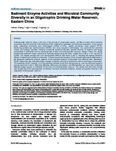
N-acetyl-beta-D-glucosaminide (2,24 mM) prepared in citrate/phosphate buffer (39 mM pH = 4.5) and incubated for 10 min at 37ºC. The reaction was stopped by addition of 100µL of 0.2M glycine buffer (pH 10.6). The reaction product was detected colorimetrically and performed at 400 nm. A standard -1 curve with p-nitrophenol (0-500 nmol ) was built and the results expressed in NAG activity (in nanograms) per sample milliliter (mL) (Figure 1). Figure 2: Levels of MPO activity in sera, ascitic fluid (asc), peritoneal lavage fluid (lav) of patients bearing EOC (n=18) or myoma (n=17). Note: The data were analyzed using one way analysis of variance (ANOVA). ***p< 0.001 for comparison between EOC serum versus EOC ascites; myoma serum versus peritoneal lavage.
A completely different profile of activity was observed for NAG activity in both groups. Thus, inflammatory enzyme activity was similar within sera or ascitic fluid in EOC group, but significantly higher than sera or peritoneal lavage fluid in the myoma group (Figure 3). Figure 1: N-ACETILGLICOSAMINIDASE (NAG) pattern curve – Pattern curve of substrate concentration (p-nitrofenilN-acetil-β-D-glicosaminida) used to calculate NAG (n-acetilglicosaminidase) concentration in the samples.
Statistical Analysis Results are presented as means±s.e.m. The data were analyzed using one-way analysis of variance (ANOVA) followed by Newman-Keuls correction factor for multiple comparisons as a post-test. The level of significance was set at p