http://journals.asm.org/site/misc/reprints.xhtml. Information about commercial ..... transitional cell cancer of the bladder, grade Il/IV ofun- known stage, with a ...
Xenorhabdus luminescens (DNA hybridization group 5) from human clinical specimens. J J Farmer 3rd, J H Jorgensen, P A Grimont, R J Akhurst, G O Poinar Jr, E Ageron, G V Pierce, J A Smith, G P Carter and K L Wilson J. Clin. Microbiol. 1989, 27(7):1594.
These include: CONTENT ALERTS
Receive: RSS Feeds, eTOCs, free email alerts (when new articles cite this article), more»
Information about commercial reprint orders: http://journals.asm.org/site/misc/reprints.xhtml To subscribe to to another ASM Journal go to: http://journals.asm.org/site/subscriptions/
Downloaded from http://jcm.asm.org/ on June 7, 2013 by guest
Updated information and services can be found at: http://jcm.asm.org/content/27/7/1594
JOURNAL OF CLINICAL MICROBIOLOGY, JUlY 1989. P. 1594-1600 0095-1137/89/071594-08$02.00/0
Vol. 27, No. 7
Xenorhabdus luminescens (DNA Hybridization Group 5) from Human Clinical Specimens J. J. FARMER 111,1* JAMES H. JORGENSEN,2 PATRICK A. D. GRIMONT,3 RAYMOND J. AKHURST,4 GEORGE O. POINAR, JR.?, ELISABETH AGERON,3 GLORIA V. PIERCE,6 JEAN A. SMITH,2 G. P. CARTER,1 KENNETH L. WILSON,' AND F. W. HICKMAN-BRENNER' Enteric Bacteriology Section, Center fjr Infèc tious Diseases, Centers fior Disease Control, Atlanta, Georgia 303331;
Received 27 January 1989/Accepted 10 April 1989
An unusual isolate from a human leg wound was identified as Xenorhabdus luminescens. This finding led to the discovery or isolation of four additional strains, two from blood and two from wounds. Three of the five strains were from patients in San Antonio, Tex. Three strains were studied by DNA-DNA hybridization (SI nuclease-trichloroacetic acid method) and were 77 to 100% related to each other, 34% related to the type strain of X. luminescens, 35 to 40% related to three of Grimont's other DNA hybridization groups of X. luminescens, and 9% related to the type strain of Xenorhabdus nematophilus. The new group of five strains was designated X. luminescens DNA hybridization group 5. All five strains were very inactive biochemically and fermented only D-glucose and D-mannose. The key reactions for recognizing this new organism are yellow pigment production, negative test for nitrate reduction to nitrite, weak bioluminescence (10 to 15 min of dark adaptation is required to see the weak light produced), and a unique hemolytic reaction on sheep blood agar plates incubated at 25°C. Two case histories of strains from wounds are given; these suggest that X. luminescens DNA hybridization group 5 may be a new bacterial agent that causes wound infections. The two cases of wound infection, along with the two blood isolates, suggest that the new organism is clinically significant.
finding led to identification of additional strains from human clinical specimens and is the subject of this report.
Organisms from unusual sources can occasionally occur in clinical specimens (9). This is not unusual if the organism is found in a broad ecological niche such as soil or water. Organisms that occupy a limited niche, such as Serratia ficaria and Proteus inyxofjaciens, are rarely if ever encountered in human clinical specimens. Until now, the genus Xenorhabdiis was thought to be an insect pathogen and a colonizer of nematodes (28) but not humans. In 1965 Poinar and Thomas described Achroinobacter nematophilus, a new bacterial species isolated from the intestinal lumen of a nematode (23). Subsequently, these authors noted that this organism did not fit well into a redefined genus Achromobacter, and they proposed Xenorhabdus as a new genus in the family Enterobacteriaceae (30). Xenorhabdus has two species, X. nemcatophilus (formerly Achromobacter nematophilus) and X. luminescens (3, 8, 30, 31). X. luminescens is very unusual because it is bioluminescent, a trait not found in any other species of the family Enterobacteriaceae (30). Over two dozen papers on the genus Xenorhabduis have been published (1-11, 13-31), and these organisms are well known as bacteria found in nematodes and as insect pathogens. In 1985 Farmer et al. (9) summarized their experience with the new species of Enterobacteriaceae and stated that Xenorhabduis was found in nematodes, but "'should be of little concern for the clinical microbiologist, since they have never been isolated from a clinical specimen." However, in 1986 one of us (J.H.J.) isolated strain 1 (Table 1) of X. lurninescens from a human would infection. This initial *
MATERIALS AND METHODS Scope of study. Originally this paper was to be a case report on a single isolate of X. lIuninescens (strain 1 in Table 1). Thus, this strain was studied in more detail than the others, which were found much later. Nomenclature. Although the genus Xenorhabdus has only two named species, each has been subdivided. Four subspecies names have been proposed in X. neinatophilus (3, 4), and four DNA hybridization groups were found by Grimont et al. (10) in a small collection of X. luminescens strains. Our clinical isolates of X. luininescens represent a fifth DNA hybridization group (Table 1). Laboratories. Six laboratories studied strain 1 and did various phenotypic tests, pathogenicity assays, and nematode association tests. We numbered the laboratories (to facilitate discussion of results) in the order in which they studied the strain: laboratory 1, University of Texas Health Science Center at San Antonio and Medical Center Hospital; laboratory 2, Texas Department of Health; laboratory 3, Enteric Bacteriology Section, Centers for Disease Control (CDC); laboratory 4, University of California, Berkeley; laboratory 5, Commonwealth Scientific and Industrial Research Organisation, Canberra, Australia; and laboratory 6, Unité des Enterobactéries, Institut Pasteur, Paris, France. Not all tests were done in each laboratory, and this is indicated in the Results section by a description such as "(laboratories 1, 3, 5)." General methods. Laboratories 1, 2, 3, and 6 normally
Corresponding author. 1594
Downloaded from http://jcm.asm.org/ on June 7, 2013 by guest
Department of Pathology, Univeersity of Texas, Health Science Center at San Antonio, and Medical Center Hospital, San Antonio, Texas 782842; Unité des Entérobactéries, Institut National de 1l Santé et de la Recherche Médicale Unité 199, Institut Pasteur, F-75724 Paris Cédex 15, France3; Division of Entornology, Coininon wealth Scientific and Industrial Research Organisation, Canberra, Australian Capital Territoiy 2600, Australia4; Department of Entomological Sciences, University of Califor-nia, Berkeley, California 94720S; (id Clinical-Enterics Section, BuOreaui of Laboratories, Texas Departinent of Health, Auistin, Texas 787566
VOL. 27. 1989
CLINICAL ISOLATES OF XENORHABDUS LUMINESCENS TABLE 1. Sources of the
1595
Xenorhabdus strains and their relatedness by DNA-DNA hybridization to the first clinical isolate (strain 1 = 3265-86) Relatedness to labeled DNA
Other designations
Strain no.
Source
from strain
Location
%/c X. lît11inlies-ens DNA group 5 1
2a 2b
il DNA group 4 12 13 X. nlefIntopIliIus 14
Xenorhahdbds 15
(ATCC 43950) (ATCC 43949)
Pretibial wound Blood Skin Blood Leg wound Abdomen. submandible
Maryland Maryland Pennsylvania Texas (San Antonio) Texas (San Antonio)
17a = Hb 17c = Hb variant 18a = HbB white colony
Nematode Nematode Nematode
South Australia South Australia
Georgia
34 34 34
17d
Nematode
South Australia
40
19 = Hm 18b = HbB red colony
Nematode Nematode
Georgia Georgia
36 37
20c = 954 20a = NC19
Nematode Nematode
North Carolina North Carolina
39 35
la = 786
Nematode
Virginia
9
9 = 286
Nematode
Brittany. France
8
(ATCC 43948) (ATCC 43951) (ATCC 43952)
Texas (San Antonio)
100 102 99 77
1
A T,,,
0
0.5
16.9
16.3
sp.
These values are the percent related of the test strains to strain 1 which was labeled.
work with human clinical isolates, and their methods are well known to clinical microbiologists (9). Laboratories 4 and 5 work with Xenorhabdus spp., insect larvae, and nematodes, and their methods have been described in detail
(3, 30). Biochemical reactions. Biochemical reactions were studied in each of the six laboratories. Laboratories 1, 2, 3, and 6 used an incubation temperature of 35 to 37°C and standard biochemical testing methods (9). Laboratory 4 used different methods, and incubation was at 30°C (16-31). Laboratory 5 used different methods. and incubation was at 28°C (1-4). Yellow pigment. Each strain was tested for yellow pigment production on Trypticase soy agar (TSA) (BBL Microbiology Systems, Cockeysville, Md.) and TSA plus 57 sheep blood (BBL) at both 25 and 36°C. These plates were observed at days 1, 2, 3 or 4 or 5, and 7. Hemolysis on sheep blood. Hemolysis was determined on blood agar (TSA plus 5% sheep blood) at 25 and 36°C. In addition, a set of plates was incubated at 36°C for 2 days and then moved to 25°C for the remaining 5 days to simulate what might occur in a clinical laboratory. Bioluminescence. Laboratories 5 and 6 measured light emissions in a scintillation counter (10). In laboratory 3 bioluminescence was detected with two methods, as follows. In method 1, the growth on a blood agar plate was observed in a darkroom (total darkness) for luminescence. A 5-min period was used for the observer's eyes to adjust to the dark. Any light seen was considered positive. In method 2 (begun in 1988), a Packard 2450 scintillation counter was used to measure light emission. Growth from a 24-h-old blood agar plate was removed (about 1.5-cm-diameter circle) with a swab, and a heavy suspension was made in 5 ml of sterile distilled water. The suspension was transferred to a 20-ml scintillation vial and was counted for 1 min with a fully
opened window setting. Two negative controls were also counted: water and an Escherichia coli suspension. Growth on MacConkey agar. A suspension of each organism was made in heart infusion broth (approximately 10' organisms per ml), and 0.01 ml of each was plated on MacConkey agar (Difco Laboratories, Detroit, Mich.) and blood agar plates. These plates were incubated at 36°C and observed at days 1, 2, 3 or 4 or 5, and 7. Pathogenicity for insect larvae and nematode association. Pathogenicity for larvae (3, 20) of the wax moth (Galleria mellonella) was determined for strain 1 in laboratories 4 and 5 by injecting between 50 and 5,000 bacterial cells into the larvae. Laboratory 4 used 15 larvae each for doses of 50, 500, and 5,000 cells (20). Laboratory 5 used a similar technique (3). The association of strain 1 with a Heterorhabditis nematode was tested in laboratory 4 by adding the bacterium to sterilized eggs of the nematode (15). This was done because the original strains of X. luiminescens formed a strong association with Heterorhabditis nematodes. The occurrence of strain 1 in the primary or secondary phase was tested in laboratories 4 and 5 by absorption of bromothymol blue and neutral red and by the presence of antibiotic activity (4, 30). DNA-DNA hybridization (laboratory 6). Laboratory 6 did DNA-DNA hybridization as follows. Strain 1 was labeled in vitro with tritium-labeled nucleotides, and hybridization was done by the SI nuclease-trichloroacetic acid method (10). The strains tested are given in Table 1. Search for additional strains of X. luminescens from human clinical specimens. The computer program Strain Matcher (9) was used to locate strains sent to CDC that are biochemically similar to strain 1. Since X. luininescens strains are very inactive biochemically, the Special Bacteriology Laboratory at CDC tested strain 1 by their methods and
Downloaded from http://jcm.asm.org/ on June 7, 2013 by guest
3 4 5 DNA group 1 6 7 8 DNA group 2 9 DNA group 3 10
3265-86 3105-77 3106-77 1216-79 2617-87 2407-88
Related at 6O'C'
1596
J. CLIN. MICROBIOL.
FARMER ET AL.
searched their card files for similar strains that had been studied previously. Strains that resembled X. luminescens biochemically were subcultured and tested for bioluminescence. Strain 5 (Table 1) was found in this manner.
g.,-
*ô
*
:
er
*46
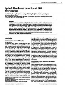
4
FIG. 1. Unusual hemolytic reaction of strain 1 on sheep blood agar (24 h, 25°C). Cell morphology and staining. Strain 1 was a gram-negative rod that varied in size and contained some filaments up to 30 ,um in length. The culture was weakly motile, with only a small portion of the population expressing motility at any time. Peritrichous flagella were seen when cells were stained with a flagella stain. No spheroplasts were detected in old cultures. Colonial morphology and growth. Strain 1 grew within 24 h at 35°C on sheep blood agar (which had Trypticase soy agar as its base) and had lactose-negative colonies on MacConkey agar (laboratory 1). It produced pale yellow, nonmucoid colonies when grown at 28°C on nutrient agar (laboratory 5). Growth appeared to be best at 30°C, but occurred in the range 19 to 37°C. Hemolysis on sheep blood agar. Strain 1 produced a very unusual type of hemolysis on sheep blood agar at 25°C (Fig. 1). There was no hemolysis immediately around the colony, but there was a thin line (about 2 mm wide) of hemolysis about 13 mm from the colony. Several experienced microbiologists indicated that they had never seen this type of hemolysis. To distinguish it from other hemolytic reactions we propose the term "Xenorhabdus luminescens hemolysis." This hemolytic reaction may be useful in recognizing strains of X. luminescens isolated from clinical specimens. Primary versus secondary phase of Xenorhabdus colonies. Cultures of Xenorhabdus often have two very distinct colony types when they are grown on agar media (30). These have been referred to as the primary and secondary phases (23, 25, 30). Cultures are typically in the primary phase on initial isolation from nematodes, but convert to the more stable secondary phase upon subculture. Strain 1 did not absorb bromothymol blue or neutral red and was negative for antibiotic production, indicating that it was in the sec-
ondary phase (3, 30). Pathogenicity for insects. Strain 1
was pathogenic for larvae of the wax moth Galleria. All larvae were killed when the strain was injected into the hemocoel. Laboratory 4 noted that the insect larvae were dead 3 days after injection and after 5 days were dark red and bioluminescent. Association with Heterorhabditis nematodes. Heterorhabtidis nematodes have been found to be colonized with certain species of Xenorhabdis, which can be essential for the development of the nematode (1, 15, 18, 23, 25, 28-30). Laboratory 4 found that strain 1 did not form this association with Heterorhabditis nematodes when bacterial cells were mixed with sterilized eggs of the nematode. The nematodes did not utilize the strain for development, in contrast to the type strain of X. luminescens, which shows this association.
Downloaded from http://jcm.asm.org/ on June 7, 2013 by guest
RESULTS Comment. Since strain 1 (3265-86) was studied first and in more detail than the other four, the results for this strain are given first. Case report: strain 1. On 19 June 1986, a 78-year-old man sought attention at the emergency room of the Medical Center Hospital, a primary teaching hospital of the University of Texas Health Science Center at San Antonio. He had an ulcer, present for about 11 days, below his left knee and had experienced upper thigh-groin pain for 5 to 6 days. There was a red streak that ran up his leg. FIe stated that the lesion began as a "pimple" just below his left knee which increased in size after he squeezed it. He denied having had any insect bites or trauma to the area. The admission history included the following information. The patient had suffered from adult-onset diabetes mellitus since 1975 that was treated with insulin (30 U of NPH insulin daily in the morning on admission). He also had coronary artery disease and had undergone coronary artery bypass grafting in 1981. He had a posttransurethral resection of the prostate for stage A2 prostate cancer and had a diagnosis of transitional cell cancer of the bladder, grade Il/IV of unknown stage, with a negative followup cystoscopy on 24 June 1986. His left testicle was removed in 1959 secondary to trauma. He has a 70-pack-year history of smoking. His physical examination and laboratory test results included blood pressure of 150/92, pulse of 90, and a temperature of 98.6°F (ca. 37°C). He had small, firm, tender, matted lymph nodes in his left groin and an ulcer (2 by 2 cm) below the left knee with surrounding erythema and a small amount of fluctuance. Erythema and induration were noted in the left thigh in a linear distribution. His leukocyte count was 9,400/ml, with 70% polymorphonuclear leukocytes, 1% band forms, 23% lymphocytes, 4% atypical lymphocytes, and a hematocrit of 41.3%. His electrolytes, erythrocyte sedimentation rate, and liver enzymes were within normal limits. X rays of the left thigh and knee indicated no evidence of osteomyelitis. Three blood cultures drawn before antibiotic therapy were negative, as was a urine culture from admission. A swab of his ulcer yielded light growth of Staphylococcus aureus and light growth of a gram-negative, lactosenegative rod. This gram-negative rod was not further characterized before the patient was discharged, but was susceptible to chloramphenicol, tetracycline, gentamicin, and cefoxitin and resistant to cephalothin and ampicillin. An aspirate of the indurated area of the left thigh, obtained in the emergency room, grew the same lactose-negative, gramnegative rod in pure culture. Two days after admission, the thigh was again aspirated and grew the same gram-negative rod in very light growth. A sonogram of the thigh was negative. The patient was treated with intraveneous cefoxitin (2 g every 6 h) and oxacillin (2 g every 4 h) for 10 days, during which the area of induration resolved and the pretibial ulcer decreased in size. The patient was then followed up as an outpatient, and all symptoms and signs of infection resolved. Since the gram-negative rod could not be identified with certainty, it was forwarded to the Texas Department of Health, where it was suspected to be Xenorhabdus sp. The isolate was further studied at laboratories 3 through 6, and each of them identified it as X. luminescens.
r
VOL. 27, 1989
CLINICAL ISOLATES OF XENORHABDUS LUMINESCENS
approximately 1 cm circumferentially. His admission blood count was 8,400 leukocytes per pi with 69% polymorphonuclear leukocytes, 2% band forms. 27% lymphocytes, 1% monocytes, and 1c eosinophils. He had 4.5 x 10⁶ erythrocytes per p.l, 425 x 10- platelets per pi.
a hematocrit of 39.4%c, a hemoglobin level of 13.5 g/dl, and normal electrolytes. After admission to the surgical service, he was taken to the operating room for debridement of the left lower extremity wounds. Immediately before surgery he was started on oxacillin (2 g every 6 h) and gentamicin (80 mg every 8 h), both intravenously, continued for 4 days. During surgery a single culture was taken from inside the cavity of the largest lesion. This produced a pure culture (light growth) of a lactose-negative, gram-negative rod which was isolated on sheep blood, chocolate, and MacConkey agars and in thioglycolate broth. One week after admission the patient was returned to the operating room, where a localized purulent pocket was drained (but not cultured) and a split-thickness skin graft was placed over the contiguous debrided areas on the left lower extremity. He was given cefoxitin (1 g every 6 h) intravenously for 3 days after surgery. He did well after surgery and was discharged 16 days after admission. He has subsequently had an uneventful recovery during followup in surgery clinic. Strain 4 was identified as a typical strain of X. luimiinescens in laboratories 1, 2, and 3 (Table 2). DNA-DNA hybridization. Table 1 gives the results of the hybridization experiments done in laboratory 6. Strain 1 was radioactively labeled and tested against strains 1, 2, and 3 (strains 4 and 5 had not been detected at this time). All three strains were highly related, and strain 3, which had the lowest value (77%), had a low àT,, value. Eight other strains of X. lininescens, representing all four DNA hybridization groups previously described by Grimont et al. (10), were only 34 to 40%, related to strain 1, with AT,,, values of over 16. The type strain of X. nencatophilus was only 9Y% related. Biochemical characterization of ail five strains of X. luminescens DNA hybridization group 5. Table 2 gives the biochemical characteristics of the five strains in media often used in clinical microbiology laboratories for biochemical characterization. At 36°C the strains were very inactive and had a pattern quite distinct from any other species or biogroup of Enterobacteriaceae. Yellow pigment. Four of the five strains produced a yellow pigment on TSA at 36°C within 2 days. At 25°C they took 2 to 5 days. Strain 1216-79 was not yellow, but produced a tan soluble pigment on TSA after 3 to 5 days (36°C). Two strains, 3107-77 and 2617-87, produced weak yellow pigment in 1 day, but were bright yellow in 2 days and were also bright yellow in TSA working stocks. Hemolysis on sheep blood. At 36°C, the strains had alpha hemolysis within 2 days, but by 3 to 5 days they had lysis and greening. At 25°C all five strains had a unique type of hemolysis (Fig. 1). It started as a tiny line of beta hemolysis some distance from the colony itself (approximately 13 mm). Eventually this line of hemolysis encircled all the growth, but did not extend up to the colonies. Four strains had this unique type of hemolysis by day 2, but strain 3 required 3 to 5 days. This type of hemolysis was not observed when strains were grown at 36°C for 7 days or when grown at 36°C for 2 days and then transferred to 25°C for the remaining 5
days. Bioluminescence. AIl of the strains were luminescent by both methods. When the blood plates were observed in the darkroom, all of the strains appeared luminescent within 6 min. This extremely weak luminescence is in contrast to most luminescent strains of Vihrio spp., which are visible within a few seconds. Strain 5 was the weakest of the strains. In the scintillation counter the strains produced various amounts of light. Although strain 5 was weak when observed in the darkroom, it had the highest count in the scintillation
Downloaded from http://jcm.asm.org/ on June 7, 2013 by guest
Bioluminescence. Laboratory 3 found strain 1 was negative after 5 min of observation (the usual amount of time allowed for the observer's eyes to adapt to the dark). Laboratories 4 and 5 found it to be weakly bioluminescent (visual observation in darkened room). Laboratory 5 noted that 10 min of dark adaptation was required to observe the faint light emission. The bioluminescence of the strain was weaker than that of a control culture (strain Cl of X. luminescens). When light emission was measured in a scintillation counter (laboratories 3 and 5), strain Cl had a value of 15,000 cpm, clinical isolate 1 had 8,000 cpm, and water and E. (oli had levels less than 50 cpm. Laboratory 4 found the culture to be bioluminescent on laboratory media and also caused the dead insect larvae to become bioluminescent 5 days after inoculation. Three other clinical isolates of X. luminescens studied at CDC. Strains 2 and 3 (Table 1) of X. luIminescens were found by a computer search (program Strain Matcher) of the records of laboratory 3. Strain 2 was isolated in 1977 from blood and skin cultures of an 80-year-old woman with endocarditis in Maryland. Strain 3 was from a blood culture of a 72-year-old woman in Pennsylvania. Strain 5 (strain no. F5197) was found in the culture collection of the Special Bacteriology Laboratory at CDC; it had been studied in 1984 and was reported "most similar to Chromobateriwun violacelon; arginine negative." It had come from two specimens labeled "abdomen and submandible" from a 36-year-old woman with a clinical diagnosis of "disseminated bacterial infection" and associated illness of "diabetes, treated with insulin." She had been seen at the Veterans Administration Hospital and also at the University of Texas Health Science Center at San Antonio. Strain 5 was found to be bioluminescent (laboratory 3) and was identified as X. luininescens. Case report: strain 4. Strain 4 of X. luminescens was isolated in November 1987 in laboratory 1. It was from a 45-year-old Hispanic man with an unremarkable past medical history who presented to the emergency room with a disrupted ulceration of the left lower extremity with active venous bleeding of 3 days duration. The patient stated that he had suffered a spider bite on the dorsum of his left foot approximately 6 weeks earlier. After apparent healing of the bite, the same lesion erupted 2 weeks after the initial incident. He then noticed some localized swelling of the posterior aspect of the distal left lower extremity and a second similar area proximal to the first erupted spontaneously at about the same time. He denied any other recent trauma to the area. The areas progressively increased in size, and the more distal of the two began to produce dark blood on the night before admission. The bleeding could be controlled with pressure. Walking had become painful and difficult for the 3 days prior to admission. His physical examination was remarkable for a blood pressure of 150/80, a pulse of 120, and respirations of 20 with a temperature of 100.2°F (ca. 38°C). There was a fluctuant mass (4 by 5 cm) on the distal aspect of the left lower extremity without cellulitis or pus, but which was covered by necrotic tissue with a central core of approximately 2 by 2 cm associated with active venous bleeding. A second, proximal inactive lesion (3 by 3 cm) was also seen. A crusting lesion (2 by 2 cm) on the dorsum of the left foot at the site of the spider bite revealed decreased sensation even
1597
1598
J. CLIN. MICROBIOL.
FARMER ET AL.
TABLE 2. Biochemical reactions (360C) of the five clinical strains of X. luininescens DNA hybridization group 5 Cumulative % Test or property
7
2
Indole production Indole production (heart infusion
O o
O
o o
20
20
20
O
O
o
O
O
O
40
60
80
+2
o
o
O
O
O
O
O
O
100 40 20
100 80 20
100 100 60
O
O
o
100
100
100
+
o
o
o
-
-
o
+2
+4
D-Glucose
Acid production Gas production Acid production from: Adonitol
o
o
o
L-Arabinose D-Arabitol
o
o
o
-
o
o
o
-
Cellobiose Dulcitol
o
o
o
-
o
o
o
-
Erythritol
o
o
o
-
D-Galactose
o
o
o
-
Glycerol
o
o
o
-
mnyo-Inositol Lactose Maltose
o
o
o
-
o
o
o
-
o
o
o
-
D-Mannitol
o
o
o
-
D-Mannose
100
100
100
+
Melibiose ot-Methyl-D-glucoside Raffinose
o
o
o
-
o
o
o
-
o
o
o
-
L-Rhamnose
o
o
o
-
Salicin
o
o
o
-
D-Sorbitol
o
o
o
-
Sucrose Trehalose
o
o
o
-
o
o
o
-
D-Xylose
o
o
o
-
Continued
counter. Although strain 4 had the lowest count, it appeared strong as the others in the darkroom (strain 4, 90,000 cpm; strain 2, 660,000 cpm; strain 3, 1,500,000 cpm; strain 1,
as
1,700,000 cpm; strain 5, 1,900,000 cpm; E. coli, 13 cpm; distilled water, 9 cpm). Strain 3 needed 2 days to produce sufficient growth for testing. Growth on MacConkey agar. All of the strains grew on MacConkey agar within 2 days; however, the plating efficiency was slightly reduced (no growth on the last quadrant of streaking as opposed to growth on blood agar). The colonies were colorless (lactose negative) and measured 0.5 to 4.0 mm in diameter. Morphology on blood agar. The strains grew well on blood agar and preferred 36°C to 25°C. After 2 days of incubation most of the colonies were 1 to 4 mm in diameter, but some were smaller. All of the strains had two colony types (the second was smaller), but biochemical reactions on both
Cumulative Y Test or property
Esculin hydrolysis Mucate fermentation Tartrate fermentation (Jordan) Acetate utilization Lipase (corn oil) DNase, 25°C DNase, 36°C Nitrate reduction to nitrite Oxidase ONPGb test Yellow pigment production, 25°C Yellow pigment production. 36°C Citrate utilization (Christensen) Tyrosine clearing Bioluminescence "Xenorhabdus hemolysis" on sheep blood, 25°C "Xenorhabdus hemolysis" on sheep blood, 36°C MacConkey agar, growth
positive on day:
Strain ATCC 43950
1
2
7
(strain 1)"
0 0 60 0 0 0 0 0 0 0 0 40 80 60 80
0 0 80 20 0 0 0
0 0 100 60 0 0 0
+ -
-
-
20 80 80 100 100
-
0
0 40 80 100 80 100 80
100
+2
0
0
0
80
100
100
+4 +2 + + +
+
' Symbols: -, negative at end of appropriate incubation temperature; +, positive at 24 h (or at 48 h for those tests not done at 24 h). Superscripts give the day the reaction became positive, if it was delayed. We propose that ATCC 43950 be designated the type strain if X. luininescens DNA hybridization group 5 is elevated to the level of species. > ONPG, o-Nitrophenyl-,B-D-galactopyranoside.
types were the same. The colonies were smooth and entire. The two strains previously described that produced a strong yellow pigment were also yellow on blood agar, but it was difficult to see the pigment of the other three strains. When Gram stains were done with 1-day-old growth from blood agar plates (36°C), the cells appeared as short-to-medium gram-negative rods with occasional filaments, granules, and bipolar staining. Antibiotic susceptibility. All five strains had large zones around each of the antibiotics except penicillin G, ampicillin, carbenicillin, and cephalothin (Table 3). DISCUSSION Xenorhabdiîs was described as a new genus in 1979 (30). It has been studied thoroughly because both X. nematophilus and X. luminescens are insect pathogens and are used in insect pest control. As more strains have been studied, the nomenclature and classification of the genus have become more complex (3, 4, 10, 31). Akhurst divided X. nematophilus into four subspecies, each of which was given a formal name (3, 4). Grimont et al. studied a small collection of X. luminescens strains and found four distinct DNA hybridization groups (10). No scientific names were given, but all four groups could be considered to be distinct species. However, until now the genus Xenorhabdus has been of little concern to the clinical microbiologist. In this paper we describe a bioluminescent organism that occurs in clinical specimens and appears to be clinically significant. The organism most resembles X. luminescens by both phenotypic characteristics and DNA hybridization. We refer to this new organism as X. luminescens DNA hybridization group 5, since it is not highly related to the previously described four DNA groups. In the future it will probably be given a scientific name. The luminescent strains of the genus Xenorhabdiis are quite distinct from the nonluminescent
Downloaded from http://jcm.asm.org/ on June 7, 2013 by guest
Malonate utilization
ATCC 43950 (strain 1)'
positive on day:
1
broth) Methyl red Voges-Proskauer (O'Meara) Voges-Proskauer (Barritt) Citrate utilization (Simmons) H2S on triple sugar iron agar H2S on peptone iron agar Urea hydrolysis (Christensen) Phenylalanine deaminase Lysine decarboxylase (Moeller) Arginine dihydrolase (Moeller) Ornithine decarboxylase (Moeller) Motility Gelatin hydrolysis (22°C) KCN test (% resistant to cyanide)
Strain
TABLE 2-Continued
CLINICAL ISOLATES OF XENORHABDUS LUMINESCENS
VOL. 27, 1989
1599
TABLE 3. Antibiotic susceptibilitY of isolates 1 through 5 Isolate no.
Zone size (mm) for":
Lab no. CL
2a 3 1 4 5
3105-77 1216-79 3265-86 2617-87 2407-88
Range Mean SD Percent susceptible
6-19 11 5.4 40
SD
NA
38 40 38 34 30
19 8 14 6 8
GM
28 25 26 25 24
20 30 18 20 9
30-40 36 4 100
9-30 10 7.5 80
24-28 26 1.5 100
S
25 15 18 20 19
15-25 10 3.6 100
K
35 32 35 29 27
27-35 32 3.6 100
TE
35 34 35 33 30
C
40 40 38 37 33
30-35 33 2.1 100
33-40 39 2.9 100
P
AM
CB
CF
6 6 6 6 6
8 15 12 6 10
27 20 18 22
6 13 10 6 8
6 6 0 0
6-15 10 3.5 20
18-27 21 3.4 20
16-13 0 3.0 0
20
CL, Colistin (10 J.g); NA, nalidixic acid (30 pLg)( SD. sulfadiazine (250 p.g); GM, gentamicin (10 p.g)( S. streptomycin (10 ,ug); K. kanamycin (30 pLg)( TE, tetracycline (30 p.g); C. chloramphenicol (30 pLg); P. penicillin (10 U); AM, ampicillin (10 p.g); CB. carbenicillin (10() p.g); CF. cephalothin (30 ,tg).
TABLE 4. Key characteristics of the bioluminescent clinical isolates as compared with the two species of Xenorhlahdus and with other species of Enterobacieriaceae Strain characteristics"
Test or property
Strains 1 through 5
X.
IbIoni-
InesCens
X. neinatophifilos
+ +
+ +
-
-
-
+
+
-
ACKNOWLEDGMENT We thank Dannie G. Hollis of the Special Bacteriology Laboratory, CDC. for searching their laboratory's collection for possible isolates of X. luzinnescens.
Most other members of the Lntero-
bacteriaceae
Bioluminescent Catalase Nitrate reduction to nitrite Yellow pigment
currently no information suggesting that strains of X. Iliinnines(ens DNA groups 1 through 4 have occurred in human clinical specimens; however, this point deserves further study. The biochemical inactivity of X. lumninies( en s may cause problems for recognition and identification in clinical laboratories. The strains listed in Table 1 could be confused with a number of different species of Enter-obac(teriac eae, particularly those that contain strains that are very inactive biochemically. However, a number of distinct properties should make it possible to recognize and identify cultures of X. Ilinines(cenis. The unusual hemolytic reaction, yellow pigment, and negative test for nitrate reduction should be helpful markers, but bioluminescence is the single characteristic that quickly defines this organism. Weak bioluminescence is easily detected in a scintillation counter, but is more difficult to detect with dark-adapted eyes. Laboratory 3 did not detect the bioluminescence of strain 1 even though this laboratory works with bioluminescent vibrios and Xeno1-habdits and used 5 min of dark adaptation before calling it negative. Cultures from clinical specimens suspected to be X. luwninesce ns should be watched in a totally dark room from 10 to 15 min or tested in an instrument. The biochemical reaction pattern given in Table 2 should be helpful in recognizing strains. Table 4 gives the tests that differentiate X. luininescens from X. neinatophillus and other members of the family Enterobaccteriac ea. We hope this report will stimulate other clinical and public health laboratories to isolate and identify this unusual organism and help define its role in human disease. Our laboratories are interested in infections that yield this organism and would be grateful for information about additional cases.
Symbols: +. most strains positive: -. most strains negative. A few species produce yellow pigment.
+ +
b
ADDENDUM IN PROOF We recently studied a sixth isolate of X. lunines(ens. It was from a groin infection of a patient at a different hospital in San Antonio, Tex. The genus Xenorhlabduis is becoming more complex. Akhurst and Boemare (R. J. Akhurst and N. E. Boemare, J. Gen. Microbiol. 134:1835-1845, 1988) recently proposed that the subspecies of X. neinatophilus be elevated to species. In this classification there are five species of Xenorhiahdus: X.
Downloaded from http://jcm.asm.org/ on June 7, 2013 by guest
ones, and it may be desirable to form a new genus to accommodate the five DNA hybridization groups now recognized in X. luininescens (3, 10, 30). Two of the five strains were from blood cultures, and strong evidence from case 5 suggests that this organism actually caused the wound infection. The evidence for clinical significance in case 1 was suggestive, but S. (il/relis was also present in the wound. We find it fascinating that two of the five isolates came from a single laboratory and that a third isolate was also from the same city (San Antonio, Tex.). The old literature sometimes mentions human wounds that glow in the dark. Luminescent wounds were sometimes noticed at battlefield hospitals in the 1800s (12), and this was thought to be a helpful rather than a harmful indication for the patient (12). Nealson and Harvey (17) commented that luminescent wounds could have been caused by the presence of X. luiminescens and that the positive prognosis could be due to known antibiotic production by Xenoi-habdus. The postulated antibiotics would presumably inhibit more harmful organisms that would cause a more severe infection. Our report gives the first evidence that X. Iluninescens can occur in human wounds and may explain the old observations of luminescent wounds. Alternatively, the luminescent wounds could have been caused by a species of Vibrio since vibrios are known to infect wounds, to occur in areas remote from the sea, and to be bioluminescent. It is interesting that our clinical strains of X. luiminescens belong to a DNA hybridization group that is different from all the strains that were isolated from nematodes. These data appear to indicate that the strain of X. lwninescens used in insect control is not important in public health. There is
1600
FARMER ET AL.
J. CLIN. MICROBIOL.
neinatophilus, X. beddingii, X. bo'ieflii, X. poinarii, and X. lumninescens (including five DNA subgroups).
16. Milstead, J. E. 1980. Pathophysiological influences of the Heterorhahditis bacrteiophora complex on fifth-instar larvae on the
LITERATURE CITED 1. Akhurst, R. J. 1980. Morphological and functional dimorphism in Xenorhabdiis spp., bacteria symbiotically associated with the insect pathogenic nematodes Neoaplectana and Heîerorhabditis. J. Gen. Microbiol. 121:303-309. 2. Akhurst, R. J. 1982. Antibiotic activity of Xenorhabdus spp., bacteria symbiotically associated with insect pathogenic nema-
18. Poinar, G. O., Jr. 1966. The presence of Acliroinobater neniatophfilus in the infective stage of a Neoapleciana sp. (Stein-
todes of the families Heterorhabditidae and Steinerneinatidae. 3. 4.
6.
aux nematodes entomophages Steinernenatidae et Heterorhabditidae. C. R. Soc. Biol. 177:107-115. 7. Dunphy, G. B., and J. M. Webster. 1984. Interaction of Xenorhabdus nenaltophilius ssp. nenaitopIilus with the hemolymph of Gai/eria inellonelil. J. insect. Physiol. 30:883-890. 8. Farmer, J. J., III. 1984. Genus Xenorhabdiis Thomas and Poinar 1979, 354AL p. 510-511. In N. R. Krieg (ed.), Bergey's manual of systematic bacteriology, vol. 1. The Williams and Wilkins Co., Baltimore. 9. Farmer, J. J., III, B. R. Davis, F. W. Hickman-Brenner, A. McWhorter, G. P. Huntley-Carter, M. A. Asbury, C. Riddle, H. G. Wathen-Grady, C. Elias, G. R. Fanning, A. G. Steigerwalt, C. M. O'Hara, G. K. Morris, P. B. Smith, and D. J. Brenner. 1985. Biochemical identification of new species and
biogroups of Enterobacteriaceae isolated from clinical specimens. J. Clin. Microbiol. 21:46-76. 10. Grimont, P. A. D., A. G. Steigerwalt, N. Boemare, F. W. Hickman-Brenner, C. Deval, F. Grimont, and D. J. Brenner. 1984. Deoxyribonucleic acid relatedness and phenotypic study
of the genus Xenorhabdus. Int. J. Syst. Bacteriol. 34:378-388. 11. Guelzow, A. 1986. Immune inhibition by the insect pathogenic nematode Neoaplectana carpocapsae Steinerneinatidae. Dev. Comp. Immunol. 10:629-630. 12. Harvey, E. N. 1957. A history of luminescence. American
Philosophical Society, Philadelphia. 13. Hotchkin, P. G., and H. K. Kaya. 1984. Electrophoresis of
soluble proteins from two species of Xenorhabdus, bacteria mutualistically associated with the nematodes Steineremna spp. and Heterorliabditis spp. J. Gen. Microbiol. 130:2725-2731. 14. Khan, A., and W. M. Brooks. 1977. A chromogenic bioluminescent bacterium associated with the entomophilic nematode Chromnonemna hefiotifidis. J. I nvertebr. Pathol. 29:253-261. 15. Lysenko, O., and J. Weiser. 1974. Bacteria associated with the nematode Neoapleciana carpocapsae and the pathogenicity of this complex for Galleria inellonella larvae. J. Invertebr. Pathol. 24:332-336.
43:496-518. erneinaiidae: Neinatoda). Nematologica 12:105-108. 19. Poinar, G. O., Jr. 1967. Description and taxonomic position of the DD-136 nematode (Sieinerneinatidue, Rhabditoidea) and its relationship to Neoaplectana(carpocapsae Weiser. Proc. Helminthol. Soc. Washington 34:199-209. 20. Poinar, G. O., Jr. 1975. Description and biology of a new insect parasitic Rhabditoid, Heterorhabdiiis bacteriophora N. Gen., N. Sp. (Rhiabditidai; Heierorilibditidae N. Fam.). Nematologica 21:463-470. 21. Poinar, G. O., Jr., R. Hess, and G. Thomas. 1980. Isolation of
defective bacteriophages from Xenorhabdus spp. (Enterobacteriaceae). Int. Res. Commun. Syst. Med. Sci. Libr. Compend. 8:141.
22. Poinar, G. O., Jr., and P. T. Himsworth. 1967. Neoaplectana parasitism of larvae of the greater wax moth, Galleria mellonella. J. Invertebr. Pathol. 9:241-246. 23. Poinar, G. O., Jr., and G. M. Thomas. 1965. A new bacterium, Achronîobacter nenal