www.nature.com/scientificreports
OPEN
Received: 23 August 2016 Accepted: 30 March 2017 Published: xx xx xxxx
DNA methylation and Transcriptome Changes Associated with Cisplatin Resistance in Ovarian Cancer Riikka J. Lund1, Kaisa Huhtinen2, Jussi Salmi1, Juha Rantala2, Elizabeth V. Nguyen1, Robert Moulder1, David R. Goodlett1,3, Riitta Lahesmaa1 & Olli Carpén2 High-grade serous ovarian cancer is the most common ovarian cancer type. Although the combination of surgery and platinum-taxane chemotherapy provide an effective treatment, drug resistance frequently occurs leading to poor outcome. In order to clarify the molecular mechanisms of drug resistance, the DNA methylation and transcriptomic changes, associated with the development of drug resistance in high-grade serous ovarian cancer, were examined from patient derived malignant ascites cells. In parallel with large-scale transcriptome changes, cisplatin resistance was associated with loss of hypermethylation at several CpG sites primarily localized in the intergenic regions of the genome. The transcriptome and CpG methylome changes in response to cisplatin treatment of both sensitive and resistant cells were minimal, indicating the importance of post-translational mechanisms in regulating death or survival of the cells. The response of resistant cells to high concentrations of cisplatin revealed transcriptomic changes in potential key drivers of drug resistance, such as KLF4. Among the strongest changes was also induction of IL6 in resistant cells and the expression was further increased in response to cisplatin. Also, several other components of IL6 signaling were affected, further supporting previous observations on its importance in malignant transformation and development of drug resistance in ovarian cancer. High-grade serous ovarian cancer (HGSOC) is the most common ovarian cancer subtype and accounts for 80% of the deaths caused by the disease. The prognosis of HGSOC is poor as most diagnosis is at late stages of the disease when the 10-year survival rate is only in the order of 15%. The main strategy for treatment involves surgical removal of the tumor tissue and chemotherapy1. Platinium compounds, such as cisplatin, in combination with taxane are typically used in chemotherapy. However, recurrence of the cancer is frequent and most of the patients will eventually become refractory to the treatment2. In order to improve the prognosis of the patients with HGSOC, new biomarkers enabling early diagnosis of the disease as well as new therapeutic strategies overcoming the drug resistance are needed1, 3. Detailed characterization of the molecular mechanisms leading to drug resistance is important for development of improved therapies. The molecular mechanisms leading to drug resistance can be heterogeneous and complex4. In addition to genetic factors, the development may involve epigenetic changes, which enable tumor cells, and possibly non-transformed cells in the microenvironment, to adapt and lose sensitivity to drug treatment. DNA methylation and transcriptional changes associated with drug resistance have been detected in several genomic sites in both cell lines and patient samples5–8. For example, methylation and transcriptional silencing of the MLH1 gene have been repeatedly associated with cisplatin resistance8, 9. Although several candidate driver genes for cisplatin resistance have been identified, further studies are required to clarify the heterogeneity of the drug resistance mechanisms and clinical significance of the findings.
1
Turku Centre for Biotechnology, University of Turku and Åbo Akademi University, Turku, Finland. 2Department of Pathology, Medicity Research Unit, University of Turku and Turku University Hospital, Turku, Finland. 3Department of Pharmaceutical Sciences, University of Maryland, Baltimore, MD, USA. Riikka J. Lund, Kaisa Huhtinen, Riitta Lahesmaa and Olli Carpén contributed equally to this work. Correspondence and requests for materials should be addressed to R.J.L. (email:
[email protected]) or O.C. (email:
[email protected])
Scientific Reports | 7: 1469 | DOI:10.1038/s41598-017-01624-4
1
www.nature.com/scientificreports/ In this study, we have further investigated the potential mechanisms associated with drug resistance by comparing cisplatin responses in sensitive and resistant patient derived HGSOC cell lines with next-generation sequencing based applications. We have used Reduced Representation Bisulfite Sequencing (RRBS) together with messenger RNA sequencing (mRNA-seq) for unbiased identification of the DNA methylation changes at single nucleotide resolution in the CpG rich regions of the genome in correlation with genome-wide transcriptome changes.
Results
Differences between cisplatin sensitive and resistant cells before drug treatment. Comparison
of cisplatin sensitive and resistant M019i cells before the drug treatment revealed large scale differences in both transcriptomes and DNA methylomes. Comparison of DNA methylomes revealed a total of 1,488 differentially methylated sites that exceeded a minimum methylation difference of 20% in each comparison (Fig. 1a, Supplementary Table S1). Interestingly, most of the differentially methylated sites (1,251 sites, 84%) were found to be less methylated in the resistant cell line. Only 237 (16%) sites were methylated at higher levels in the resistant cell line and had lower methylation levels in the sensitive line. Most of the differentially methylated sites were in the non-coding regions of the genome (Fig. 1b). Of the sites 90 (6.0%) were in exons and 26 (1.7%) in the TSS. The majority of differentially methylated sites (76%) were located within 100 Kbp distance from a TSS (Fig. 1c) and nearly all sites (1,479) were within 1 Mbp distance from a TSS of a gene. The genes close to the differentially methylated sites were associated with canonical pathways such as cAMP-mediated signaling (32 molecules, p = 7.14E-04), G-protein coupled receptor (GPCR) signaling (37 molecules, p = 8.33E-04), WNT/ beta-catenin signaling (25 molecules, p = 1.92E-03) and human embryonic stem cell pluripotency (22 molecules, p = 2.11E-03), see Supplementary Table S2 for the complete list of functional enrichment results. Consistently, the top putative upstream regulators included such as POU5F1 (57 targets, p = 2.21E-10), CTNNB1 (95 targets, p = 1.42E-09), SOX2 (54 targets, 3.05E-09), KLF4 (41 targets, 6.03E-07) and TP53 (151 targets, p = 6.42E-07). However, no transcription counts were detected for well-known pluripotency factors POU5F1 and SOX2. The top molecular functions of the genes included differentiation of cells (422 molecules, 4.08E-27), proliferation of cells (604 molecules, p = 4.45E-23) and the strongest disease association was cancer (1,585 molecules, p = 2.98E-51). Considerable differences in the transcriptome were also observed between cisplatin sensitive and resistant cells. Altogether 587 differentially expressed genes were detected. Out of these 346 showed increased expression and 241 decreased expression in the resistant cells (Fig. 1d, Supplementary Table S3). As patient characteristics may influence the results10, the expression changes of selected genes were further validated using qRT-PCR in two platinum sensitive cell lines M019i and OC002 and their resistant variants (Fig. 1e). Pathway analysis revealed functional enrichment of the molecules to several canonical pathways, the most prominent ones were involved in oxidative metabolism and stress, such as “production of nitric oxide and reactive oxygen species in macrophages” (19 molecules, p = 9.77E-07), and inflammatory signaling, such interferon signaling (8 molecules, p = 3.61E-06). See Supplementary Table S2 for the complete list of enrichment results. Among the most significant putative upstream regulators were TNF (132 targets, 1.19E-34), beta-estradiol (118 targets, 1.19E-34) and IFNL1 (26 targets, 4.14E-24). The genes differentially expressed by the cisplatin sensitive and resistant cells were enriched in the top functional categories including cell death (222 molecules, p = 1.72E-18) and cellular movement (160 molecules, p = 4.59E-18). Cancer was again among the most significant disease enrichments (516 molecules, p = 9.74E-15).
Responses of sensitive and resistant cells to low cisplatin treatment. We next examined the epigenetic and transcriptional perturbations in sensitive and resistant cell lines in response to 0.6 μM cisplatin treatment. Interestingly, although cisplatin treatment led to CpG methylation changes in both sensitive and resistant cells, these alterations did not trigger changes in the gene expression. In the sensitive line, changes in the methylation of 59 sites (FDR 5%), 28 with increased and 31 with decreased methylation, were detected in response to cisplatin treatment (Supplementary Table S1). The genes closest to the altered sites were functionally linked to neuronal signaling pathways, such as development of neurons (18 molecules, p = 6.27E-06) and canonical pathways, such as synaptic long term depression (6 molecules, p = 4.11E-02). Consistently, the putative upstream regulators included APP (13 targets, p = 1.15E-04) and APOE (5 targets, p = 3.54E-03). However, no changes in gene expression were observed with the chosen filtering criteria (minimum absolute FC = 1.5, FDR ≤ 0.05 or unadjusted pval ≤ 0.05) indicating that the detected DNA methylation changes did not cause changes in transcriptional activity in sensitive cells. In the resistant cell line changes were observed in 74 sites (FDR 5%): 34 with increased and 40 decreased methylation in response to 0.6 μM cisplatin treatment. The genes closest to the altered sites were linked to the WNT/beta-catenin canonical pathway (5 molecules, p = 2.96E-02) and, similarly to sensitive cells, in molecular functions associated with neuronal development and functions, such as development of neurons (21 molecules, p = 4.9E-06). The putative upstream regulators included REST (5 targets, p = 6.38E-04), FGFR2 (5 targets, p = 7.39E-04), CTNNB1 (11 targets, p = 1.77E-03) and KLF4 (6 targets, p = 2.72E-03). Interestingly, none of the sites differentially methylated in response to cisplatin in resistant cells overlapped with those detected in sensitive cell line. Also, as was the case for the sensitive cells, no changes in gene expression were observed in response to 0.6 μM cisplatin treatment of resistant cells (minimum absolute FC = 1.5, FDR ≤ 0.05 or unadjusted pval ≤ 0.05). Response of resistant cells to high cisplatin treatment. We also examined the response of the resist-
ant cell line to a higher concentration of cisplatin (7 μM, IC50), which was not tolerated by the sensitive cells. In contrast to the lower concentration, changes were now detected in both CpG methylomes and transcriptomes. Decreased methylation of 28 sites was observed, whereas 49 sites become more methylated (Supplementary Table S1). The genes closest to the altered sites were associated with molecular functions, such as development
Scientific Reports | 7: 1469 | DOI:10.1038/s41598-017-01624-4
2
www.nature.com/scientificreports/
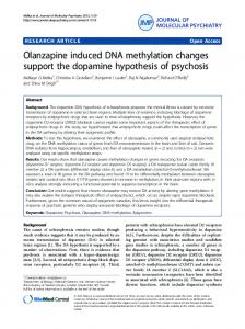
Figure 1. CpG methylome and transcriptome differences between cisplatin sensitive and resistant ovarian cancer lines. DNA methylomes of the cells sensitive or resistant to cisplatin were profiled with Reduced Representation Bisulfite Sequencing and transcriptomes with mRNA-sequencing. In (a) are the CpG sites with coverage ≥ 10 and minimum methylation difference of 20% (qval ≤ 0.05) in M019i cells, (see also Supplementary Table S1), (b) the distribution of differentially methylated cites in genomic regions, (c) distance of the differentially methylated sited from the closest transcription start sites, (d) the transcriptome differences (minimum absolute FC = 1.5, FDR ≤ 0.05) between cisplatin sensitive and resistant ovarian cancer cells (M019i), (e–h) qRT-PCR validation of AKR1C1, CYP4F11, CYP24A1, MIR205HG, and SLC6A14 differences in M019i and OC001 cells (y-axis: relative expression level).
of embryo, tissues and cells. The most prominent disease function was cancer (p ≤ 0.05), and notably, among the most significant subclasses was epithelial cancer (106 molecules, p = 3.38E-06). The canonical pathway enrichments included WNT/beta-catenin signaling (6 molecules, p = 3.54E-04). With the harsher cisplatin treatment (7 μM) changes in transcription of 387 genes were detected (327 upregulated and 60 downregulated genes). The strongest functional and disease enrichment categories for the altered genes included cell cycle progression (61 molecules, p = 1.66E-10) and cancer (351 molecules, p ≤ 2.91E-03). The top canonical pathway enrichments indicated changes in DNA damage and cell cycle control and included such as “Role of BRCA1 in DNA damage Response” (10 molecules, p = 9.81E-07) and “Role of CHK Proteins in Cell Cycle Checkpoint Control” (7 molecules, p = 4.48E-05). The putative upstream regulators included such as let-7 (21 targets, p = 1.76E-14), TP53 (61 targets, p = 8.76E-13) and many other factors (Supplementary Table S2).
Scientific Reports | 7: 1469 | DOI:10.1038/s41598-017-01624-4
3
www.nature.com/scientificreports/
Figure 2. Integrative analysis of DNA methylome, transcriptome and functional enrichment data. (a) In the figure are the numbers of differentially expressed genes (GE), differentially methylated CpG sites (meCpG) and their overlap (both) between cisplatin sensitive and resistant cells (M019i) before cisplatin treatment (0 μM), and changes detected in response to drug treatment. The putative upstream regulators with gene expression changes and canonical pathway enrichments common for both DNA methylome and transcriptome data are shown in the (b) Gene expression changes, DNA methylation changes or both are shown for the known direct target genes of KLF4, in different comparisons as indicated by the color codes in (a). (c) Comparison of differences observed before cisplatin treatment and in response to 7 μM cisplatin treatment of resistant cells as indicated in the figure. *indicates the number of differentially methylated sites: in integrative comparisons, the number of overlapping genes closest to differentially methylated sites is shown. The functional analyses and networks in the figure were generated by using Ingenuity Pathway Analysis (IPA , Qiagen).
®
Identification of putative drug resistance genes through integrative analysis. In order to obtain
deeper insights in to the molecular mechanisms of drug resistance we carried out an integrative analysis of the RRBS and RNA-seq data as well as functional enrichment data throughout the conditions (Fig. 2a). Comparison of the data sets from the cisplatin sensitive and resistant cells before drug treatment revealed overlap in 50 differentially expressed genes with a total of 90 differentially methylated CpG sites in the close genomic proximity (Supplementary Table S4). Of these CpG sites 71 (79%) were localized in functional genomic elements including exons, introns or regulatory elements, such as enhancers (annotated in ovary by Roadmap Epigenomics Project, http://egg2.wustl.edu/). Pathway analysis revealed that 22 of these differentially expressed genes associated with DNA methylation changes were involved in cell death (p = 1.00E-03) and 46 were associated with abdominal cancer (p = 7.19E-04). Comparison of the functional enrichment results, determined separately for DNA methylome and transcriptome data, revealed overlaps in the canonical pathway enrichments including WNT/beta-catenin, protein kinase A (PKA), relaxin, epithelial adherence junction, ERK/MAPK, prolactin and glucocorticoid receptor (GRC) signaling. Putative upstream regulators common for both transcriptome and DNA methylation changes, and showing changes also in gene expression between cisplatin sensitive and resistant cells, included IL6, IL6ST, SMAD3, KLF4, TGFBR1, EGF, JUN, PPARG, PPARGC1A and AR (p