Clinical Orthopaedics and Related Research®
Clin Orthop Relat Res (2012) 470:91–98 DOI 10.1007/s11999-011-2121-6
A Publication of The Association of Bone and Joint Surgeons®
SYMPOSIUM: PAPERS PRESENTED AT THE ANNUAL MEETINGS OF THE KNEE SOCIETY
Does a Modified Gap-balancing Technique Result in Medial-pivot Knee Kinematics in Cruciate-retaining Total Knee Arthroplasty? A Pilot Study Wolfgang Fitz MD, Sonal Sodha, William Reichmann, Tom Minas MD, MS
Published online: 8 October 2011 Ó The Association of Bone and Joint Surgeons1 2011
Abstract Background Normal knee kinematics is characterized by posterior femorotibial rollback with tibial internal rotation and medial-pivot rotation in flexion. Cruciate-retaining TKAs (CR-TKAs) do not reproduce normal knee kinematics. Questions/purposes We hypothesized a more anatomic reconstruction of the medial femoral condyle, simultaneously preserving the tension of the PCL and medial collateral ligament, resulted in (1) medial-pivot rotation The institution of one or more of the authors (WF, TM) has received funding from Conformis Inc, Burlington, MA, USA. Drs. Fitz and Minas are members of the scientific advisory board, hold consultancies, have stock and stock option ownership, and receive royalties for intellectual property from Conformis Inc. Dr. Fitz has received research support from Oped Inc (Framingham, MA, USA) and iGetBetter Inc (Waltham, MA, USA). Dr. Minas is a consultant for Genzyme Corp (Cambridge, MA, USA). Each author certifies that he or she, or a member of their immediate family, has no commercial associations that might pose a conflict of interest in connection with the submitted article. Clinical Orthopaedics and Related Research neither advocates nor endorses the use of any treatment, drug, or device. Readers are encouraged to always seek additional information, including FDA approval status, of any drug or device before clinical use. Each author certifies that his or her institution approved the human protocol for this investigation, that all investigations were conducted in conformity with ethical principles of research, and that informed consent for participation in the study was obtained. W. Fitz (&), S. Sodha, W. Reichmann, T. Minas Department of Orthopedic Surgery, Brigham and Women’s Hospital, Harvard Medical School, Boston, MA, USA e-mail:
[email protected] W. Fitz, T. Minas Brigham and Women’s Orthopedic and Arthritis Center, Brigham and Women’s Cartilage Repair Center, 850 Boylston Street, Chestnut Hill, MA 02467, USA
and tibial internal rotation, (2) lateral femoral rollback, and (3) reduced liftoff. Patients and Methods We compared 10 patients who underwent CR-TKA using the new technique at their 1-year followup to a matched control group of nine patients using a traditional gap-balancing technique at their 2- to 4-year followup. All patients received lateral radiographs in extension and flexion, which we utilized for threedimensional implant matching to calculate tibial internal rotation, lateral rollback, and lateral liftoff in extension and flexion. Results The new gap-balancing technique resulted in a median of 3.5° tibial internal rotation with 2.7-mm rollback of the lateral femoral condyle relative to the medial condyle in flexion, which was different from the control group. We found no differences in liftoff between the groups. Conclusions The new technique resulted in tibial internal rotation with flexion and lateral rollback comparing the lateral to the medial condyle in flexion, but no differences in condylar liftoff. These preliminary results were comparable to published kinematic results of an asymmetric CR-TKA or medial-pivot CR-TKA but not to symmetric CR-TKA. Level of Evidence Level IV, therapeutic study. See Guidelines for Authors for a complete description of levels of evidence.
Introduction Normal knees show large variability in different knee kinematics, but generally the lateral femoral condyle sits slightly more anteriorly compared to the medial femoral condyle in extension. During flexion, both condyles translate posteriorly. The lateral femoral condyle translates more compared
123
92
Fitz et al.
to the medial femoral condyle, resulting in lateral femoral rollback and tibial internal rotation with flexion [4, 13]. Normal knee kinematics, such as medial-pivot rotation, tibial internal rotation, lateral femoral rollback, and posterior femorotibial translation with flexion, are rarely observed after cruciate-retaining TKA (CR-TKA) [4–7, 21, 23, 24]. One medial-pivot TKA design has demonstrated up to 10° tibial internal rotation at 100° flexion and lateral femoral rollback of 7.3 mm [16], which was confirmed by Schmidt et al. [21] who measured rollback of 3 mm. One asymmetric CR-TKA design demonstrated tibial internal rotation of 1.4° and lateral rollback [2]. Medial-pivot TKAs have comparable good long-term results [15], but patients prefer this design over others [19, 20]. Implant design and surgical technique affect kinematics; Bull et al. [3], using navigation in a cadaveric model before and after implantation of a TKA, observed loss of normal knee kinematics, which is also known as the tibial screwhome mechanism. They thought this was related to the insertion of a symmetric femoral component. We wondered whether a more anatomic restoration of the medial condyle distally and posteriorly would result in closer-to-normal knee kinematics with tibial external rotation with flexion, anterior translation with flexion, and lateral condylar rollback [4, 7, 24]. A more anatomic restoration of the medial condyle is achieved by taking less than the implant thickness off the affected medial distal condyle in varus knees, taking the estimated bone and cartilage loss into account, which results in a more anatomic joint line restoration of the distal medial condyle. In valgus knees, the femoral implant thickness is resected off the distal, not affected medial condyle. In flexion, for varus and valgus knees, only the implant thickness is removed off the posterior medial condyle to more anatomically restore the posterior medial condylar offset. PCL tension is restored by taking additional bone off the tibia if necessary and eliminating partial PCL releases to increase the flexion gap. We determined whether this modified gap-balancing technique, based on anatomic restoration of the medial femoral geometry and restoration of the PCL tension in flexion, would result in closer-to-normal knee kinematics compared to a more traditional surgical technique using a symmetric, CR-TKA implant. We asked specifically whether these modifications would result in knee kinematics characterized by (1) more tibial internal rotation with flexion, (2) more lateral condylar rollback with flexion, and (3) less condylar liftoff.
Patients and Materials From January to April 2010, we enrolled a total of 22 knees in 20 patients during their routine followup visit. We
123
Clinical Orthopaedics and Related Research1
excluded three patients for misfit of implant template matching or missing data, leaving 10 patients and 10 knees in treatment group at 1-year followup and seven patients with nine knees in the historical control group at 3–4 years after surgery. Both groups were comparable in regard to number, age, operated site, sex, preoperative diagnosis, deformity, contracture, and preoperative ROM. No patient in either group preoperatively presented with either a medial collateral ligament deficiency or posterolateral instability. In the control group, polyethylene (PE) insert thicknesses were higher, flexion was higher on both lateral radiographs (median flexion of 100° in the control group versus 90° using the new technique), and calculated flexion between implants was higher, but the femoral component was more flexed in the treatment group (8° versus 2°). The median PE insert thickness was 10 mm in the control group and 8 mm in the group using the new technique (Table 1). The new technique evolved gradually from the traditional gap-balancing technique over a period of 1 year in 2008. Patients undergoing the new technique were operated on between January and August 2009 and the control group between August 2005 and September 2007. Thus, we included no patients who underwent surgery while we were developing this technique. The change in knee kinematics was noted intraoperatively when consistent lateral rollback in flexion was observed with flexion. All patients underwent a tissue-sparing short trivector approach and received a PFC1 Sigma1 CR-TKA (DePuy Orthopedics, Inc, Warsaw, IN, USA) using a curved insert. One surgeon (WF) performed all surgeries. The patella was not everted, and no synovial or lateral releases were needed in any case. The surgeon performed aggressive removal of osteophytes but no medial releases. The balancing techniques were based on the principles of John Insall’s classic gap-balancing technique [9]. In the control group, the distal femur and proximal tibia were cut in a standard fashion, removing 8 to 11 mm off the distal medial femur and 3 mm off the medial and 9 mm off the lateral tibial plateau in varus knees. In valgus knees, 7 mm was resected off the distal medial femur and an equal amount (5–6 mm) was taken off the medial and lateral tibial plateau [22]. Using spacer blocks, the extension gap was balanced, and medial ligament releases performed if necessary. In 90° of flexion, using anterior referencing, a double-piston tensiometer [12] with a distraction force of 90 N was inserted. We followed the recommendation of Kesman et al. [12] to rely on distraction distance rather than tension. Posterior resection was based on tensiometer metric scale to match extension gap, and tight PCL portions in flexion were routinely released to match extension gap [11]. The new balancing technique focused on the restoration of the medial condylar J curve. In varus knees, the difference of calculated cartilage and bone loss, not the implant
Volume 470, Number 1, January 2012
Comparison of Two Gap-balancing Techniques
Table 1. Descriptive statistics for the two groups Parameter
Study group (new technique)
Number of patients/knees
10/10
Age (years)
65.5 (60.0, 83.0)
Control group (traditional technique) 7/9 60.0 (53.0, 86.0)
Sex Female
8 (80%)
5 (56%)
Male
2 (20%)
4 (44%)
Right
5 (50%)
6 (67%)
Left
5 (50%)
3 (33%)
Full extension
8 (80%)
9 (100%)
Less than full extension
2 (20%)
Side
Preoperative extension
Contracture
2 (2° and 10°)
0 (0%) No
Alignment Varus Valgus Tibiofemoral angle (°) ROM (°)
9 (90%) 1 (10%) 4.0 ( 12.0, 6.0)
5 (56%) 4 (44%) 3.0 ( 15.0, 10.0)
120.0 (90.0, 135.0) 120.0 (115.0,125.0)
Polyethylene thickness (mm)
8.0 (8.0, 10.0)
Calculated angle between both components in extension (°)
5.4 ( 24.4, 10.8)
6.2 ( 20.5, 2.0)
Calculated angle between both components in flexion (°)
81.8 (61.4, 100.7)
95.3 (79.1, 105.0)
8.5 ( 9.0, 14.0)
3.0 ( 8.0, 13.0)
Angle on radiograph in extension (°) Angle on radiograph in flexion (°)
10.0 (8.0, 15.0)
90.0 (67.0, 107.0) 100.0 (85.0, 115.0)
Flexion of femoral component (extension radiograph) (°)
8.0 (5.0, 12.0)
2.0 (0.0, 8.0)
Flexion of tibial component (extension radiograph) (°)
5.0 (5.0, 5.0)
5.0 (5.0, 6.0)
Flexion of femoral component (flexion radiograph) (°)
7.0 (5.0, 10.0)
3.0 (0.0, 7.0)
Flexion of tibial component (flexion radiograph) (°)
5.0 (5.0, 6.0)
5.0 (5.0, 6.0)
Values are expressed as median, with 95% CI in parentheses, for continuous variables and as number of knees, with percentage in parentheses, for categorical variables.
thickness of 9 mm, was taken off the affected medial condyle. This resulted in 4 to 6 mm of bone resection off the distal medial femoral condyle (Fig. 1). The amount of resection off the distal lateral condyle varied depending on the valgus angle of the distal femoral cut (in our series,
93
between 5° and 7° of valgus) and the distal posterior condylar offset. In valgus knees, 9 mm was resected off the distal medial condyle with a valgus angle of 5°. The larger the valgus deformity was or the more hypoplastic the lateral distal femoral condyle was, the less bone was taken off, sometimes resecting only 1 to 2 mm of bone of the distal lateral condyle. To compensate for the reduced distal femoral resection in varus knees, about 2 to 3 mm more of bone was taken off the tibia but less in valgus [22]. This resulted in 3- to 5-mm medial tibial resections off the deepest point of the medial tibia in mild varus knees and 1- to 2-mm resections in severe varus knees. After removal of the femoral and tibial osteophytes, the medial flexion gap was measured in millimeters to assess the appropriate PCL tension. No PCL releases were performed in any of the knees. The tibial composite thickness of the thinnest PE insert and the tibial tray was 8.5 mm in the PFC1 system; a gap of 10 mm was required to provide a joint laxity of 1.5 mm. If the gap measured less than 10 mm, an additional 1 mm or 2 mm was taken off the tibia until the medial flexion gap measured 10 mm. If the gap was larger, for example 13 mm, a thicker PE insert was chosen, which had to be matched to the extension gap using a thicker spacer block in extension; however, this did not occur in any of our patients. If the extension gap was too tight, the surgeon removed additional bone from the distal femur until gaps were equal. Femoral rotation was measured using a double tensiometer [12], which was inserted in 90° flexion. After distraction to 10 mm on the metric scale medially and 13 to 14 mm laterally, the posterior femoral component thickness of 8 mm was taken off the posterior medial condyle. The distraction laterally to 13 to 14 mm resulted in less bone resection off the posterior lateral condyle, which was between 4 and 5 mm of cartilage and bone. This unequal resection of medial and lateral condyles resulted in external rotation of the femoral component. Femoral sizing was performed using anterior referencing, not off the anterior femoral cortex, but off the deepest point of the trochlear groove (Fig. 2). If measurements were between sizes, the smaller AP femoral size was chosen. The minimum anterior trochlear resection was based on the femoral implant thickness at the trochlea, which was 4.5 mm in the PFC1. All patients received preemptive analgesia, intraoperative periarticular injections of Marcaine1 (AstraZeneca LP, Wilmington, DE, USA) with epinephrine, Toradol1 (Roche Pharmaceuticals, Nutley, NJ, USA), and morphine, if there was no contraindication. All patients received intraarticular catheters for 36 hours, delivering 2 mL/hour of 0.125% plain Marcaine1, and underwent an accelerated rehabilitation program. All patients were mobilized with assistance and walked with weightbearing as tolerated within 6 hours after surgery. All attended two supervised
123
94
Fitz et al.
Clinical Orthopaedics and Related Research1
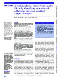
Fig. 1A–D Radiographs show how (A) a standing lateral film in extension and (C) a flexed supine lateral film are uploaded in JointTrack and (B, D) 3D templates are matched with implants.
physical therapy session per day. Additionally, all patients received an active assisted continuous motion device twice a day for 30 minutes each. We saw all patients at 2 weeks, 12 weeks, 12 months, and then every other year for routine clinical examinations and radiographic followup consisting of one bilateral standing AP film, lateral standing films in extension, and supine films at 90° flexion, including bilateral skyline films. We utilized lateral films in extension and flexion for the three-dimensional (3D) matching technique. No patient in either group had a minor or major complication, such as superficial or deep infection, deep venous thrombosis, pulmonary embolism, falls, or wound healing problems. We saved all digital radiographs as .jpeg files and uploaded them with their calibration files containing the
123
source-to-image distance, X and Y values of the center point, and inches to pixels ratio. We uploaded 3D .stl implant files in Rhinoceros1 (McNeel, Seattle, WA, USA). We oriented implant models to a coordinate system and mirrored right femoral models to create left femoral models. We then uploaded 3D implant files of tibial and femoral components and radiographs in an open-source software program (JointTrack; jointtrack.sourceforge.net). We matched implants with radiographs (Fig. 3) and calculated the relative position of femoral and tibial components. A postprocessing program of JointTrack calculated the deepest points of the medial and lateral condyles in relationship to the tibial component. Variables, such as the AP position of the medial and lateral condyles, angle between both condyles and the long axis of the tibia,
Volume 470, Number 1, January 2012
Comparison of Two Gap-balancing Techniques
relative position of the lateral condyle compared to the medial condyle, and liftoff of the medial and lateral condyles, were calculated. A previous study described and validated this method [1]. We computed descriptive statistics for demographic and preoperative characteristics. Percentages were calculated for categorical variables, while the median and range were calculated for continuous variables. We calculated the median tibial internal/external rotation, contact-contact line angle, AP position of the medial condyle, AP position of the lateral condyle, distance between the medial and lateral condyles, liftoff of the medial condyle, liftoff of the lateral condyle, and varus/valgus angle for both groups and calculated 95% CIs for the medians. We used the nonparametric
Fig. 2 This intraoperative image shows the modification of anterior referencing used in the new gap-balancing technique. Instead of referencing off the anterior femoral cortex, the deepest point of the trochlea groove is utilized using a depth gauge. The minimum resection represents the prosthetic trochlea thickness, which is 4.5 mm using a PFC1 CR-TKA.
95
Wilcoxon rank-sum test to compare the groups with respect to kinematic outcomes. A Bonferroni correction was used to adjust for multiple comparisons. We used SAS statistical software (Version 9.2) to conduct all analyses (SAS Institute, Inc, Cary, NC, USA).
Results In flexion, the new gap-balancing technique resulted in tibial internal rotation of +3.5° (95% CI: 0.1°, 9.0°), which was different (p = 0.005) from the control group, where we observed tibial external rotation of 4.6° (95% CI: 6.6°, 0.3°) (Table 2). Nine of 10 knees in the treatment group had tibial internal rotation, but seven of nine knees in the control group had tibial external rotation. In extension,
Fig. 3 This radiograph shows how distal and posterior resection is calculated based on implant thickness. If implant thickness (a) is 8 mm posterior and 9 mm distally, cartilage and bone loss (c) is 3 mm; thus, total bone resection (b) is implant thickness (a) minus cartilage and bone loss (c): b = a c.
Table 2. Results of the exact two-sample Wilcoxon test for knee kinematic measurements with the leg in flexion after 1 year of followup Parameter
Study group (new technique)
Control group (traditional technique)
p value
Tibial internal (+)/external ( ) rotation (°)
3.54 (0.13, 8.95)
4.59 ( 6.66, 0.29)
0.005
Contact-contact line angle (°)
3.51 (0.13, 8.95)
4.59 ( 6.66, 0.29)
0.008
AP position medial condyle (mm)
2.59 ( 4.51, 3.22)
8.20 ( 8.80,
AP position lateral condyle (mm)
4.35 ( 5.38,
4.52 ( 5.42, 0.70)
2.14)
2.52)
0.223 [ 0.999
Distance medial/lateral condyle (mm)
2.72 (0.10, 6.65)
3.47 ( 5.07, 0.22)
0.008
Liftoff medial condyle (mm)
7.18 (5.32, 9.06)
7.81 (5.37, 11.78)
[ 0.999
Liftoff lateral condyle (mm)
6.45 (5.74, 8.65)
10.40 (8.95, 12.08)
0.017
Varus/valgus angle (°)
0.33 ( 1.77, 2.37)
2.11 ( 7.40, 1.19)
0.522
Values are expressed as median, with 95% CI in parentheses; p values were adjusted for multiple comparisons using a Bonferroni correction.
123
96
Clinical Orthopaedics and Related Research1
Fitz et al.
Table 3. Results of the exact two-sample Wilcoxon test for knee kinematic measurements with the leg in extension after 1 year of followup Parameter
Study group (new technique)
Tibial internal (+)/external ( ) rotation (°)
1.00 ( 4.64, 2.16)
Control group (traditional technique)
p value
1.06 ( 2.25, 5.50)
[ 0.999
Contact-contact line angle (°)
0.94 ( 2.40, 1.86)
1.43 ( 2.49, 5.19)
[ 0.999
AP position medial condyle (mm)
5.46 ( 7.88,
3.35)
4.49 ( 7.14,
1.79)
[ 0.999
AP position lateral condyle (mm)
5.96 ( 7.13,
4.27)
5.28 ( 6.90,
3.36)
Distance medial/lateral condyle (mm)
0.72 ( 1.85, 1.47)
1.06 ( 1.87, 3.93)
[ 0.999
Liftoff medial condyle (mm)
6.46 (5.54, 7.33)
9.24 (5.94, 13.34)
0.348
Liftoff lateral condyle (mm) Varus/valgus angle (°)
6.45 (4.73, 7.72) 0.02 ( 0.69, 1.67)
7.78 (6.25, 12.32) 0.64 ( 1.64, 3.13)
0.223 [ 0.999
[ 0.999
Values are expressed as medians, with 95% CI in parentheses; p values were adjusted for multiple comparisons using a Bonferroni correction.
we found no differences in the tibial internal/external rotation between the two groups (Table 3). In flexion, the new gap-balancing technique resulted in lateral rollback of +2.7 mm (95% CI: 0.1 mm, 6.7 mm), which was different (p = 0.008) from the control group, where the lateral condyle was 3.5 mm (95% CI: 5.1 mm, 0.2 mm) more anteriorly compared to the medial condyle (Table 2). In extension, we found no differences in the lateral rollback between the two groups (Table 3). The two gap-balancing techniques did not result in different liftoffs comparing the medial and lateral condyles in extension (Table 3), but there were differences in flexion (Table 2). The lateral liftoff was larger (p = 0.017) in the control group than in the treatment group: 10.4 mm (95% CI: 9.0 mm, 12.1 mm) and 6.5 mm (95% CI: 5.7 mm, 8.7 mm), respectively.
Discussion Normal knee kinematics is characterized by posterior femorotibial rollback with tibial internal rotation and medial-pivot rotation in flexion. CR-TKAs do not reproduce normal knee kinematics. We therefore described the kinematic changes using a modified gap-balancing technique in a symmetric CR-TKA. We asked whether a more anatomic restoration of the distal and posterior medial femoral condyle would result in closer-to-normal knee kinematics compared to a more traditional surgical gapbalancing technique. We specifically sought to determine whether these modifications would result in knee kinematics characterized by more tibial internal rotation in flexion, more lateral rollback in flexion, and less condylar liftoff with flexion. There are several limitations of this study. First, lateral radiographs were taken in different degrees of flexion measured on anatomic landmarks on the femur and tibia (100° versus 90°) in both groups and may not represent comparable positions to observe kinematics. The ease of
123
bending the knee 3 years postoperatively compared to 1 year may be the reason for the different positions. In vivo fluoroscopic studies avoid this problem by selecting the frames in 10° to 30° increments [2, 4, 5, 13, 16, 17, 23, 24] at certain flexion angles. Second, our radiographs in flexion were nonweightbearing. Most but not all [18] published kinematic results are weightbearing, making it difficult to compare our nonweightbearing findings to prior literature. Johal et al. [10] observed tibiofemoral movement in 10 weightbearing and nonweightbearing Caucasian knees using MRI and found tibial internal rotation with flexion was present in both groups but was a magnitude of rotation greater and occurred earlier with weightbearing. We may have seen even larger degrees of tibial internal rotation and lateral rollback in weightbearing films, and we will investigate this further. Third, we report preliminary kinematic data without clinical results and cannot conclude whether these differences have a clinically important impact on patient function or implant durability. Fourth, we do not know whether this new technique can be reproduced by other surgeons. The results of this pilot study suggest a role for a randomized prospective study comparing this new technique with a traditional surgical technique. Our preliminary findings show similarity to published kinematic results using an asymmetric CR-TKA or a medial-pivot TKA under weightbearing but less similarity to symmetric CR-TKA designs (Table 4). Bertin et al. [2] looked at asymmetric femoral components of a CR-TKA during deep-knee bending in 20 patients and observed internal rotation of 1.4°. Only 10 of his 20 patients had internal rotation. We observed tibial internal rotation in nine of 10 patients in our treatment group and only one had external rotation of 2°. Our control group showed seven of nine patients with tibial external rotation. Schmidt et al. [21] studied five patients with a medial-pivot CR-TKA design and found a medial-pivot motion in three of the five knees with tibial internal rotation of 2.2° during gait. Miyazaki et al. [16] reported on 29 medial-pivot CR-TKA and observed postoperative flexion of 130.6° with an
Volume 470, Number 1, January 2012
Comparison of Two Gap-balancing Techniques
97
Table 4. Comparison of our findings with published results of tibial internal rotation and femoral rollback between full extension 0° and 90° of flexion Study
Implant
Tibial internal rotation
Lateral rollback (mm)
Bertin et al. [2]
Asymmetric CR-TKA
1.4°
Dennis et al. [4]
Curved CR-TKA
0
Johal et al. [10]
Normal, nonweightbearing males, neutral tibial rotation
14° (from 0° to 120°)
Miyazaki et al. [16]
Advance1 medial-pivot TKA
5.5°
2
Miyazaki et al. [16]
Advantim1
8°
6
Miyazaki et al. [16]
Fine
10°
6
1
Nozaki et al. [17]
Advantim
Current study
CR-TKA (new technique)
Current study
CR-TKA (traditional technique)
(2 different surgeons)
1.3 0
2° and 3° 3.5° 4.6° (external rotation)
18
\1 2.7 3.5
CR-TKA = cruciate-retaining TKA.
average of 10° of tibial internal rotation. Mannan and Scott [15] reported on 10-year results of 228 medial-pivot TKAs using a different design and found no improvement in flexion compared to other published CR-TKA results. Our kinematic observations in the treatment group were similar to the medial-pivot CR-TKA regarding tibial internal rotation with flexion. Stiehl et al. [24] compared CR-TKA with normal knees using fluoroscopy and found anterior translation of the condyles with flexion. They thought the absence of physiological rollback likely reflected ACL deficiency, alteration of the normal joint line, or other subtle changes modifying kinematics. In an in vivo multicenter study of CR-TKA and posterior-stabilizing TKA, Dennis et al. [5] observed anterior femoral translation in the majority of CR-TKAs medially and laterally of greater than 3 mm but in some knees even more than 9 mm. Another study [23] found lateral- but not medial-pivot motion, which we observed in our control group. Hill et al. [8] and others [14] observed anterior translation of the medial condyle of 4 mm in 90°, which was slightly more than the findings by Johal et al. [10]. We would advise caution concerning our liftoff results since this pilot study was performed without 3D templates of the PE inserts. Schmidt et al. [21] measured distances from the medial and lateral condyles to the tibial plateau to assess condylar liftoff. If the difference was greater than 1 mm, liftoff was denoted. Bertin et al. [2] used the same technique, but it remained unclear whether both authors measured the distance of the lowest point of the medial and lateral condyles to the closest point on the tibial PE insert or to the tibial plateau. Dennis et al. [7] rotated the 3D image of the matched femoral and tibial components to a frontal view and measured the distance of both condyles to the tibial tray. They also described only the distance of the lowest point of the medial and lateral condyles to the origin
of the 3D Cartesian coordinate system but not to the projected curved surface of the tibial insert. Dennis et al. [7] observed liftoff in 75% of CR-TKAs and cruciatesubstituting TKAs. Bertin et al. [2] reported decreased liftoff laterally, with increased flexion (50% of subjects at 80° and 35% at 100°) and a reduction of lateral liftoff in the medial-pivot TKA: only one of five had liftoff. Whether the general lack of liftoff in our study was the result of the comparison of two different gap-balancing techniques remains unclear and warrants further investigation. Our findings are interesting but need further investigation. The suggested closer-to-normal reconstruction of the distal and posterior medial condyle, combined with the preservation of the PCL, may result in more reproducible kinematics characterized by tibial internal rotation and lateral rollback with flexion. To perform more accurate liftoff calculations, kinematic studies in the future should include 3D models of individual PE insert thicknesses, in addition to tibial plateau templates to calculate separation between PE surface and the most inferior points of both condyles. This new gap-balancing technique in a symmetric CR-TKA may have the potential to achieve closerto-normal knee kinematics characterized by tibial internal rotation and lateral condylar rollback in flexion without changing the implant design. Acknowledgments The authors thank Scott Banks, PhD, for assistance in JointTrack and Karen Aneshansley for proofreading the manuscript.
References 1. Banks SA, Hodge WA. Accurate measurement of threedimensional knee replacement kinematics using single-plane fluoroscopy. IEEE Trans Biomed Eng. 1996;43:638–649. 2. Bertin KC, Komistek RD, Dennis DA, Hoff WA, Anderson DT, Langer T. In vivo determination of posterior femoral roll-back for
123
98
3.
4.
5.
6.
7.
8.
9.
10.
11.
12.
Fitz et al. subjects having a NexGen posterior cruciate-retaining total knee arthroplasty. J Arthroplasty. 2002;17:1040–1048. Bull AM, Kessler O, Alam M, Amis AA. Changes in knee kinematics reflect the articular geometry after arthroplasty. Clin Orthop Relat Res. 2008;466:2491–2499. Dennis DA, Komistek RD, Colwell CE Jr, Ranawat CS, Scott RD, Thornhill TS, Lapp MA. In vivo anteroposterior femorotibial translation of total knee arthroplasty: a multicenter analysis. Clin Orthop Relat Res. 1998;356:47–57. Dennis DA, Komistek RD, Scuderi G, Argenson JN, Insall J, Mahfouz M, Aubaniac JM, Haas B. In vivo three-dimensional determination of kinematics for subjects with a normal knee or a unicompartmental or total knee replacement. J Bone Joint Surg Am. 2001;83-A(suppl 2):104–115. Dennis DA, Komistek RD, Walker SA, Cheal EJ, Stiehl JB. Femoral condylar lift-off in vivo in total knee arthroplasty. J Bone Joint Surg Br. 2001;83:33–39. Dennis DA, Mahfouz MR, Komistek RD, Hoff W. In vivo determination of normal and anterior cruciate ligament-deficient knee kinematics. J Biomech. 2005;38:241–253. Hill PF, Vedi V, Williams A, Iwaki H, Pinskerova V, Freeman MA. Tibiofemoral movement 2: the loaded and unloaded living knee studied by MRI. J Bone Joint Surg Br. 2000;82:1196–1198. Insall J, Ranawat CS, Scott WN, Walker P. Total condylar knee replacement: preliminary report. Clin Orthop Relat Res. 1976; 120:149–154. Johal P, Williams A, Wragg P, Hunt D, Gedroyc W. Tibiofemoral movement in the living knee: a study of weight bearing and non-weight bearing knee kinematics using ‘‘interventional’’ MRI. J Biomech. 2005;38:269–276. Kadoya Y, Kobayashi A, Komatsu T, Nakagawa S, Yamano Y. Effects of posterior cruciate ligament resection on the tibiofemoral joint gap. Clin Orthop Relat Res. 2001;391:210–217. Kesman TJ, Kane PH, Kaufman KR, Trousdale RT. Caution required when using an intraoperative knee balancer in total knee arthroplasty. J Arthroplasty. 2010;25:829–831.
123
Clinical Orthopaedics and Related Research1 13. Komistek RD, Dennis DA, Mahfouz M. In vivo fluoroscopic analysis of the normal human knee. Clin Orthop Relat Res. 2003;410:69–81. 14. Kurosawa H, Walker PS, Abe S, Garg A, Hunter T. Geometry and motion of the knee for implant and orthotic design. J Biomech. 1985;18:487–499. 15. Mannan K, Scott G. The Medial Rotation total knee replacement: a clinical and radiological review at a mean follow-up of six years. J Bone Joint Surg Br. 2009;91:750–756. 16. Miyazaki Y, Nakamura T, Kogame K, Saito M, Yamamoto K, Suguro T. Analysis of the kinematics of total knee prostheses with a medial pivot design. J Arthroplasty. 2010 December 6 [Epub ahead of print]. 17. Nozaki H, Banks SA, Suguro T, Hodge WA. Observations of femoral roll-back in cruciate-retaining knee arthroplasty. Clin Orthop Relat Res. 2002;404:308–314. 18. Pinskerova V, Johal P, Nakagawa S, Sosna A, Williams A, Gedroyc W, Freeman MA. Does the femur roll-back with flexion? J Bone Joint Surg Br. 2004;86:925–931. 19. Pritchett JW. Patient preferences in knee prostheses. J Bone Joint Surg Br. 2004;86:979–982. 20. Pritchett JW. Patients prefer a bicruciate-retaining or the medial pivot total knee prosthesis. J Arthroplasty. 2011;26:224–228. 21. Schmidt R, Komistek RD, Blaha JD, Penenberg BL, Maloney WJ. Fluoroscopic analyses of cruciate-retaining and medial pivot knee implants. Clin Orthop Relat Res. 2003;410:139–147. 22. Schnurr C, Cse´csei G, Nessler J, Eysel P, Ko¨nig DP. How much tibial resection is required in total knee arthroplasty? Int Orthop. 2011;35:989–994. 23. Stiehl JB, Komistek RD, Dennis DA. Detrimental kinematics of a flat on flat total condylar knee arthroplasty. Clin Orthop Relat Res. 1999;365:139–148. 24. Stiehl JB, Komistek RD, Dennis DA, Paxson RD, Hoff WA. Fluoroscopic analysis of kinematics after posterior-cruciateretaining knee arthroplasty. J Bone Joint Surg Br. 1995;776:884– 889.