171-176.qxd
2/6/2009
12:14 ÌÌ
™ÂÏ›‰·171
ONCOLOGY REPORTS 22: 171-176, 2009
171
Does dexamethasone inhibit anticancer activity of cetuximab in squamous cell carcinoma cell lines of the head and neck? JENS WAGENBLAST1, MEHRAN BAGHI1, SUSANNE MÖRTEL1, DANIEL HIRTH1, LAURA THRON1, CHRISTOPH ARNOLDNER2, WOLFGANG GSTÖTTNER2, ANGELIKA MAY1 and MARKUS HAMBEK1 1
ENT Department, Goethe University Medical School, Frankfurt am Main, Germany; 2ENT Department, Medical University of Vienna, Austria Received February 23, 2009; Accepted April 27, 2009 DOI: 10.3892/or_00000421
Abstract. Glucocorticoids such as dexamethasone are widely used as comedication in the treatment of head and neck cancer, e.g., to improve appetite and decrease weight loss and fatigue in patients with advanced disease or as antiallergic and antiemetic prophylaxis during anti-EGFR therapy. However, the literature suggests that dexamethasone induces resistance to antineoplastic agents in many solid tumor models in vitro and in vivo. Since this phenomenon has never been investigated in head and neck cancer, the present study was conducted to investigate the effect of dexamethasone on the antiproliferative activity of cetuximab in vitro in squamous cell carcinoma of the head and neck (SCCHN) cell lines. The antiproliferative effect of the anti-EGFR agent cetuximab alone and in combination with increasing concentrations of dexamethasone was examined in eight SCCHN cell lines at three different time-points (24, 48 and 72 h). Cell growth inhibition and viability were measured quantitatively using WST and LDH assays. Absolute tumor cell numbers were determined by cell counting in a Rosenthal chamber. Cetuximab alone inhibited the growth of all eight SCCHN cell lines significantly (p=0.008). In some cases the addition of dexamethasone reduced the antiproliferative activity of cetuximab (p≤0.038) but remained significant in all of the eight SCCHN cell lines compared with untreated controls (p≤0.028) at each drug concentration and each time-point. In contrast to the results reported for other tumor models, in our study dexamethasone showed in the majority of the evaluated dexamethasone drug concentrations and timepoints no inhibition of the cytotoxic activity of cetuximab. The reasons for these discrepant findings are unclear but may be related to the degree of tumor cell differentiation or
_________________________________________ Correspondence to: Dr Jens Wagenblast, ENT Department, University Hospital, Theodor-Stern-Kai 7, D-60590 Frankfurt/ Main, Germany E-mail:
[email protected]
Key words: cetuximab, dexamethasone, squamous cell carcinoma of the head and neck cell lines, resistance, apoptosis
proliferation rate. Thus, further studies are required to elucidate the molecular mechanisms underlying the interaction between dexamethasone and cetuximab in different tumors. Introduction Cancer of the head and neck most frequently affects the oral cavity, pharynx or larynx and accounts for more than 5% of all malignancies worldwide. In 2002, more than 500,000 new cancer diagnoses and more than 300,000 deaths were attributable to this disease (1). Most head and neck cancers are squamous cell carcinomas (SCCHN). The prognosis depends primarily on disease stage and performance status at the time of diagnosis (2,3). Treatment options are limited for metastatic and/or recurrent disease, and there is an urgent need for new, well tolerated therapies (4). Molecular targeted therapies, especially those targeting the epidermal growth factor receptor (EGFR) have recently attracted attention as promising candidates for the treatment of head and neck cancers (5). EGFR overexpression in head and neck cancer has been correlated with a poor prognosis (6,7), and the potential therapeutic value of modulating the EGFR signalling pathway is reflected by the broad range of molecular EGFR inhibitors developed in recent years. Cetuximab is an immunoglobuline G1 monoclonal antibody with a higher affinity to the extracellular domain of the receptor compared with its natural ligands. The drug is under active investigation as a promising anticancer agent in general (8) and especially in SCCHN (9). A significant activity has been demonstrated in clinical trials of EGFR-blocking antibodies (10). Skin toxicity is the most important side effect of treatment with EGFR inhibitors including cetuximab. Severe infusion reactions have also been reported in rare cases and described as hypersensitivity (10), anaphylactic (11) or allergic reactions (12), although the mechanism of this adverse effect has not yet been identified. In most cases the cetuximab infusion was stopped after an infusion reaction occurred. Melichar et al (13), however, reported the successful continuation of cetuximab infusion after an infusion reaction when glucocorticoids were added and the patients were monitored in an intensive care unit. Apart from antiallergic prophylaxis during anti-EGFR therapy, glucocorticoids such as dexamethasone are widely used as a comedication in the treatment of head and neck cancer, e.g., to improve appetite or decrease weight
171-176.qxd
2/6/2009
12:14 ÌÌ
™ÂÏ›‰·172
WAGENBLAST et al: DEXAMETHASONE AND CETUXIMAB IN SCCHN CELL LINES
172 A
B
C
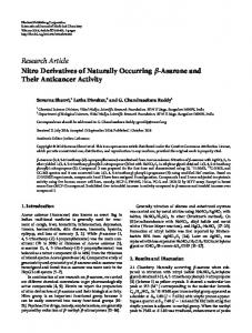
Figure 1. (A-C) The growth-inhibitory effect of cetuximab at two concentrations (C1=0.5 and C2=5.0 μmol/l), administered either alone or in combination with one of four concentrations of dexamethasone (D1=1.0 μmol/l, D2=2.5 μmol/l, D3=5.0 μmol/l, D4=10.0 μmol/l) in the SCCHN cell line PJ-34 which was representative for the eight tumor cell lines investigated (yellow columns). The corresponding untreated tumor cell line (black column) served as a control and was incubated only with the cell-type specific medium Quantum 263 with L-glutamine. The absolute tumor cell numbers in treated and control cell lines were determined in a Rosenthal chamber at 24, 48 and 72 h after treatment or incubation with Quantum 263 (controls), respectively. Mean values of three independent experiments with standard deviation are shown. Similar results were obtained in all eight tumor cell lines investigated. Compared with the control group, single agent cetuximab had a highly significant (p=0.008) antiproliferative effect at both concentrations (0.5 and 5.0 μmol/l) and at all time-points (24, 48 and 72 h) in all eight squamous cell carcinoma cell lines. Cetuximab was also found to be significantly active at both concentrations and at every time-point when it was given in combination with dexamethasone at all concentrations compared with untreated controls using the Wilcoxon test for matched pairs samples (p≤0.021 at 24 h, p≤0.028 at 48 h and p≤0.008 at 72 h).
171-176.qxd
2/6/2009
12:14 ÌÌ
™ÂÏ›‰·173
ONCOLOGY REPORTS 22: 171-176, 2009
173
loss, fatigue or nausea in patients with advanced disease. However, in the past few years several publications have discussed the potential of glucocorticoids like dexamethasone to induce resistance to antineoplastic agents (14,15). Thus, the question arises if dexamethasone used to prevent or treat allergic reactions to cetuximab may also lead to resistance to the agent in the treatment of SCCHN. The primary objective of our study was to investigate the interaction of dexamethasone and cetuximab in several SCCHN cell lines.
cells were subsequently incubated for 30 min at room temperature. During the incubation period, the micro-plates were protected from light. The optical density of each well was determined using a microplate reader (Dynatech Laboratories, Chantilly, VA, USA) at a wavelength of 490 nm with a reference wavelength of 630 nm. Each experiment was done in triplicate. For statistical analysis, a Wilcoxon test for matched pairs (dependent samples) was performed using SPSS 13.0 software for Windows.
Materials and methods
Results
Eight different SCCHN cell lines were used in this study. PE/CA-PJ-15, PE/CA-PJ-34, PE/CA-PJ-41 and PE/CA-PJ-49 cells were obtained from ECACC (European Collection of Cell Cultures, Salisbury, Wiltshire, UK), and Cal-27 and Kyse-140 cells were purchased from DSMZ GmbH (Braunschweig, Germany). CLS-354 and UM-SCC-14C were obtained from CLS Cell Line Service (Eppelheim, Germany). The fibroblast cell line was a gift of the Department of Dermatology, University Hospital, Frankfurt/Main, Germany. Cetuximab was obtained from Merck (Darmstadt, Germany) and dexamethasone from Sigma-Aldrich (Munich, Germany). The cell lines were cultivated according to the instructions of the suppliers without antibiotics at 37˚C in the cell-type specific medium Quantum 263 with L-glutamine (PAA Laboratories GmbH, Pasching, Austria). Cells were seeded in 96-multiwell plates (1x100,000 cells/well), and after incubation for 24 h, the cells were treated with cetuximab alone or in combination with dexamethasone for 24, 48 or 76 h, respectively. In all experiments described in this publication, cetuximab was used in two different concentrations (0.5 and 5.0 μmol/l), while dexamethasone was used at four increasing concentrations ranging from 1 to 10 μmol/l; these concentrations are comparable with the clinically achievable tissue concentrations of the drug (16). The number of cells was determined in a Rosenthal chamber after 24, 48 and 72 h of treatment. Cell viability and cell killing were determined by a WST and lactate dehydrogenase (LDH) assay, respectively. For the WST assay, 1x10⁵ cells per well were cultivated in a 96-well plate for 24 h and then treated with the aforementioned concentrations of cetuximab and dexamethasone for 24, 48 or 72 h, respectively. WST (10 μl) at 5 g/l (Roche Diagnostics, Mannheim, Germany) were added to the medium in triplicate at each dose and incubated for 1 h at 37˚C. Absorbance was measured at 450 nm using a microplate reader. LDH activity in the culture medium was measured with the Cytotoxicity Detection Kit plus purchased from Roche Mannheim, Germany. Briefly, cells were incubated in a 96-well microplate (Falcon, Franklin Lakes, NJ, USA), with 5,000 cells in 200 μl seeded per well with Quantum 263 PAA. After 24 h, the medium was removed and replaced either by the same medium containing cetuximab with or without dexamethasone at the concentrations specified above or drug-free medium (low controls), or medium containing 1% Triton X-100 (Sigma Chemical Co.) to determine total cellular LDH (high controls). After 24, 48 or 72 h of treatment, 100-μl samples were removed from the wells and transferred to another well-plate, 100 μl of the LDH assay reaction mixture were added to each well, and
Cetuximab alone showed a highly significant (p=0.008) antiproliferative effect compared with the control group at both concentrations (0.5 and 5.0 μmol/l) and at all timepoints (24, 48 and 72 h) in all eight cell lines. Since the present study was conducted to investigate the effect of dexamethasone on the antiproliferative activity of cetuximab in vitro in squamous cell carcinoma of the head and neck (SCCHN) cell lines, cetuximab was combined with four different concentrations of dexamethasone. When cetuximab was given in combination with dexamethasone at four different concentrations, the drug also exerted significant antiproliferative activity compared with untreated controls at both concentrations and each time-point considered (p≤0.021 at 24 h, p≤0.028 at 48 h and p≤0.008 at 72 h), although there was a significantly (p