[Downloaded free from http://www.annalspc.com on Wednesday, September 28, 2016, IP: 185.96.46.2]
CASE REPORT
Double-outlet left atrium: Ventriculo-atrial malalignment defect Sudhir S Shetkar, Shyam S Kothari Department of Cardiology, All India Institute of Medical Sciences, New Delhi, India
ABSTRACT A four‑month‑old female infant presented with heart failure and cyanosis. On echocardiography, there was no outlet to the right atrium except an atrial septal defect and the left atrium drained into the respective ventricles by two atrioventricular valves. The case is reported for its rarity, with a brief literature review. Keywords: Congenital heart defects, double‑outlet atrium, double‑outlet left atrium
INTRODUCTION Double‑outlet left atrium (DOLA) is a very rare congenital heart defect, wherein, the left atrium drains into both the ventricles, and the only outlet for the right atrium is the atrial septal defect (ASD). The status of the left atrioventricular valve (AV) can be variable in this setting, and the term DOLA has been used somewhat differently by authors. Only a few isolated cases are reported in the literature.[1‑5] We report a patient with DOLA in view of its rarity.
CASE REPORT A four‑month‑old, female infant, weighing two kilograms, presented to us with failure to thrive and easy fatigability during feeding, since birth. She had two episodes of lower respiratory tract infection in the past and had a history of mild cyanosis on crying. The physical examination revealed a heart rate of 124 beats/minute, blood pressure of 84/52 mmHg, respiratory rate of 32/minute with oxygen saturation (SaO2) of 92% in room air. A pansystolic murmur at the left lower sternal border and a split second heart sound were heard on auscultation. Mild hepatomegaly was noted. Video available on www.annalspc.com Access this article online Quick Response Code:
Website: www.annalspc.com DOI: 10.4103/0974-2069.115266
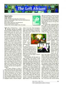
The electrocardiogram (EKG) showed left atrial enlargement, with left axis deviation, without ventricular hypertrophy. The chest radiograph showed cardiomegaly and increased pulmonary vascularity. A transthoracic echocardiography [Figure 1 and Video 1] revealed situs solitus, levocardia, and d‑loop ventricles, with normal pulmonary and systemic venous drainage. Well‑defined right and left atria were present. There was right atrial outlet Atresia, with the only outlet for the right atrium being a secundum ASD. The left atrium emptied into the right and left ventricles through two AV valves. The right‑sided AV valve had septal attachments and opened into the trabeculated right ventricle, while the left AV valve opened into a smooth‑walled left ventricle. Thus, morphologically the right and left AV valves resembled the tricuspid and mitral valves, respectively. There was no significant AV regurgitation. There was a restrictive inlet ventricular septal defect (VSD) shunting left to right with a gradient of 40 mmHg [Figure 1e and 1f]. The great vessels were normal in origin and position. There was no right ventricular outflow obstruction. The left ventricular outflow tract (LVOT) was somewhat elongated [Figure 1d]. The coronary sinus and aortic arch anatomy were normal. She was planned for an initial pulmonary artery banding followed by total repair at a later date. However, she died at home from a possible chest infection few weeks after presentation to the hospital.
DISCUSSION Double‑outlet atrium has been initially described as a condition, wherein, either the right or left atrium drains into both the ventricles. The anomaly is quite rare. Double‑outlet left atrium (DOLA) is even rarer than double‑outlet right atrium (DORA). Only few isolated
Address for correspondence: Dr. Shyam S Kothari, Department of Cardiology, All India Institute of Medical Sciences, New Delhi ‑ 110 029, India. E‑mail:
[email protected] 158
Annals of Pediatric Cardiology 2013 Vol 6 Issue 2
[Downloaded free from http://www.annalspc.com on Wednesday, September 28, 2016, IP: 185.96.46.2] Shetkar and Kothari: Double outlet left atrium
a
b a
c
e
d
f
Figure 1: (a) Echocardiography in the subcostal view shows right atrium outlet atresia with a large secundum ASD. Left atrium opens into the right and left ventricle through two AV valves. (b) OS‑ASD shunting right to left. (c) Left atrial appendage confirms morphological LA. (d) Parasternal long axis view shows elongated LVOT. (e) Subcostal view shows inlet ventricular septal defect, (f) Shunting left to right, with a gradient of 40 mm Hg. ASD: Atrial septal defect, LVOT: Left ventricular outflow tract, AO: Aorta, MPA: Main pulmonary artery, RPA and LPA: Right and left pulmonary artery
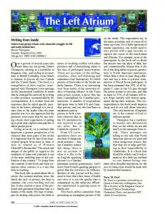
cases of DOLA are reported. Von Mierop [6] initially described it as a variant of the endocardial cushion defect, with the assumption that the AV valve represents a common AV valve and the abnormality results from the extreme malalignment of the atrial septum. However, subsequently, different viewpoints and cases with variations in the pathoanatomy in both DORA and DOLA have been described. The unique hearts with double outlet atrium, thus present difficulties in nosologic characterization and precise terminology. An overtly simplistic scheme for the pathogenesis of double‑outlet atrium is presented in Figure 2. Normally the planes of the atrial and ventricular septa intersect each other at an angle of about 15 degrees.[7] Deviations from this normal relationship have been reported in cases with a common AV canal.[7] An extreme dextroposition of the atrial septum to the right rather than growing to the center of the AV canal, leading to its fusion with the right margin of the AV canal, typifies cases having right atrial outlet atresia [Figure 2a].[4,6] When the right atrial outlet is atretic, the ASD remains the only outlet for the right atrium. DOLA, if occur in the setting of a common AV junction, such outlet would be an ostium Annals of Pediatric Cardiology 2013 Vol 6 Issue 2
b
Figure 2: Schematic diagram showing the anatomical spectrum of double‑outlet left atrium with proposed morphogenetic hypotheses (a) Fusion of the atrial septum with the right lateral atrial border leads to right atrial outlet atresia; the spectrum of the AV valve ranges from straddling AV valve to two AV valves. (b) Malalignment between the IAS and IVS will lead to an anomaly, without right atrial outlet atresia with three AV valves. (Adapted from reference 8) AV: Atrioventricular valve, IAS: Interatrial septum, IVS: Interventricular septum
primum ASD. Malalignment between the interatrial septum (IAS) and interventricular septum (IVS) explains those cases without right atrial outlet atresia.[8,9] These cases can then present with three AV valves [Figure 2b], one guarding the right atrial outlet and two guarding the left atrial outlet.[9] Such has actually been reported with double‑outlet right atrium (DORA).[9] To the best of our knowledge the clinical entity of DOLA comprising three AV valves has been reported only once in the setting of a l‑loop topology and the third valve being an overriding and straddling left AV valve.[10] Thus, ventriculoatrial malalignment may be one of the reasons for such an anomaly. This type of malalignment need not be only malalignment between the atrial and ventricular septal tissues, but may involve the entire ventricle loop. It may be remembered that the atria are fixed in the fetus because of the systemic and pulmonary venous drainage, whereas, the ventricle mass undergoes much rotation and movement. Ventriculoarterial malalignment is well recognized for the pathogenesis of congenital heart lesions, but the importance of ventriculoatrial malalignment is less well appreciated.[11] The reason/s for such a malalignment remains unknown, and the role of the endocardial cushion is speculative and seems unlikely. Recent studies of cardiac development suggest that the failure of ingrowth into the developing heart from the dorsal mesenchymal tissues is responsible for this malformation and not the endocardial cushion defect, as considered previously.[12] Double‑outlet atrium in the presence of atresia of one AV valve poses other thematic problems. Since the right AV valve is atretic, the left AV valve cannot be described as guarding a common AV junction, and hence, it is not a common AV valve. It is the left AV valve that has been embryologically divided into the right and left AV valves (morphologically like the tricuspid and 159
[Downloaded free from http://www.annalspc.com on Wednesday, September 28, 2016, IP: 185.96.46.2] Shetkar and Kothari: Double outlet left atrium
Table 1: Reported cases of double-outlet left atrium with common/two AV valves and their associated lesions Cases
Age
Sex
Atrial communication
LA outlet
Associated lesions
Case 1[1] Case 2[2] Case 3[3] Case 4[4] Case 5[5] Case 6[5] Case 7[5] Case 8 (present case)
‑ Six months Eight months 25 years One month 31 years 32 years Four months
‑ F F F F F M F
ASD OS‑ASD OS‑ASD SV‑ASD OS‑ASD ASD OS‑ASD OS‑ASD
Common AV valve Two AV valves Two AV valves Two AV valves Two AV valves Two AV valves Two AV valves Two AV valves
L‑looped DORV, Pulmonary stenosis Anteriorly malaligned VSD, Pulmonary atresia Pulmonary stenosis Mild left AV regurgitation Pulmonary atresia LSVC to LA Peri‑membranous VSD, Mild left AV regurgitation Inlet VSD, Mild left AV regurgitation
OS‑ASD: Ostium secundum atrial septal defect; SV‑ASD: Sinus venosus atrial septal defect; DORV: Double‑outlet right ventricle; AV: Atrioventricular; VSD: Ventricular septal defect, #,-.#All cases except case 2 had two adequate ventricles
mitral valves). Kiraly et al.,[13] suggested that in such a uniatrial biventricular connection, the valves might not be described as mitral and tricuspid valves. In a study of straddling AV valves with atresia of one AV valve, similar to our patient in some ways, they have shown that the valves are often abnormal and may influence the long‑term surgical results. However, in our patient the AV valves were phenotypically quite like tricuspid and mitral valves, without significant regurgitation. Hearts with straddling of AV valves and one AV valve atresia often have one ventricle that is hypoplastic, and consequently, are amenable for univentricular repair only. [13,14] However, DOLA with two well‑developed ventricles like the present case have been described previously [Table 1]. The conduction system anatomy in such hearts might vary compared to the normals. Some authors suggest that the term double‑outlet atrium (DORA/DOLA) should be restricted to hearts with common or well‑defined AV valves and not to hearts having straddling AV valves with atresia of one atrial outlet.[7,14] Others argue, as noted above, that the patent AV valve is not a common AV valve, and irrespective of the details, this valve is a straddling valve.[13] A pragmatic classification proposed for DORA may be applicable for DOLA patient management. [9] Notwithstanding these differing points of view, a systematic stepwise approach of segmental analysis for the three cardiac segments (atria, ventricles, great vessels) and the two junctions (atrioventricular, and ventriculoarterial), as always recommended for congenital heart defects, will clarify the anatomy and decide the treatment. The clinical presentation of this condition depends on the size of the atrial septal defect (ASD) and its associated anomalies. With a non‑restrictive ASD, the clinical findings are similar to those of a large atrial septal defect; however, with restrictive ASD, the signs and symptoms are those of congestive heart failure. Cyanosis may be due to a complete mixing of blood at the atrial level or associated pulmonary stenosis or atresia. Other reported associations with DOLA include pulmonary stenosis or atresia, left superior vena cava (LSVC) to left atrium, ventricular septal defect, and AV regurgitation [Table 1]. 160
The clinical diagnosis can be established by echocardiography, and angiography is rarely required. The treatment depends on the status of ASD and the ventricles. A restrictive ASD may require intervention to establish an interatrial connection. When the ASD is non‑restrictive, the clinical course is more benign and the management depends on the status of the ventricles and the associated anomalies. Surgical treatment involves complete excision and reconstruction of the abnormal IAS, with patch closure of the defect to separate the left and right AV valves. A simple patch closure of the ASD, if the anomaly is not identified correctly, would be catastrophic.[3] Associated anomalies are common and need management appropriately. In conclusion, we report an infant with right outlet atresia, in whom the left atrium opened into normal‑sized ventricles. The DOLA may represent a ventriculoatrial malalignment lesion.
ACKNOWLEDGMENT We thank Professor Robert Anderson and Professor Richard Van Praagh for their advice regarding the pathoanatomy.
REFERENCES 1. Horiuchi T, Saji K, Osuka Y, Sato K, Okada Y. Successful correction of double outlet left atrium associated with complete atrioventricular canal and l‑loop double outlet right ventricle with stenosis of the pulmonary artery. J Cardiovasc Surg 1976;17:157‑61. 2. Bini RM, Pellegrino PA, Mazzucco A, Gallucci V, Milanesi O, Maddalena F, et al. Tricuspid atresia with double‑outlet left atrium. Chest 1980;78:109‑11. 3. Susuki Y, Hamada Y, Haneda K, Horiuchi T, Ogata H. Double outlet left atrium with intact ventricular septum. Ann Thorac Surg 1988;45:332‑4. 4. Naik AM, Nabar AA, Dalvi BV. Double outlet left atrium. Indian Heart J 1997;49:418‑20. 5. Espinola‑Zavaleta N, Vargas‑Barrón J, Hernández‑Morales G, Keirns C, Romero‑Cárdenas A, Roldán J. Clinical and echocardiographic aspects of double‑outlet left atrium. J Am Soc Echocardiogr 2002;15:86‑9. Annals of Pediatric Cardiology 2013 Vol 6 Issue 2
[Downloaded free from http://www.annalspc.com on Wednesday, September 28, 2016, IP: 185.96.46.2] Shetkar and Kothari: Double outlet left atrium 6. Van Mierop LH. Pathology and pathogenesis of endocardial cushion defect: Surgical implication. In: Dávila JC, editor. Second Henry Ford Hospital International Symposium on cardiac surgery. New York: Appleton‑Century‑Crofts; 1977. p. 201‑7. 7. Coto EO, Calabro R, Marsico F, Lopez Arranz JS. Right atrial outlet atresia with straddling left atrioventricular valve. A form of double outlet atrium. Br Heart J 1981;45:317‑24. 8. Beroukhim RS, Geva T. Echocardiographic features of double‑outlet right atrium and straddling tricuspid valve with intact ventricular septum: A rare cardiac anomaly associated with pulmonary atresia and single coronary artery ostium. J Am Soc Echocardiogr 2010;23:580.e5‑10. 9. Edwin F, Kinsley RH, Mamorare HM, Govendrageloo K. The spectrum of double‑outlet right atrium including hearts with three atrioventricular valves. Eur J Cardiothorac Surg 2012;41:947‑9. 10. Gnanapragasam JP, Houston AB, Anderson RH. Double outlet left atrium with double inlet left ventricle and discordant atrioventricular connection. Cardiol young 1991;1:155‑7. 11. Van Praagh R. The importance of ventriculoatrial
Annals of Pediatric Cardiology 2013 Vol 6 Issue 2
malalignment in anomalies of the atrioventricular valves, illustrated by “mitral atresia” and congenital mitral stenosis with large left ventricle. In: Doyle EF, Engle MA, Gersony WM, Rashkind WJ, Talner NS, editors. Pediatric Cardiology. Proceedings of the Second World Congress. New York: Springer‑Verlag; 1986. p. 901‑3. 12. Anderson RH, A Wessels, Vettukattil JJ. Morphology and morphogenesis of atrioventricular septal defect with common atrioventricular junction. World J Pediatr Congenit Heart Surg 2010;1:59‑67. 13. Kiraly L, Hubay M, Cook AC, Ho SY, Anderson RH. Morphologic features of the uniatrial but biventricular atrioventricular connections. J Thorac Cardiovasc Surg 2007;133:229‑34. 14. Westerman GR, Norton JB, Van Devanter SH. Double‑outlet right atrium associated with tetralogy of Fallot and common atrioventricular valve. J Thorac Cardiovasc Surg 1986;91:205‑7. How to cite this article: Shetkar SS, Kothari SS. Double-outlet left atrium: Ventriculo-atrial malalignment defect. Ann Pediatr Card 2013;6:158-61. Source of Support: Nil, Conflict of Interest: None declared
161