International Journal of
Molecular Sciences Article
Downregulation of Runx2 by 1,25-Dihydroxyvitamin D3 Induces the Transdifferentiation of Osteoblasts to Adipocytes Jung Ha Kim 1,† , Semun Seong 1,2,† , Kabsun Kim 1 , Inyoung Kim 1 , Byung-Chul Jeong 1,2 and Nacksung Kim 1,2, * 1
2
* †
Department of Pharmacology, Chonnam National University Medical School, Gwangju 61469, Korea;
[email protected] (J.H.K.);
[email protected] (S.S.);
[email protected] (K.K.);
[email protected] (I.K.);
[email protected] (B.-C.J.) Department of Biomedical Sciences, Chonnam National University Medical School, Gwangju 61469, Korea Correspondence:
[email protected]; Tel.: +82-62-220-4418 These authors contributed equally to this work.
Academic Editor: Charles J. Malemud Received: 7 March 2016; Accepted: 16 May 2016; Published: 19 May 2016
Abstract: 1,25-Dihydroxyvitamin D3 (1,25(OH)2 D3 ) indirectly stimulates bone formation, but little is known about its direct effect on bone formation. In this study, we observed that 1,25(OH)2 D3 enhances adipocyte differentiation, but inhibits osteoblast differentiation during osteogenesis. The positive role of 1,25(OH)2 D3 in adipocyte differentiation was confirmed when murine osteoblasts were cultured in adipogenic medium. Additionally, 1,25(OH)2 D3 enhanced the expression of adipocyte marker genes, but inhibited the expression of osteoblast marker genes in osteoblasts. The inhibition of osteoblast differentiation and promotion of adipocyte differentiation mediated by 1,25(OH)2 D3 were compensated by Runx2 overexpression. Our results suggest that 1,25(OH)2 D3 induces the transdifferentiation of osteoblasts to adipocytes via Runx2 downregulation in osteoblasts. Keywords: 1,25(OH)2 D3 ; transdifferentiation; osteoblast; adipocyte
1. Introduction Bone is an important organ for supporting the body and regulating mineral homeostasis. Bone is continuously remodeled by two cell types: osteoclasts and osteoblasts [1]. Osteoclasts resorb old bone and osteoblasts produce the bone matrix [2,3]. Various factors, such as the paratyroid hormone and 1,25-dihydroxyvitamin D3 (1,25(OH)2 D3 ), regulate bone homeostasis by modulating bone cells [2,4]. The steroid hormone 1,25(OH)2 D3 is a key regulator of calcium homeostasis and skeletal health [5]. In bone, 1,25(OH)2 D3 regulates mineralization both in an indirect and a direct manner. 1,25(OH)2 D3 increases calcium absorption from the intestines, which indirectly stimulates bone formation [6]. It can also directly influence bone formation via the regulation of osteoblast differentiation and function. Various studies have demonstrated the direct effects of 1,25(OH)2 D3 on osteoblast differentiation and function in vitro. However, the results of these studies are highly controversial. Several studies have shown that 1,25(OH)2 D3 stimulates osteoblast differentiation by increasing alkaline phosphatase (ALP) activity and osteocalcin expression in human primary osteoblast-like cells [7–9]. However, several lines of evidence suggest that 1,25(OH)2 D3 negatively regulates osteoblast differentiation. For example, 1,25(OH)2 D3 downregulates osteocalcin expression in mouse calvaria 3T3 (MC3T3)-E1 osteoblasts [10]. Lieben et al. [11] have also reported that 1,25(OH)2 D3 impairs mineralization by upregulating the expression of mineralization inhibitors in mouse primary osteoblasts. Osteoblasts, which are responsible for bone formation, are derived from mesenchymal stem cells by the action of several transcription factors, including Runx2, osterix, and β-catenin [12]. In particular, Int. J. Mol. Sci. 2016, 17, 770; doi:10.3390/ijms17050770
www.mdpi.com/journal/ijms
Int. J. Mol. Sci. 2016, 17, 770
2 of 11
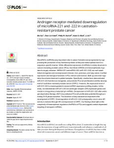
Runx2, a cell-specific member of the Runt family of transcription factors, is essential for mesenchymal cell differentiation into osteoblasts. Runx2 promotes the acquisition of an osteoblastic phenotype by mesenchymal stem cells by inducing the expression of genes encoding major bone matrix proteins, e.g., Col1a1, osteopontin, bone sialoprotein (BSP), and osteocalcin [11,12]. Runx2´/´ mice exhibit a complete lack of intramembranous and endochondral ossification in vivo and Runx2´/´ calvarial cells cannot differentiate into osteoblasts, even in the presence of osteogenic factors in vitro [12–14]. Multiple factors that play an important role in osteoblast differentiation via the regulation of Runx2 expression or activation have been identified. Adipocytes as well as osteoblasts are derived from mesenchymal stem cells. Various transcription factors, such as CCAAT/enhancer binding protein-α (CEBP-α), CEBP-β, and peroxisome proliferator-activated receptor-γ (PPAR-γ), are essential for mesenchymal cell differentiation into adipocytes [15]. Increased adipose tissue in the bone marrow of osteoporotic patients and individuals with age-dependent bone loss may be associated with the transdifferentiation of osteoblasts to adipocytes [16]. While the effect of 1,25(OH)2 D3 on the transdifferentiation of skeletal muscle cells to adipose cells is known, it is not clear if 1,25(OH)2 D3 is involved in the transdifferentiation of osteoblasts to adipocytes in the bone marrow [17]. To elucidate the direct effect of locally produced 1,25(OH)2 D3 on bone formation, we investigated the effect of 1,25(OH)2 D3 on osteoblast differentiation, Runx2 expression, and the transdifferentiation of osteoblasts to adipocytes. 2. Results 2.1. 1,25-Dihydroxyvitamin D3 (1,25(OH)2 D3 ) Inhibits Osteoblast Differentiation and Runx2 Expression in Primary Osteoblasts To elucidate the direct effect of 1,25(OH)2 D3 on bone formation, we examined its role in osteoblast differentiation. When mouse primary osteoblasts were cultured in osteogenic medium including bone morphogenic protein 2 (BMP2), ascorbic acid, and β-glycerophosphate, bone nodule formation was dramatically induced (Figure 1a,b). However, supplementation with 1,25(OH)2 D3 significantly inhibited osteoblast differentiation induced by the osteogenic medium in a dose-dependent manner (Figure 1a,b). The negative effect of 1,25(OH)2 D3 on osteogenesis was confirmed by the expression of osteoblast differentiation-related genes. As shown in Figure 1c, 1,25(OH)2 D3 inhibited the expression of osteoblastic genes, such as Runx2, ALP, and BSP. Therefore, 1,25(OH)2 D3 may negatively regulate osteoblast differentiation by inhibiting the expression of Runx2 and its downstream targets ALP and BSP. 2.2. 1,25(OH)2 D3 Induces the Transdifferentiation of Osteoblasts to Adipocytes Intriguingly, 1,25(OH)2 D3 treatment during osteoblast differentiation resulted in lipid droplet formation with the inhibition of bone nodule formation. Therefore, we examined the effect of 1,25(OH)2 D3 on adipocyte differentiation during osteoblast differentiation. Osteoblasts were cultured in osteogenic medium either without or with 1,25(OH)2 D3 . Positive Oil Red-O staining confirmed the presence of lipid droplets, which were primarily observed in osteoblasts treated with high concentrations of 1,25(OH)2 D3 (Figure 2a,b). Additionally, 1,25(OH)2 D3 significantly increased the expression of adipocyte marker genes, including CEBP-α, PPAR-γ, and adipocyte protein 2 (aP2), compared with control cells. These results indicated that 1,25(OH)2 D3 can induce the transdifferentiation of osteoblasts to adipocytes during osteoblast differentiation.
Int. J. Mol. Sci. 2016, 17, 770
3 of 11
Int. J. Mol. Sci. 2016, 17, 770
3 of 11
Int. J. Mol. Sci. 2016, 17, 770
3 of 11
Figure 1. 1,25-dihydroxyvitamin D3 (1,25(OH)2D3) inhibits osteoblast differentiation. (a–c) Primary
Figure 1. 1,25-dihydroxyvitamin D3 (1,25(OH)2 D3 ) inhibits osteoblast differentiation. (a–c) Primary osteoblasts were cultured in osteogenic medium (OGM) with vehicle or increasing concentrations of Figure were 1. 1,25-dihydroxyvitamin D3 (1,25(OH) 3) inhibits osteoblast Primary of osteoblasts cultured in osteogenic medium2D(OGM) with vehicledifferentiation. or increasing (a–c) concentrations 1,25(OH)2D3. (a) Cultured cells were fixed and stained with Alizarin red; (b) Alizarin red staining was osteoblasts were cultured in osteogenic medium (OGM) with vehicle or increasing concentrations of 1,25(OH) cells at were and stained with Alizarin red; (b) in Alizarin red staining 2 D3 . (a) quantified byCultured densitometry 562 fixed nm; (c) Primary osteoblasts were cultured osteogenic medium was 1,25(OH) 2D3. (a) Cultured cells were fixed and stained with Alizarin red; (b) Alizarin red staining was quantified densitometry 562 nm; Primary osteoblasts were cultured in osteogenic medium (OGM)bycontaining eitheratvehicle or (c) 1,25(OH) 2D3 (10−8 M). The mRNA levels of Runx2, alkaline quantified by densitometry at 562 (c) Primary osteoblasts cultured levels in osteogenic medium ´8 M). were (OGM) containing eitherand vehicle ornm; 1,25(OH) The mRNA of Runx2, alkaline 2 D3 (10 phosphatase (ALP), bone sialoprotein (BSP) were analyzed by real-time polymerase chain (OGM) containing either vehicle or 1,25(OH)2D3 (10−8 M). The mRNA levels of Runx2, alkaline phosphatase analyzed bycontrols. real-time polymerase chain reaction reaction (ALP), (PCR). #and p < bone 0.05; *sialoprotein p < 0.01; ** p