Optimising Homing Endonuclease Gene Drive Performance in a Semi-Refractory Species: The Drosophila melanogaster Experience a Yuk-Sang Chan1., David S. Huen1.¤ *, Ruth Glauert1¤b, Eleanor Whiteway1¤c, Steven Russell1,2
1 Dept. of Genetics, University of Cambridge, Cambridge, Cambridgeshire, United Kingdom, 2 Cambridge Systems Biology Centre, Cambridge, Cambridgeshire, United Kingdom
Abstract Homing endonuclease gene (HEG) drive is a promising insect population control technique that employs meganucleases to impair the fitness of pest populations. Our previous studies showed that HEG drive was more difficult to achieve in Drosophila melanogaster than Anopheles gambiae and we therefore investigated ways of improving homing performance in Drosophila. We show that homing in Drosophila responds to increased expression of HEGs specifically during the spermatogonia stage and this could be achieved through improved construct design. We found that 39-UTR choice was important to maximise expression levels, with HEG activity increasing as we employed Hsp70, SV40, vasa and bTub56D derived UTRs. We also searched for spermatogonium-specific promoters and found that the Rcd-1r promoter was able to drive specific expression at this stage. Since Rcd-1 is a regulator of differentiation in other species, it suggests that Rcd-1r may serve a similar role during spermatogonial differentiation in Drosophila. Contrary to expectations, a fragment containing the entire region between the TBPH gene and the bgcn translational start drove strong HEG expression only during late spermatogenesis rather than in the germline stem cells and spermatogonia as expected. We also observed that the fraction of targets undergoing homing was temperature-sensitive, falling nearly four-fold when the temperature was lowered to 18uC. Taken together, this study demonstrates how a few simple measures can lead to substantial improvements in the HEG-based gene drive strategy and reinforce the idea that the HEG approach may be widely applicable to a variety of insect control programs. Citation: Chan Y-S, Huen DS, Glauert R, Whiteway E, Russell S (2013) Optimising Homing Endonuclease Gene Drive Performance in a Semi-Refractory Species: The Drosophila melanogaster Experience. PLoS ONE 8(1): e54130. doi:10.1371/journal.pone.0054130 Editor: Shree Ram Singh, National Cancer Institute, United States of America Received August 27, 2012; Accepted December 10, 2012; Published January 18, 2013 Copyright: ß 2013 Chan et al. This is an open-access article distributed under the terms of the Creative Commons Attribution License, which permits unrestricted use, distribution, and reproduction in any medium, provided the original author and source are credited. Funding: This work was funded via a FNIH grant to Austin Burt (Imperial College). The FNIH funding originated from the generous support of the Bill & Melinda Gates Foundation. The funders had no role in study design, data collection and analysis, decision to publish, or preparation of the manuscript. Competing Interests: The authors have declared that no competing interests exist. * E-mail:
[email protected] ¤a Current address: School of Applied Sciences, University of Wolverhampton, Wolverhampton, West Midlands, United Kingdom ¤b Current address: Dept. of Veterinary Medicine, University of Cambridge, Cambridge, Cambridgeshire, United Kingdom ¤c Current address: Faraday Institute for Science and Religion, St. Edmund’s College, Cambridge, Cambridgeshire, United Kingdom . These authors contributed equally to this work.
specificity have recently been developed for gene therapy [3,4]. Burt proposed that such methods could be applied to engineer HEGs that recognise and cleave sequences within coding sequences of genes in insect genomes, with the subsequent invasion of these HEGs into a population leading to the inactivation of target genes and the subsequent decline in fitness of the targeted population [2]. In particular, HEG gene drive could be particularly effective if activity was restricted to the male germline to target genes required for female fertility/viability or engineered to destroy the X-chromosome by cutting at multiple Xspecific sites [5,6]. Natural homing endonucleases are restricted to fungal genomes and have not been identified in any metazoans to date, thus it is possible that metazoans are inherently refractory to HEG spread. Recently, the spread of HEGs in vivo has been demonstrated experimentally in both Anopheles and Drosophila using the model HEG, I-SceI [7,8]. However, the ease with which efficient homing was achieved in Anopheles was in sharp contrast to the difficulty in establishing homing in Drosophila. In particular, the homologous
Introduction Some arthropods pose serious threats to human and animal health as well as to agriculture. Such threats may be direct, as in the case of agricultural pests, or indirect, as with vectors for disease-causing organisms. Because currently deployed approaches appear to have been ineffective in controlling some arthropods, genetically-based approaches have been increasingly investigated as an alternative route to the control or eradication of arthropod threats [1]. The homing endonuclease (HEG) gene drive system is one proposed genetic strategy [2]. Homing endonucleases differ functionally from the more well-known restriction endonucleases in that they possess longer recognition sequences of 18–22 base pairs in length. When a HEG is integrated into its recognition sequence in the genome, its protein product acts to cleave its cognate site on the homologous chromosome and gene conversion or homologous recombination can result in a new copy of the HEG being inserted. Techniques for engineering HEG target PLOS ONE | www.plosone.org
1
January 2013 | Volume 8 | Issue 1 | e54130
Optimising Homing Endonuclease Gene Drive
recombination activity in the Drosophila testis necessary for efficient homing was shown to be restricted to the spermatogonia [8]. In this paper, we describe how improvements in homing performance, on which the HEG gene drive depends, can be achieved. We also investigated the role of 39-UTR choice, the use of spermatogonially-directed promoters, and the relationship between homing and HEG activity. We investigated factors that could potentially influence HEG drive performance, including genome context and ambient temperature and show that the latter, but not the former, has a strong influence on gene drive performance. While we initially developed the HEG system in Drosophila as a model for its use in the malaria mosquito, the increasing importance of controlling more closely related pest species such as Drosophila suzukii or the Mediterranean fruit fly Ceratitis capitata, suggest the development of more efficient HEGbased homing strategies could be more widely applicable in pest control [9,10].
Methods Constructs All genomic coordinates are from Flybase Release 5.46 [11]. Only constructs novel to this work are described here. Earlier constructs are described in [8]. Promoter fragments were chosen such that they extended to and abutted the start codon with the intent of including any upstream translational-regulatory sequences that may modulate expression. The bgcn promoter used was an 817 bp fragment extending upstream of the start codon (2R:19747036.19746220). The Rcd1-r (CG9573) promoter was a 937 bp fragment extending upstream of the start codon (2L:9014859.9013923). The bam 39-UTR was a 545 bp fragment extending from bam into the neighbouring overlapping 39-end of the CG11854 transcribed region (3R:21069230.21068686). The vas 39-UTR was a 318 bp fragment extending across the stop codon and beyond the end of the transcribed region (2L:15074153.15074470). The bgcn 39-UTR used was a 387 bp fragment spanning the entire bgcn 39-UTR and part of the gbb 39-UTR (2R:19741086.19740700). The Rcd-1r-K227M-bTub56D nickase construct was created by mutating I-SceI K227codon in Rcd-1r-HEG-2-bTub56D to encode methionine instead.
Homing Assay
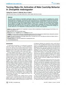
Figure 1. Homing assay. In this assay, donor and target constructs were placed at the same QC31 insertion site on homologous chromosomes (the donor and target chromosomes marked black and blue respectively). The target construct contains a GFP open reading frame (ORF) driven by an eye-specific promoter where the GFP ORF is split with an in-frame homing endonuclease recognition site (represented by adjacent green boxes). Transgenics bearing an intact target construct therefore exhibit GFP fluorescence in the eye. The donor construct has a homing endonuclease transcription unit is inserted into the HEG recognition site disrupting the GFP ORF and abolishing GFP fluorescence in the eye (loss of fluorescence represented by the GFP ORF being filled in white). Most constructs also include an RFP marker to allow the HEG insert to be tracked. Expression of the HEG in the germline causes cleavage of its recognition site in the target construct and subsequent repair leads to a number of different outcomes that can be differentiated by fluorescence and phenotypic markers as shown in the figure. The donor and target chromosomes are distinguished either with the linked cu marker (applicable with males only because of recombination) or a very closely linked mini-white marker within the donor construct (which is applicable to both sexes). It should be noted that NHEJ repair results in loss of GFP fluorescence in approximately two-thirds of cases only. The remaining third of NHEJ lesions can only be distinguished from unmodified targets by PCR and cleavage with ISceI. doi:10.1371/journal.pone.0054130.g001
Our homing assay has previously been described in detail [8]. A summary is shown in figure 1. Both target and donor constructs were inserted into specific attP locations within the Drosophila genome using the QC31 integrase method such that they could be homologously juxtaposed in vivo [12,13]. The donor and target constructs were differentiated by the use of linked chromosomal marker(s) (principally cu) and/or the presence of the eye colouration conferred by the presence of a functional mini-white marker on donor but not target constructs. We elected to report the majority of results in terms of the directly observed metrics, GFP loss (fraction of all targets where GFP fluorescence is lost) and homed fraction (fraction of GFPnegative targets repaired via homologous recombination), using these as proxies for the fraction of total targets modified by DNA repair and the fraction of modified events attributable to homologous recombination (HR). A discrepancy arises between these measures because while HR invariably results in loss of GFP reporter fluorescence, non-homologous end-joining (NHEJ) repair can lead to in-frame lesions that are GFP-positive. This, in turn, results in the fraction of targets modified by repair being underestimated and the fraction of repaired targets arising from
HR being correspondingly overestimated. In the case of pure NHEJ with in-frame events constituting a third of all repair events, the GFP loss would be a third lower than the true fraction of targets cleaved and repaired. While NHEJ in-frame lesions can be unambiguously identified by molecular biology, cost and labour constraints precluded its use with the large number of assays performed in this work. It is possible to estimate the number inframe NHEJ events as a proportion of the number of out-of-frame NHEJ events but the accuracy of these estimates is doubtful. Since we are primarily interested in comparing related homing constructs, we reason that this caveat is of relatively low importance. When comparing the effect of 39-UTRs on performance, the promoter and consequently the propensity to generate in-frame NHEJ events is unchanged and GFP loss is then a valid proxy for HEG activity. Similarly, when comparing the effect of genome location, the constructs are identical and NHEJ propensity remains fairly similar at the different locations. For constructs with radically different NHEJ propensities, e.g. when comparing Rcd-1r- and Mst87F-driven constructs, meaningful comparisons of HEG activity are impossible since precise religation dominates in the
PLOS ONE | www.plosone.org
2
January 2013 | Volume 8 | Issue 1 | e54130
Optimising Homing Endonuclease Gene Drive
= w;; attP2{bgcn-HEG-2-bTub56D } cu/attP2{wDarkLime} x R y w;;attP2 cu = w;; attP2{Rcd-1r-HEG-2-bTub56D} cu/attP2{wDarkLime} x R y w;;attP2 cu = w;; attP2{CG9576-HEG-2-bTub56D} cu/attP2{wDarkLime} x R y w;;attP2 cu The twin transgene cross presented in Table 2 was: = w;attP40 { Rcd-1r-HEG-2-bTub56D }; attP2{Rcd-1r-HEG2-bTub56D} cu/attP2{wDarkLime} x R y w;;attP2 cu Additional crosses performed for Table 3 were: = w; attP40{Rcd-1r-HEG-2-bTub56D}/attP40{wDarkLime} x R y w;;attP2 cu = w; attP51D{Rcd-1r-HEG-2-bTub56D}/attP51D{wDarkLime} x R y w;;attP2 cu = w;; attP86Fb {Rcd-1r-HEG-2-bTub56D} cu/attP86Fb {wDarkLime} x R y w;;attP2 cu The additional cross in Table 4 is: = w; attP40{Rcd-1r-HEG-2-bTub56D}; attP2{bgcn-HEG-2bTub56D } cu/attP2{wDarkLime} x R w;;attP2 cu All crosses were performed at 25uC unless otherwise stated. The ectopic homing assay in Table 5 was performed with single male crosses of type: = w; attP40{Rcd-1r-HEG-2-bTub56D}/+; attP2{wDarkLime} x R w[1118] = w;; attP2{wDarkLime} x R w; attP40 {Rcd-1r-HEG-2bTub56D}
mitotic stages while double-strand break repair is greatly reduced at the later stages of spermatogenesis when NHEJ events appear to dominate [8,14,15]. A lower HEG activity is therefore required to generate a scorable repair lesion late in spermatogenesis than in the spermatogonial cells where HR occurs. Finally, the metric that of greatest import when comparing construct performance in HEG gene drive is the fraction of total targets homed which has the advantage of being directly measurable and immediately relevant. To ensure comparability, all of the results in Table 1 were obtained with integrants at the attP2 site [12]. Crosses unique to this work presented in this table are: = w;; attP2{bam-HEG-1} cu/attP2{pDarkLime} x R y w;;attP2 cu = w;; attP2{bam-HEG-2-bam} cu/attP2{pDarkLime} x R y w;;attP2 cu = w;; attP2{vas-HEG-1} cu/attP2{pDarkLime} x R y w;;attP2 cu = w;; attP2{vas-HEG-2-SV40} cu/attP2{pDarkLime} x R y w;;attP2 cu = w;; attP2{vas-HEG-2-vas} cu/attP2{pDarkLime} x R y w;;attP2 cu = w;; attP2{Act5C-P-HEG-1} cu/attP2{pDarkLime} x R y w;;attP2 cu = w;; attP2{aly-HEG-2-bTub56D } cu/attP2{pDarkLime} x R y w;;attP2 cu = w;; attP2{bgcn-HEG-1} cu/attP2{pDarkLime} x R y w;;attP2 cu = w;; attP2{bgcn-HEG-2-bgcn} cu/attP2{wDarkLime} x R y w;;attP2 cu
Table 1. Summary of results of various promoter/39-UTR combinations for transgenes at attP2.
Fraction of targets homed
Promoter
39-UTR
Construct
GFP loss
Homing fraction
bTub85D
Hsp70Ab
bTub85D-HEG-1
70% (688/985)1
1% (1/94)
,1%
Mst87F
Hsp70Ab
Mst87F-HEG-1
61% (638/1041)1
0% (0/94)
Nil
Hsp70Ab
Hsp70Ab
Hsp70Ab-HEG1
26% (78/296)1
78% (225/287)
20%
bam
Hsp70Ab
bam-HEG-1
0% (0/55)
ND2
ND2
Native 3’-UTR
bam-HEG-2-bam
2.8% (66/2326)
64% (42/66)
1.8%
1
bTub56D
bam-HEG-2-bTub56D
9.1% (357/3910)
Hsp70Ab
vas-HEG1
1.9% (7/361)
SV40 early
vas-HEG-2-SV40
11% (210/1873)
52% (110/210)
5.9%
Native 3’-UTR
vas-HEG-2-vas
36% (328/911)
48% (157/328)
17%
bTub56D
vas-HEG-2-bTub56D
33% (1234/3764)1
42% (523/1234)
14%
Hsp70Ab
Act5C-P-HEG-1
5.5% (44/793)
34% (15/44)
1.9%
bTub56D
Act5C-P-HEG-2bTub56D
53% (671/1272)1
38% (252/671)
20%
Act5C-P (males), 18uC
bTub56D
Act5C-P-HEG-2bTub56D
19.0% (609/3198)
31.5% (192/609)
6.0%
aly
Hsp70Ab
aly-HEG-1
38% (417/1094)1
2% (2/94)
1.8%
bTub56D
aly-HEG-2-bTub56D
70% (754/1083)
6.2% (47/754)
4.3%
Hsp70Ab
bgcn-HEG-1
58% (676/1162)
0.3% (1/282)
,0.1%
62% (1486/2385)
vas
Act5C-P (males)
bgcn
69% (245/357)
6.3%
ND2
ND2
Native 3’-UTR
bgcn-HEG-2-bgcn
0.3% (4/1486)
0.2%
bTub56D
bgcn-HEG-2-bTub56D 65% (642/992)
0.5% (3/642)
0.3%
Rcd-1r
bTub56D
Rcd-1r-HEG-2-bTub56D 37% (1273/3422)
61% (782/1273)
23%
Rcd-1r, 18uC
bTub56D
Rcd-1r-HEG-2-bTub56D 15% (395/2614)
38% (152/395)
5.8%
CG9576
bTub56D
CG9576-HEG-2bTub56D
12% (27/232)
1.7%
14% (232/1612)
1
previously reported in [8]. ND: not done. doi:10.1371/journal.pone.0054130.t001 2
PLOS ONE | www.plosone.org
3
January 2013 | Volume 8 | Issue 1 | e54130
Optimising Homing Endonuclease Gene Drive
Table 2. Homing performance is expression-limited.
Transgene copy number 1 copy
1
2 copies2
GFP loss
Home/GFP-
Fraction of targets homed
37% (1273/3422)
61% (782/1273)
23%
77% (901/1170)
71% (636/901)
54%
1
attP2{Rcd-1r-HEG-2-bTub56D }. attP40{ Rcd-1r-HEG-2-bTub56D }/+; attP2{Rcd-1r-HEG-2-bTub56D}/attP2{wDarkLime}. doi:10.1371/journal.pone.0054130.t002 2
= w;; attP2{wDarkLime} x Rw; attP40{Rcd-1r-HEG-2bTub56D}/+; attP2{wDarkLime}/+ Flies of the genotype w; attP40{Rcd-1r-HEG-2-bTub56D}/+; attP2{wDarkLime} were identified by increased GFP fluorescence as a result of homozygosity for wDarkLime. It was necessary to transmit the wDarkLime insertion via the female line to avoid HEG expression mutating the I-SceI GFP target prior to creating the transheterozygote. Scoring for increased GFP fluorescence was a difficult procedure and potentially error-prone. To control for this, the transheterozygotes were evaluated in single male crosses so those involving a transheterozygote hemizygous for wDarkLime could be readily distinguished by an anomalously high proportion of GFP-negative progeny (.50% GFP loss). The observed GFP losses for each of the 42 crosses showed all but one result yielding GFP losses scattered around the average GFP loss of 21% with one well-separated outlier at 60%. That outlier was excluded from further analysis.
The vas promoter was chosen because it has small but detectable homing activity with the Hsp70Ab 39-UTR and it was coupled to several other 39-UTRs to investigate the impact of 39-UTR choice on testis HEG activity. These UTRs included the SV40 early intron/39-UTR combination deployed in the extensively-used pUAST vectors [18], the vas native 39-UTR and the bTub56D 39UTR. The first was selected because it contained an intron and splicing has previously been reported to be required for strong transgene expression in Drosophila [19]. The latter was chosen because bTub56D is known to be expressed at high levels at the early stages of spermatogenesis [20]. From Table 1, it is evident that when coupled with promoters active during early stages of spermatogenesis (bam, vas, Act5C-P), the Hsp70Ab 39-UTR performed particularly poorly in comparison to the other 39-UTRs (Hsp70Ab,,SV40, vas < bTub56D). In contrast, for promoters driving expression during later stages (aly, bgcn), the Hsp70Ab 39-UTR-mediated activity was only modestly reduced with the aly promoter and approached the bTub56D 39UTR in performance with the bgcn promoter. While the 39-UTRs of genes known to be expressed in the testis performed equally well, it was surprising that the popular SV40 early intron/39-UTR combination only yielded HEG activity at 30% of that observed with the former 39-UTRs. Since the results showed no notable advantage in using vas native 39-UTR, we based our subsequent HEG-2 design around the bTub56D 39-UTR [8]. We also investigated whether native 39-UTRs raised expression. The original bam promoter-driven transgene with the Hsp70Ab 39UTR had negligible HEG activity. When it was coupled with the bam 39-UTR HEG activity, as expressed by the loss of GFP at the target site, rose to ,3% but this was considerably lower than the 9% achieved with the bTub56D 39-UTR without appreciable change in homing efficiency (Table 1). In the ovary, RBP9 acts to downregulate bam transcripts via sites within the bam 39-UTR [21]. It is possible that the reduced expression with the bam 39-UTR may also arise from this mechanism: according to the Spermpress microarray data RBP9 is expressed in the mitotic cell population of the testis (see below) [22].
Fluorescence Microscopy Flies were scored for their fluorescence status with a MZ16F microscope (Leica) using the GFP2 and TXR filter sets.
in situ Hybridisation I-SceI transcripts were detected with a PCR-generated anti-sense probe against a part of the I-SceI coding region using the protocol described in [16]. A sense probe to the same region was used as control. Details of this probeset were previously published in [8].
Results The 39-UTR Strongly Influences Level of HEG Expression Our original HEG-1-based constructs used the Hsp70Ab 39UTR derived from the 70I-SceI construct [8,17]. We observed that while this vector yielded high levels of HEG activity with promoters expressing later in spermatogenesis (e.g. aly, bTub85D, Mst87F), there was little or no activity when used with promoters targeted to the early germline stem cell and spermatogonial stages (i.e. bam, vas) (see Table 1). Other 39-UTRs were therefore investigated as a means of improving expression.
Table 3. Genome location and Rcd-1r-HEG-2-bTub56D transgene performance.
Homing (as fraction of GFP-negative targets)
Homing (as fraction of all targets) Nearest genes
Chromosomal band
GFP loss
2L; 25C6 (attP40)
39% (1467/3733)
58% (846/1467)
23%
Msp-300: ubiquitous
2R; 51D (attP51D)
53% (1541/2884)
71% (1094/1541)
38%
CR43622, CG33467:male-specific
3L; 68A4 (attP2)1
37% (1273/3422)
61% (782/1273)
23%
CG6310, Mocs: ubiquitous
3R; 86Fb (attP86Fb)
58% (2671/4573)
58% (1538/2671)
34%
Clc: ubiquitous
1
extracted from Table 1. doi:10.1371/journal.pone.0054130.t003
PLOS ONE | www.plosone.org
4
January 2013 | Volume 8 | Issue 1 | e54130
Optimising Homing Endonuclease Gene Drive
Table 4. Co-expression of Rcd-1r-HEG-bTub56D and bgcn-HEG-bTub56D.
Construct attP40{Rcd-1r-HEG-2-bTub56D attP2{bgcn-HEG-2-bTub56D}
2
3
Both
GFP-negative events arising from HR1
GFP-negative events arising from NHEJ1
GFP-positive1
23% (846/3733)
17% (621/3733)
61% (2266/3733)
0.3% (3/992)
64% (639/992)
35% (350/992)
27% (296/1082)
47% (507/1082)
26% (279/1082)
1
as fraction of all targets. restated from Table 3. 3 restated from Table 1. doi:10.1371/journal.pone.0054130.t004 2
than bam but showed the same temporal expression profile, while recognising that the Spermpress categories only approximate the biological categories of spermatogonia/spermatocytes/spermatids. Further, a search of the tissue specific gene expression data was performed to restrict the candidates to those that were only expressed in the adult gonad as reported in FlyAtlas [25]. Two candidates were examined further: Rcd-1r (CG9573) and CG9576 (Table 1). Of these, only the Rcd-1r promoter showed significant HEG activity and homing in our assay (Table 1). The Rcd-1r promoter resulted in fourfold more HEG activity (37%) than the bam promoter (9%) that it was intended to replace. Although GFP loss achieved with Rcd-1r (37%) was lower than that of the Act5C promoter/P-intron combination (Act5C-P; 53%), it combined with a much higher homing fraction (61% vs 38%) resulting in a comparable fraction of total target chromosomes repaired via homing. in situ hybridisation showed that the Rcd-1r promoter-driven transgene expressed specifically in spermatogonia (see figure 2). This promoter, in combination with the bTub56 39UTR, was therefore chosen for our subsequent constructs.
Identification of Promoters that can Mediate HEG Drive Efficiently We previously reported that efficient homing in the testis requires HEG expression at the spermatogonial stage [8]. Although large increases in HEG activity as evidenced by GFP loss were secured by the use of the bTub56D 39-UTR, the highest levels of HEG activity did not correlate with similarly high rates of homing (Table 1). Promoters that had the potential to raise spermatogonial expression further were therefore sought. Genetic evidence indicates that bgcn is functional in the germline stem cell and during spermatogonial stages of spermatogenesis [23]. While previous workers fused the 2 kb upstream of the transcription start site to a bgcn cDNA/GFP fusion to achieve expression and phenotypic rescue, such a fragment would have extended deep into the neighbouring TBPH coding region [24]. Instead, we used a 817 bp fragment as the promoter sequence since that extended upstream from the bgcn start codon and included all intergenic space between bgcn and TBPH as well as the entire 39-UTR of TBPH. However, the homing results we obtained were contrary to our expectations. Although high levels of HEG activity were achieved with the Hsp70Ab 39-UTR, NHEJ dominated, suggesting that the promoter was not driving in the spermatogonial cells. We considered the possibility that the Hsp70Ab 39-UTR suppressed earlier expression and investigated the effect of using the native bgcn 39-UTR and the bTub56 39UTR. Although we achieved a further increase in HEG activity with these 39-UTRs, homing activity remained extremely low. We therefore sought further promoters with the desired pattern of expression and undertook a bioinformatics search of the Spermpress microarray data [22]. Vibranovski et al dissected the testis into three regions termed the mitotic, meiotic and postmeiotic zones and performed microarray expression analysis on each region. A gene like bam that expressed solely in spermatogonia should show declining expression during progression through spermatogenesis and that was observed in the Spermpress data. We therefore sought genes that were expressed more strongly
Performance is Expression-limited The fraction of total targets repaired as homing events is dependent on both the fraction of targets cut by the HEG and the homing fraction, that is, the proportion of events repaired via homologous recombination. It was therefore surprising to find that the fraction of total targets homed with the Act5C-P promoter (20%) and previously reported results with the Rcd-1r promoter (23%) and the Hsp70 promoter (21%) were all very close to each other, which might suggest an inherent biological limitation to homing in Drosophila. We therefore investigated whether higher levels of I-SceI expression could raise the fraction of total targets homed. To achieve this, we supplied a further copy of the Rcd-1rdriven transgene on chromosome 2 in addition to that on chromosome 3. The fraction of total targets homed doubled, suggesting that the homing was limited by expression of the HEG rather than any intrinsic biological limitation (Table 2).
Table 5. Ectopic homing.
Construct vas-HEG-2-bTub56D
2 2
Target class
Donor
Acceptor
GFP loss1
Homed events
Unpaired
attP1 (55C4)
attP2 (68A4)
29% (302/1057)
5 3
Unpaired
attP1 (55C4)
attP2 (68A4)
55% (181/327)
Rcd-1r-HEG-2-bTub56D
Unpaired
attP40 (25C6)
attP2 (68A4)
67% (1810/2685)
20
Rcd-1r-HEG-2-bTub56D
Paired
attP40 (25C6)
attP2 (68A4)
21% (628/3011)
21 (10 of 41 crosses)
Act5C-P-HEG-2-bTub56D
1
as fraction of all targets. Figures for actual GFP-negative and total target counts follows. previously reported in [8] and included here for ease of comparison. doi:10.1371/journal.pone.0054130.t005 2
PLOS ONE | www.plosone.org
5
January 2013 | Volume 8 | Issue 1 | e54130
Optimising Homing Endonuclease Gene Drive
converted by NHEJ events by the presence of bgcn-driven HEG expression (Table 4). Thus we conclude that the bgcn promoter fragment we used only drives strong post-spermatogonial expression.
The I-SceI Nickase does not Mediate Homing in the Testis Previous work suggested that, like double strand breaks, singlestrand nicks could also induce HR [27,28]. To test this, an Rcd-1r promoter-driven transgenic stock, attP2{Rcd-1r-K227MbTub56D}, was generated using the K227M variant of I-SceI that has previously been shown to have a strong preference for nicking the target on a specific strand [29]. In all other respects, the construct used was was identical to its progenitor, attP2{Rcd-1rHEG-2-bTub56D}. No HR or NHEJ events were observed in our assay after scoring 418 target chromosomes, suggesting that this variant is not active in our assay or that the single strand breaks are not recombinogenic in our HEG assay.
Figure 2. in situ hybridisation of I-SceI expression in Rcd-1rdriven transgenics. The I-SceI transcript is clearly detected in the spermatogonial population of the testis (marked by adjacent black asterisk). In situ hybridisation was performed as described in [8]. doi:10.1371/journal.pone.0054130.g002
HEGs can Home to Paired Ectopic Sites Rather than employ homologous recombination to insert a HEG construct at its correct target location in the genome, which may be technically challenging with some species, ectopic homing may be used to move a randomly integrated HEG donor construct to its recognition site in the genome. Our previous work established that homing between a donor HEG construct at the attP1 site on chromosome 2 to a target construct at the attP2 site on chromosome 3 occurred frequently enough that one could be confident that an ectopic jump could be isolated by screening several thousand progeny [8]. However, that experiment only employed a hemizygous target construct at the attP2 site: in the natural configuration, a pair of targets would be present at cognate sites on the homologous chromosomes. HR could potentially favour the use of homologous template to the extent that ectopic homing becomes an exceedingly rare outcome. To allay this concern, we performed an ectopic homing experiment using both paired and unpaired wDarkLime targets at attP2 and a Rcd-1rHEG-bTub56D donor at attP40 (Table 5). Surprisingly, we found that the frequency of ectopic homing in the paired case was of a similar magnitude to that observed in the unpaired case. The modest number of ectopic jumps observed is too low to precisely determine the ectopic homing rate but is adequate to demonstrate that ectopic jumps can readily be identified on screening an acceptable number of progeny.
Genome Context Influences Homing via Level of HEG Expression Genome context could have a role in determining the propensity for homologous recombination and to investigate this, we examined the performance of homing at three additional autosomal attP locations (Table 3) [13]. GFP loss varied between approximately 39% (attP40) to 58% (attP86Fb) at different sites while the homing fraction varied from approximately 58% to 71%, combining to yield a roughly two-fold variation in fraction of total targets homed, ranging from approximately 23% to 38%. The bulk of observed variation in the fraction of total targets homed appears to have arisen from genome context effects on HEG expression leading to variation in HEG activity, rather than from changes in the homing efficiency. This is in line with previous work reporting that the same transgene integrated at different attP integration sites can result in widely varying expression in a tissuedependent manner [26]. Since the fraction of total targets homed at some of the alternative attP sites we assayed was significantly higher than the best achieved level at our original attP2 inserts, it provided further evidence that homing performance in Drosophila scales with the level of HEG expression.
Low temperature Reduces HEG Spread
bgcn Promoter Expression Occurs after the Spermatogonial Stage
As the natural environment within which HEG gene drive is required to act is subject to diurnal and seasonal changes, we also determined the effect of ambient temperature on homing performance. When the attP2{Rcd-1r-HEG-2-bTub56D} cross was performed at 18uC rather than the usual 25uC, both HEG activity (as evidenced by loss of GFP fluorescence) and the efficiency of homing fell sharply, with the fraction of total targets homed declining nearly fourfold. We repeated this experiment with a different transgenic, attP2{ Act5C-P-HEG-2-bTub56D}, which also showed declines in both HEG activity and homing efficiency leading to a very similar overall decline in the fraction of total targets homed. The data suggest ambient temperature could be an important determinant of HEG gene drive performance. (Table 1).
The anomalous results obtained with our version of the bgcn promoter could potentially be attributed to expression in germline stem cells (GSCs) that are refractory to HR. Since the GSC stage precedes the HR-responsive spermatogonial stage during differentiation, one would expect that when I-SceI is simultaneously supplied from an Rcd-1r-driven transgene and a bgcn-driven transgene, NHEJ should dominate the observed repair events. Conversely, if the NHEJ events due to HEG activity driven by the bgcn promoter arose from expression subsequent to the spermatogonial stage, the majority of repair events should be attributable to HR. When tested experimentally using an Rcd-1r driven HEG at the second chromosome attP40 site and measuring homing between the bgcn driven HEG and its target at the third chromosome attP2 site, homing rates were very similar to those observed with Rcd-1r driven HEG alone at attP40. We suggest the difference is due to targets that were unmodified with Rcd-1rdriven HEG expression subsequently being almost wholly PLOS ONE | www.plosone.org
Discussion Previous studies establishing the feasibility of HEG-based gene drive in Diptera indicated that the process was more efficient in 6
January 2013 | Volume 8 | Issue 1 | e54130
Optimising Homing Endonuclease Gene Drive
enhancers are not uncommon [32]. In addition, if post-spermatogonial expression is a natural feature of the promoter, it suggests that bgcn may have further uncharacterised roles during later stages of spermatogenesis that are currently masked by its mutant phenotype at the spermatogonial stage. The absence of homing associated with the expression of the ISceI nicking mutant is consistent with previous work showing that HR occurs less frequently when induced by nicks rather than DSBs, presumably because nick repair is rapid [27,28,33,34]. The reduction was particularly pronounced when insertions are desired and homing unavoidably requires a large insert in the template [27]. In our experiment, we would expect approximately 96 homing events from the 418 chromosomes surveyed if wild type ISceI were used; the absence of any homing with the I-SceI nickase suggests that nicks are at least two orders of magnitude less efficient in inducing HR in the Drosophila testis. While nickases do have the advantage of much lower NHEJ rates, and with that potentially slower development of HEG resistance due to the accumulation of NHEJ-induced sequence changes to the target site, the loss in homing activity is an excessive price to pay in the context of a HEG-based gene drive system. The ability of an ectopic template to compete against a template at the homologous site was initially unexpected. However, since homing is restricted to fast-cycling transit-amplifying spermatogonia in these experiments, a large fraction of the genome will be in a post-replicative state regularly and sister chromatid repair is thereby enabled. Indeed, a large proportion of repair events may occur at this stage since HEG access to DNA, and consequently HEG cleavage, is restricted by chromosome condensation during M phase. From this perspective, an ectopic template will be frequently competing against a homologously-located template even in the unpaired case. This observation suggests that, at least in Drosophilids, it could be relatively easy to generate a stock with the correctly-homed transgene via normal transposon-mediated transgenesis followed by ectopic homing rather than requiring a sophisticated targeted insertion system that is unlikely to be easily accessible in non-laboratory pest species. The reduced homing performance at low temperatures observed in our experiments could have arisen from any of a variety of causes, including lower enzyme activity, lower expression of the HEG or reduced propensity towards HR. However, the strong dependence of I-SceI-driven homing activity on temperature suggests that the temperature-activity profile of deployed HEGs is a relevant factor when modelling HEG spread. Habitats where a HEG-based control strategy could be envisaged may exhibit significant seasonal temperature variation. Where a cold-sensitive HEG insert exerts a fitness cost, its population frequency may be adversely affected in an environment where, for example, the peak breeding season coincides with a wet, cool season. It was observed that efficient HEG drive was readily achieved in Anopheles gambiae but rather less so in Drosophila melanogaster, and this variation in response may suggest that HEG drive is an insect control strategy applicable only to a limited number of species [7,8]. It appears likely that the difference arises from achiasmy in Drosophila males: since crossovers are absent in this species, the HR machinery is no longer required during meiotic stages and homing is consequently restricted to earlier, transit amplifying cells [8]. Achiasmy is widespread in higher Diptera [35], an order to which many insect pests belong, and the utility of HEG drive will depend on it being usable even in these less favourable circumstances. We have shown here that even a semi-refractory species such as Drosophila is not inherently inferior in its ability to support homing: with sufficient HEG activity in the correct cell type, high levels of
Anopheles gambiae than in Drosophila melanogaster. In this study, we have shown that homing in Drosophila scales with the level of HEG expression raising the question of whether poor performance relative to Anopheles is solely due to lower expression or is also affected by other constraints. First, unlike Anopheles, Drosophila spermatogenesis proceeds via an achiasmate mechanism and crossovers are absent from the male germline. We speculate that this has the effect of restricting homologous recombination to the transit-amplifying mitotic spermatogonial stage. In contrast, with Anopheles, HR may still be operational during the early spermatocyte stage prior to the first meiotic division since it mediates crossover events in the germline. The longer period during which HR is available is expected to allow higher rates of homing for a given level of HEG activity. We have also shown that optimisation of promoter and 39-UTR choice can raise HEG activity levels very substantially. We were initially surprised that 39-UTR choice had such a pronounced effect in our homing assays. Even though the Hsp70 39-UTR has been used in a number of Drosophila constructs, there is experimental evidence that it contributes toward Hsp70 induction by destabilising its mRNA in the absence of heat stress [30]. However, mRNA destabilisation does not fully explain our observations: our previously described Hsp26 promoter-driven HEG construct achieved modest levels of homing (11% homing fraction) when combined with the Hsp70Ab 39-UTR under unstressed conditions. One may speculate that this promoter may have the ability to override the destabilising effect of the Hsp70Ab 39-UTR, perhaps through stabilising factors bound to the polymerase complex being transferred to the nascent transcript. We also observed that the widely-used SV40 early intron/39-UTR sequence performed poorly in our assays, consequently its use in constructs where high levels of expression during early stages of spermatogenesis is not recommended. Our current study suggests that the bTub56D 39-UTR is able to support robust expression in the male germline. We identified and tested the promoter region from CG9573 as a potentially suitable spermatogonial promoter prior to the identification of the gene as Rcd-1r [31]. Drosophila melanogaster has three paralogues of Rcd-1r, related to a regulator of differentiation in yeast. We speculate that Rcd-1r is a regulator of spermatogonial differentiation and is expected to have a malesterile phenotype. Indeed, the nearest male-sterile, ms(2)29F, was originally associated with a P-element insertion, Rcd-1r07717, however this association has since been excluded and the location of Rcd-1r, within 39UTR region of the overlapping gene CG13102 makes further study of the gene difficult [31]. We were surprised that the bgcn promoter constructs resulted in very low levels of HEG activity during early stages of spermatogenesis but high activity during post-spermatogonial stages. We believe this is most likely due to the loss of distal control elements in the truncated promoter fragment we used or be due to an unanticipated interaction between the bgcn control elements and other elements within our constructs. Since our bgcn fragment includes the entire region between bgcn and its upstream neighbour, TBPH, as well as the entire bgcn 59-UTR, we expected it would contain all key regulatory sites. bgcn intronic enhancers can be excluded since a genomic fragment containing the bgcn 59 region and a substantial portion of the 39 end of the adjacent TBPH gene was able to drive a bgcn cDNA to rescue bgcn mutants [17]. Therefore if the apparent low expression level of our bgcn driven HEG is the result of loss of regulatory elements necessary for gonial cell expression, these elements must reside within the TBPH gene or within the bgcn coding region. We note that evidence has been recently advanced to suggest that exonic PLOS ONE | www.plosone.org
7
January 2013 | Volume 8 | Issue 1 | e54130
Optimising Homing Endonuclease Gene Drive
homing can be achieved. We also demonstrate that appropriate HEG activity can be achieved with judicious choices of 39-UTRs and promoters. Moreover, chromosomal sites do not appear to vary much in their ability to support homologous recombination: rather, they act indirectly by influencing the expression of the HEG transgenes. HEG drive could therefore be potentially extended to genes that exhibit repressive chromatin in the testis by the use of insulator elements in transgene constructs [26]. The
combination of these measures could allow HEG drive to be applied even in the most recalcitrant species.
Author Contributions Conceived and designed the experiments: DSH SR. Performed the experiments: YC DSH RG EW. Analyzed the data: DSH SR. Contributed reagents/materials/analysis tools: YC EW DSH SR. Wrote the paper: DSH SR.
References 1. Thomas DD, Donnelly CA, Wood RJ, Alphey LS (2000) Insect population control using a dominant, repressible, lethal genetic system. Science 287: 2474– 2476. 2. Burt A (2003) Site-specific selfish genes as tools for the control and genetic engineering of natural populations. Proc R Soc Lond B Biol Sci 270: 921–928. 3. Takeuchi R, Lambert AR, Mak AN-S, Jacoby K, Dickson RJ, et al. (2011) Tapping natural reservoirs of homing endonucleases for targeted gene modification. Proc Natl Acad Sci U S A 108: 13077–13082. 4. Ulge UY, Baker DA, Monnat RJ (2011) Comprehensive computational design of mCreI homing endonuclease cleavage specificity for genome engineering. Nucleic Acids Res 39: 4330–4339. 5. Deredec A, Burt A, Godfray HCJ (2008) The population genetics of using homing endonuclease genes in vector and pest management. Genetics 179: 2013–2026. 6. Deredec A, Godfray HCJ, Burt A (2011) Requirements for effective malaria control with homing endonuclease genes. Proc Natl Acad Sci U S A 108: E870– E880. 7. Windbichler N, Menichelli M, Papathanos PA, Thyme SB, Li H, et al. (2011) A synthetic homing endonuclease-based gene drive system in the human malaria mosquito. Nature 473: 212–215. 8. Chan Y, Naujoks D, Huen D, Russell S (2011) Insect Population Control by Homing Endonuclease-Based Gene Drive: An Evaluation in Drosophila melanogaster. Genetics 188: 33–44. 9. Lee JC, Bruck DJ, Curry H, Edwards D, Haviland DR, et al. (2011) The susceptibility of small fruits and cherries to the spotted-wing drosophila, Drosophila suzukii. Pest Manag Sci 67: 1358–1367. 10. Malacrida AR, Gomulski LM, Bonizzoni M, Bertin S, Gasperi G, et al. (2007) Globalization and fruitfly invasion and expansion: the medfly paradigm. Genetica 131: 1–9. 11. McQuilton P, St Pierre SE, Thurmond J (2011) FlyBase 101 - the basics of navigating FlyBase. Nucleic Acids Res 40: 1–9. 12. Groth A, Fish M, Nusse R, Calos M (2004) Construction of transgenic Drosophila by using the site-specific integrase from phage QC31. Genetics 166: 1775–1782. 13. Bischof J, Maeda RK, Hediger M, Karch F, Basler K (2007) An optimized transgenesis system for Drosophila using germ-line-specific QC31 integrases. Proc Natl Acad Sci U S A 104: 3312–3317. 14. Gong WJ, Golic KG (2003) Ends-out, or replacement, gene targeting in Drosophila. Proc Natl Acad Sci U S A 100: 2556–2561. 15. Rong YS, Golic KG (2003) The homologous chromosome is an effective template for the repair of mitotic DNA double-strand breaks in Drosophila. Genetics 165: 1831–1842. 16. Morris C, Benson E, White-Cooper H (2009) Determination of gene expression patterns using in situ hybridization to Drosophila testes. Nat Protoc 4: 1807–1819. 17. Rong YS, Golic KG (2000) Gene targeting by homologous recombination in Drosophila. Science 288: 2013–2018. 18. Rørth P, Szabo K, Bailey A, Laverty T, Rehm J, Rubin GM, Weigmann K, Milan M, Benes V, Ansorge W, Cohen SM (1998) Systematic gain-of-function genetics in Drosophila. Development 125: 1049–1057.
PLOS ONE | www.plosone.org
19. Duncker BP, Davies PL, Walker VK (1997) Introns boost transgene expression in Drosophila melanogaster. Mol Gen Genet 254: 291–296. 20. Buttgereit D, Renkawitz-Pohl R (1993) Expression of b-1-tubulin (bTub56D) in Drosophila testis stem cells is regulated by a short upstream sequence while intron elements guide expression in somatic cells. Mol Gen Genet 241: 263–270. 21. Kim-Ha J, Kim J, Kim Y-J (1999) Requirement of RBP9, a Drosophila Hu Homolog, for Regulation of Cystocyte Differentiation and Oocyte Determination during Oogenesis. Mol. Cell. Biol. 19: 2505–2514. 22. Vibranovski MD, Lopes HF, Karr TL, Long M (2009) Stage-Specific Expression Profiling of Drosophila Spermatogenesis Suggests that Meiotic Sex Chromosome Inactivation Drives Genomic Relocation of Testis-Expressed Genes. PLoS Genet 5: 13. 23. Go¨nczy P, Matunis E, DiNardo S (1997) bag-of-marbles and benign gonial cell neoplasm act in the germline to restrict proliferation during Drosophila spermatogenesis. Development 124: 4361–4371. 24. Li Y, Minor NT, Park JK, Mckearin DM, Maines JZ (2009) Bam and Bgcn antagonize Nano-dependent germ-line stem cell maintenance. Proc Natl Acad Sci U S A 106: 9304–9309. 25. Chintapalli VR, Wang J, Dow JAT (2007) Using FlyAtlas to identify better Drosophila melanogaster models of human disease. Nat Genet 39: 715–720. 26. Markstein M, Pitsouli C, Villalta C, Celniker SE, Perrimon N (2008) Exploiting position effects and the gypsy retrovirus insulator to engineer precisely expressed transgenes. Nat Genet 40: 476–483. 27. Metzger MJ, McConnell-Smith A, Stoddard BL, Miller AD (2011) Single-strand nicks induce homologous recombination with less toxicity than double-strand breaks using an AAV vector template. Nucleic Acids Res 39: 926–935. 28. McConnell Smith A, Takeuchi R, Pellenz S, Davis L, Maizels N, et al. (2009) Generation of a nicking enzyme that stimulates site-specific gene conversion from the I-AniI LAGLIDADG homing endonuclease. Proc Natl Acad Sci U S A 106: 5099–5104. 29. Niu Y, Tenney K, Li H, Gimble FS (2008) Engineering variants of the I-SceI homing endonuclease with strand-specific and site-specific DNA-nicking activity. J Mol Biol 382: 188–202. 30. Petersen R, Lindquist S (1988) The Drosophila hsp70 message is rapidly degraded at normal temperatures and stabilized by heat shock. Gene 72: 161–168. 31. Quezada-Dı´az JE, Muliyil T, Rı´o J, Betra´n E (2010) Drcd-1 related: a positively selected spermatogenesis retrogene in Drosophila. Genetica 138: 925–937. 32. Birnbaum RY, Clowney EJ, Agamy O, Kim MJ, Zhao J, et al. (2012) Coding exons function as tissue-specific enhancers of nearby genes. Genome Res 22: 1059–1068. 33. Kim E, Kim S, Kim DH, Choi B-S, Choi I-Y, et al. (2012) Precision genome engineering with programmable DNA-nicking enzymes. Genome Res 22: 1327– 1333. 34. Wang J, Friedman G, Doyon Y, Wang NS, Li CJ, et al. (2012) Targeted gene addition to a predetermined site in the human genome using a ZFN-based nicking enzyme. Genome Res 22: 1316–1326. 35. John B (2005) Meiosis. Cambridge: Cambridge University Press.
8
January 2013 | Volume 8 | Issue 1 | e54130