ofthese pores increased cell polarization and migration compared with cells on ...... defined as narrow annular con- nections between the main cell body.
Dynamic imaging of neutrophil migration in three dimensions: mechanical interactions between cells and matrix John
T. H. Mandeville,
Department
Abstract: to
Pathology,
of
Fluorescence
obtain
and
a spatial
through a temporal
Neutrophils tant gradient
migrating within
of
Univeisity
microscopy (3-D)
resolution
A. Lawson,
Columbia
confocal
three-dimensional
neutrophils migrating motic membrane with 5
Moira
College
was
images
of
used
in response a 3-D matrix
in the were
z-dimension.
to a chemoattracapparently able
to generate traction by use of lateral pseudopods serted into footholds in the matrix as evidenced matrix distortion. Similar anchored pseudopods seen
in
branes increased
cells with
with cells ofpseudopods trix
and
migrating
on porous
filters
Contact fibrils
matrix show
that
neutrophils
Leukoc.
Key
Words:
observed
ofneutrophils observed. These in 3-D
migrating
structures to generate 61:
Riot.
constriction
appeared
to
crawling along observations are
in the matrix, traction for
188-200;
ring
.
able
to the
polymerization,
motors,
[2,
12],
and
the
ECM.
2-D
their net migration number of cells
and
there
through crossing
studies of how cells actually Migrating cells undergo
have
been
3-D substrates a barrier. There
many
studies
of
by counting have been
the few
crawl through 3-D matrices. cycles of adhesion, generation
.
Journal
of
Leukocyte
Biology
must
it
[8-li] signaling. by a combination
traction
to differences
lead
ECM.
Third, that the
cell
that on by the
and
surface
architecture may on the substrate
allow with
receptor-mediated adhesion. Secare not homogeneous, and they dimensions, orientations, compo-
and mechanical properties. These to contact guidance of migration,
can
the
differ from is enveloped
in cytoskeletal of a 3-D purchase
pnes-
between
migration in 3-D matrices First, a cell in a matrix
leading
as
the inter-
on hydrostatic
involve
less reliance on specific ond, biological matrices contain fibers of differing sitions,
such
a 3-D matrix can present cell must either deform
inhomogeneities directed by the
a barrier or degrade
to migrain order
It has
been
3-D
may
[16], able
monocytes to migrate
proposed
differ
that
the in
mechanism
2-D
of traction
[13-15].
in
Lymphocytes
[17], and neutrophils [14, 17] that are unon collagen-coated glass are able to migrate of collagen.
In addition, neutrophils lackeither through genetic defects or by treatment with 132 function-blocking integnins,
132
display but
that
from
no
profound
deficits
significant
in migration
differences
on
on
3-D
2-D
substrates
as measured by the distance penetrated by the advancing front of cells [18].
into Macno-
(polymonphonuclear (3-D) extracellu-
of force, and de-adhesion [3]. Adhesion to the ECM is largely mediated by members of the integrin superfamily I4] Integnins are heterodimenic transmembrane glycopro-
188
actin
but
How might surfaces?
of collagen, the matrix
[2],
via molecules
of myosin sure
through matrices ing functional in the I2 subunit
calcein
lan matrix (ECM) is critical for host defense against bacteria and is an important component of pathological inflammation. Neutnophils are rapidly locomoting cells that exhibit chemotaxis toward a chemoauractant source [1]. Much has been learned about neutnophil migration on two-dimensional surfaces
cytoskeleton
to pass.
INTRODUCTION
(2-D)
actin
York
7J and inside-out may be mediated
surfaces
migration of neutrophils through the three-dimensional
New
talin, and vinculin [5]. The link between the cytoskeleton is regulated by a complex
antibodies,
The directed leukocytes)
Surgeons,
action of outside-in [6, The generation offonce
to uti-
not present locomotion.
amnion
.
linked
a-actinin, ECM and
tion
1997.
foothold
and
morphology. The presence the cell to gain mechanical
Expansion in the ma-
and
R. Maxfield
of Physicians
substrate,
mem-
ofthese pores compared
through the openings. Neutroofthe elastic amnion matrix durpermanently altering the sub-
guidance was also
lize mechanical on 2-D surfaces,
J.
was
inby were
polycarbonate
the presence and migration
membranes without pores. distal to narrow constrictions
be used to pull cells phils deformed parts ing migration without strate.
across
0.8-.tm pores; cell polarization
Frederick
teins
human
a 3-D matrix of amresolution of 30-60 2 im
and
Volume
61,
February
1997
Abbreviations:
2-D,
acetoxymethyl
ester;
two-dimensional;
indacene-sphingomyelin;
ECM,
tg/ml
PBS,
extracellular
and
10%
contrast;
saline;
(v/v)
carboxytetramethyb-hodamine, interference
matrix;
FWHM,
phosphate-buffered
saponin
calf
full
fMLP,
width
PBS-S.
serum;
AM,
PBS
ester;
dimethyl
containing
250
5-(and DIC,
sulfoxide;
N-formyl-L-
at half-maximal
rhodamine/SE,
succinimidyl DMSO,
three-dimensional;
4,4-difluoro-4-bora-3a,4a-diaza-s-
methionyl-L-leucyl-L-phenylalanine; intensity;
3-D,
BODIPY-SM,
PET,
6-)-
differential polyethylene
terephthalate. Correspondence: try,
Cornell
NY
10021.
ber
Frederick
University
Received July 22, 1996.
29,
Medical 1996;
R.
Maxfleld,
College, revised
Department 1300
October
York 18,
of BiochemisAvenue,
1996;
New
accepted
York, Octo-
phages
migrating
served
into
fibrin
matrices
have
by scanning electron microscopy into the surrounding matrix that may
tions
to generate
traction
[19].
Contact
guidance,
or
also
been
ob-
shown to exhibit directed [23, 24]. Although contact ing through 3-D biological
137
alignment
and
migration
the
(3)
move
the
ically, or (4) stop moving. Without migrating cells within the matrix, can
only
be
inferred.
studied on texhave also been
phils
as
dynamic the final
Neutrophils
carry
roles [25]. Cell motility studies in three on experimental methods such says that do not permit direct [18,
26-28],
electron
often based migration asof cells during that
only static images [14, 17, 19], on differential contrast (DIC) and phase images of live cells stnicted
to a single
of the
matrix
of fluonescently phils during cally
focal
plane
[14-161.
We
labeled chemotactic
cells
relevant
3-D
The matrix of the structure
with
have
little
used
of
are
microscopy
provides
to no
to directly observe migration through
re-
resolution
[31],
enogeneous
matrix
in which
both
permissive
study,
and
and
contact
cuss
the
similarities
2-D
and
3-D.
to study the
guidance
from and
amnion a range
to cell
nature
Neutrophils
were
previously
described
matrix
structures,
form
differences
between
tumbled
Human
and
sodium lated lution
heparin by
blood
was
obtained placed
M
(Becton-Dickinson,
centrifugation
(1-Step-Polymorphs.
for
30
San
Jose,
mm at 400
Accurate
from
Chemical
CA).
g over and
tubes
F. R. Maxfield,
following
am-
in situ
im-
unpublished
fluorescent
Inc.,
markers
used
acetoxymethyl
Eugene,
temperature calf
pluronic
using
within
buffer 0.2%
were
stored
(AM)
cells
were
[incubation
Molecular
Cells
and
ester
Briefly,
serum,
F-127;
as indicated. medium,
OR).
in loading
fetal w/v
calcein/AM
then
over
dimethyl
sul-
Probesj
con-
rinsed
ice
in PBS,
until
use.
Cells
Probes
and
5 h of loading.
label prepared
suspended Lipids,
argon,
and
was
by the
in a 1:1
From
solution
Neutrophils
stock and
were
solution,
vesicles
in cold
PBS,
resuspended
after
labeling.
Ethanol a 100
for
BODIPY-SM (Avanti
was
tM
evaporated
then
and
mm.
were
solution
PBS,
ice
30
in cold
labeling
1351.
vesicles
against
to 95%
as described
GIBCO-BRL,
fJ.
7.4)
KCI,
contained
diameter)
then
washing
munofluorescence
pH
(PBS,
morphology.
amniotic inner
was
Amnions
preparations
lysis
saline
5 mM
Preparations
within
by gentle
ig/mL;
nion
NaCl,
prepared
(22-mm
U/mL;
hypotonic
membranes
was
extensive
(500
medium
matrix.
migration
rings
NH4OH.
(500
Na2HPO4,
mM
of human
epithelium
and
icillin
foxide
parallel to the basement membrane, which is itself composed primarily of a dense meshwork of laminin, fibronectin, and collagens type IV and V [13]. The amniotic basement membrane has been reported to be a barrier to
In this
ing
mM
characteristic
(obtained
2 h, followed
microscopy
we used was human amnion, which is typical of loose fibno-connective tissue that neutno-
neutrophil
tissue
Teflon
were
phils encounter in vivo [29]. The stromal amnion is composed primarily of fibnils collagens type I, III, and V [30] that are
ions,
for
8.1
7.4).
their
pieces
amniotic
by a 30-s
(150
of amniotic
Neutrophils
interference that are
confocal
across
liceman
a path through elastase, cato play major
dimensions as trans-filter observation
by
removed
in phosphate-buffered
N-2-hydroxyethylpiperazine-N’-2-ethanesulfonic pH
membranes
The
KC1,
mM
glucose,
amniotic
cental
rinsed
medium
assessed
Human
were
then mM
20
mM
To summarize,
mechan-
armament
2.7
Preparation
observation of three options
an
NaC1,
CaCl2, 10
obstacle
were
in incubation
acid,
pnoteolytic enzymes well-suited for digesting a noncompliant matrix, of which neutrophil thepsin G, and proteinase 3 are considered
migration
mM
of
migration in aligned matrices guidance of neutrophils migrattissues has been inferred from
obstacle,
erythrocytes
Cells
pended
these studies, it has not been observed directly. Neutnophils that encounter a physical barrier in a matrix have four options: (1) remove the obstacle chemically, (2) around
Contaminating
1 mM
the
cells along anisotnopic substrates, has been tuned or grooved surfaces [20-22]. Cells
move
NY).
procedure.
to insert projecserve as anchors
isoso-
Westbury,
Mandevilte
Chemotaxis Amnions 1 h before
et at.
Neutrophil
were use.
chambers rinsed
and
Membranes
migration
soaked were
within
in incubation then
suspended
a three-dimensional
medium over
at 37#{176}Cfor> a smaller
Teflon
matrix
189
0-ring
(16-mm
nM
inner
St. t.ouis, with
MO)
medium,
in the
culture
and
fresh
then
medium were
used
into
rinsed
from
were
transferred
for
the
upper
for
for
to a humidified In some
imaging
of live
microscope
cases,
cells
stage
were
fixed
unlabeled
were
cells, the
were
were
previously
plated
the chemoattractant after
IMLP
placed
the
oil immersion Migrating
and
initial
tail
from
each
IWctifl,
dish
phils
fixed.
dishes
substrate
images
disk
of confocal
micros(’ope
(Dialux
CA) WaS
Leitz
an image
with
(CCD-72:
Dag--MTI,
(DSP-200:
Washington.
.
Inc.,
Dage-MTI).
images approa(-hing light source. Cells
j((tiV(
fieI(l
cording
ntxt
fO(LIS knob To obtain
were
obtained
moving sections,
of the
for
Zeiss, was
planes.
with
images
fitted
artifact
would
an
with
a spinning
upright Co.
confocality.
The
through
gelatin intensity
beads
Rasband,
by
available
use
of
to Internet
in the
z-axis
confocal
Briefly,
10%
half-maximal
zippy.nimh.nih.gov
disk
±
at
in
directory
resolution.
microscope
Optical
had
a FWHM
SE).
San
,
Inter-
video
video
camera
frame
permitted
Oberkochen, optically
Moving
step
sizes
per
they
acquired disks
190
of
to image was
from
one
ranging 25
roughly
little
a
10-.tm
have
moved
volume
frame. out
of the
1-s
intervals
and
(PMO-650;
Leukocyte
Pinnacle
Biology
tm
0.8
arc ob-
dichroic
mirror
a 525-
hence
of view.
was
digitized Irvine,
61,
disk.
uously
were
focal
dual
plane
The
confocal used
in these
confocal
0.1
.tm
0.3
±
im
(mean
±
in the
interference
and
using
a z-axis
plane
were
four
scans
on a magnetostepper
motor.
acquired
Occasional
cells
mode.
over
stored
contin-
adjustments
in the
using for
field
of view
or
beads
as
line
steps.
scans
range taken
Optical
ranged
fluorescent
the
of pinhole through
sections
from
aper-
individual
acquired
a FWHM
SE) for 3-D reconstructions, SE) for time-series acquired
±
detection,
Pores
averaging
calibrated
microscope
(mean
for red
detection.
reflection
calculated
z-axis
filter
green
by a 560-nm
of view.
from
at 0.2-.tm
scanning ±
was
tubes
images.
field
experiments
acquired
and
to keep
was
FWHM
mirror,
focal
moving
of the
microscope
above.
dichroic
acquired
necessary out
fluo-
a 527-nm
section)
a single
color
were
structures
described
were
through
were
filter;
Kalman
optical
laser.
matrix
excitation
for
with
each
Zeiss).
argon
bandpass
using
obtained
infinity1.2;
=
a 25-mW
emission
visualized
a 40x NA
from
scan-
Cambridge,
using
photomultiplier
filter
a laser
rhodamine-labeled
long-pass
frames
to construct
of the
and
two separate
s to acquire Volume
line
with
bandpass
images
on the
of 1.8 and
0.2
±
a FWHM
at a single
optical
plane.
Image processing Digitized
3-D
volume
images
passed
and
used
nal
noise
sity
was
viewed
inherent used
to
tions
1.5
cells, could
tions.
pixel’s
a summed-pixel
optical
the
following
onto
1997
the
color tial
stereo relationships
rotated
were
then
and
background.
to arbitrary
intenWe
surface
viewing These
a 3-D
sig-
a threshold
a solid
conditions.
first
to diminish
and
cells
to form
were
rendered
used image
angles
volume
animated
slice. monitor
images,
and projec-
sequence
Pairs
next
algorithm, Image
image
of
vertical
the
each
were Inc.
dual-labeled
slice
,
viewed Beaverton, samples.
(Vicom were
was
was
on OR)
to
Maynard,
compared
intensity pixel
projec-
Vis-
made
algorithm,
was
maximum
where
stereo transferred
processor
projections
(1) a brightest-pixel
stacks
(Tektronix,
were
Corporation,
of 2-D
either
and
as 3-D
data
Equipment IP9000
CA).
in the
between
for viewing
stereo (Digital
using
projection
(2)
filter
to generate
a Gould-Vicom
intensity
existing
be
of paired
ofimages
could
using
between
Fremont,
stack
imaging,
concatenated
with
of the
level
as serial
Images
cells.
MA)
each
directly
be
minicomputer
optically
light
ways: (Indec
acquired
1331.
convolution
different program
images
x
then
images
equipped
from
3 Gaussian
two
microVoxel
described
lighting/shading
generation
Computing,
volumes
algorithm
could
different then
3-D
3-D
following
as previously
rendering
of migrating For
3
a microVAX-Il ual
optical
x
in low
Alternatively,
per
The
to discriminate
under
a field
in the
to render
a 3
which
of the
visualized
stereo-viewing.
sections
through
plane
were
rendering
each
Each
February
514-nm
selected
were
Zeiss)
a 514-nm
a 600-nm
images 5-10
Alternatively,
coarse
cells
CA)
and
3-D
z-axis).
fewer fewer
were
filters
Digital optical
with
to 535-nm
polycarbonate
at a rapid
being
desired,
the
with
Microscience,
(C-Apochromat,
cells
between
a first-pixel
morphology
moving
Micro,
Volume
divided
were
by re-
frames
in the
as it was
field
they
was
frames
of cell
was and
migrated at
60
I tm
emissions
optical
volume
cell
resolution
rescence
confocal
focal
from
was
imaged
Systems)
to the
volume
distortion
diameter
simultaneously
obtained
(Axiovert; objective
or BODIPY-SM-labeled
averager
Germany).
neutrophils.
approximately
Calcein-
were Bio-Rad
microscope source
us to ac-
sectioned
attached
an inverted
illumination
and
images (MRC-600;
micro-
Videoscope
and
of migrating
temporal
acquired
magneto-optical
Journal
there (e.g..
higher
before was
mm
(1 speed
When
imaged
(mean
width
to measure
spinning
water-immersion
of 2.9
amniotic
used Instrument
motor
on
to 2.3
fluorescence
through
confocal
Carl
MA)
laser
device
frame, stepper
with
resolutions
were
server used
fluorescence
corrected,
tures
using a conventional mercury with a 2Sx water-immersion
offocal
se(Juentially,
of 5 jtm/min
seetiofls
tm
full
(Molec-
[38J.
suspended
by Wayne
FTP it was
attachment
to identify
phalloidin.
to acquire
IN),
disk
by a z-axis 3-D
Ex-
on three neutro-
the
confocal
beads,
(KS-1381;
City,
spinning
micros(’ope.
moving
and
1371
rhodamine
maximum
Michigan
0.8;
fibro-
preparations
Technical
intensifier
volume
a series
acquisition
sectioned).
0.2
±
(requiring
Control
described
migration
charge-coupled
=
or
achieved
to motion
speed
Cap-
beads
thickness
cross-sections
(written
anonymous
acquired
ning
their
recorded
image- series. A typical 3-D volume frame consisted of 60 optical which we- found to be a suitable compromise between temporal
spatial
At this
from
was
)lm.
NA
of (‘ells,
images
to tlw
Bio:
video rates were imaged
(Plan-Neofluar,
Iach
from
10 jig/mL
we primarily
DC), The
on
leading
condition.
amnion
Germany)
(K2
equipped lid.
the
of neutrophils of these cells.
during
Wetzlar,
apertures
each
with
were
1 h with
used
images,
attachment
5(1 to pinhole
natft)nal.
(ltiir( lamp
20;
optical
7 tm
with
were
moving
a Zeiss
recorded
at least
for
minutes
microscopy
microscopes 3-D
were
previously
confocal
chamber
both
for
stained
neutrophils
To obtain
(115k confocal
Systems,
fluorescent
vertical
of beads
The
from
program
on the
The
5 mm.
using
fields
in our
been
and
ofcalcein-labeled
scot)e
present has
permeabilized.
membrane.
JOSt.
used
as
calcein/AM
in which
sequential
coated
dishes
Germany)
images
to move
were
(Petri
with
monitored
as those
dishes
without
beneath,
Oberkochen.
resultant
Three
or
experimental
was
Zeiss.
observed
motility
spinning
tYpes
determined
of 3.6
at 37#{176}C.Five
were repeated with fresh preparations days. To determine the morphology
Nipkow Two
three
The
motility
35g.
on coverslip
neutrophil
were
(Indec
as de-
at 37#{176}C.After
maintained
defined
were s 18.
a physiological
p(-riments separate
was Image
slice
a field
steps.
to 37#{176}Cwith
with
loaded
added.
The
were
cell
and
plated
which
cell
objective.
in 200
were
was
(Carl
cells
of the
position
((‘115 were
users
z-axis
on the
mounted
for 5 mm
stage
microscope
x
e(Ige
software
300-nm
image
through
at 0.2-tm NIH
using
the
sections
dishes
coverslips
filters
(10 nM)
of fMLP.
inverted
videotape.
a 63
the
on a microscope
application
Axiomat
and
Neutrophils
onto
CA)
coverslip-bottom
bottom
1361).
and
Livermore,
onto
in the
described
was then
taken
Dual-wavelength (Poretics.
mounted
punched
as described
were
(w/v)
calibrated
chambers
in 2-D
membranes
holes
was
to determine
and
amnion
Constructor
Laser scanning confocal microscopy
pores
with
Volume
/pub/nih-image),
the
warmed
within
to
cells
images
the
allowed
unattached
confocal
Probes)
(FWHM)
or BODIPYand
Cells
time
with
available
CA).
ular of
replaced
studies,
itola, The
incubated
a gradient
was
commercially
chamber
was
Calcein/AM-
migration
at which
For
Boyden
chamber
staining.
mm,
20
Co.,
to establish
neutrophils.
chamber.
assays
0.8-.tm
section
106
40
Chemical
chamber
tray
immunofluorescent
matrix
Polycarbonate
due
The
upper
used
containing
below.
Motility
and
x
were
the
curtain.
scribed
in the
5
neutrophils
migrate
an air
medium
dish Sigma
a modified
chamber.
37#{176}Cwarm
containing
SM-labeled cells
the
creating
lower
mm on a humidified
30
fMLP.
l)(
in a tissue
(fMLP;
in incubation
chemoattractant
for
on
diameter)
formyl-methionyl-leucyl-phenylalanine
of
where with
that
retained,
added
a Tektronix to observe
or
to that
of
SGS625 the
spa-
Time-series ferred
sequences
to the
taken
from
microVAX-II/IP-9000
a single and
optical
viewed
plane
were
as dual-colored
trans-
grating
cells
moving
viously
described
images.
cells
degradation
amnions
were
a cocktail 60
(Sigma). ig/mL
by secreted
for
2 h in incubation
(Sigma).
without
rinsing
described
aprotinin
This
by
laser
the fol-
.tM
motile
100
is effective
against
directly
to mem-
added
Cells
and
migrating
in the pres-
confocal
microscopy
scanning
the
5%
±
calcein-labeled
spinning cells with
use
of pre-
motile cells
disk 5-10
further to 1 tM calcein. extensions were visible
cell volume
with
56
control (mean
±
confocal microscopy, RM calcein allowed
excitation
microscopy,
tration plasmic
antipain
(Sigma),
cocktail were
inhibitors.
imaged
10
focal
[37]:
light levels. For laser scanning conwe were able to reduce the dye concen-
us to minimize
containing
including
(Sigma),
Neutrophils
of proteinase
were
proteinases,
medium
inhibitors,
9 .tg/mL
inhibitor
of proteinases.
of inhibitors
neutrophil
fluoride
(Sigma),
trypsin
spectrum
branes
proteinase
leupeptin
soybean
ence
matrix
phenylmethylsulfonyl
10 .tM
a broad
as
of the
pre-incubated
tM
8%
±
For imaging using we found that loading
of low-molecular-weight
lowing:
60
glass
methods
SE).
Proteinase inhibition To block
vs.
on fibronectin-coated
marker,
we also
To see whether using calcein,
imaged
cells
using
fine which
cytois a
the cell mem-
brane marker BODIPY-SM. Confocal imaging and 3-D rendening showed that all surface structures of cells labeled with BODIPY-SM were also visible with calcein labeling (not
above.
shown).
Antibody The
and
following
anti-as Inc.
phalloidin
antibodies
integrin (Ab1928;
lular
domain
(A2543;
CA).
rabbit
Co.
(Pierce).
and
body
integrin
rabbit
rhodammne-goat
polclonal
(Chem-
(Pierce
Chem-
secondary
anti-mouse
secondary
integrins
and
a 3-D
matrix,
through
(in
some
for
20
cases
mm
the
and
in the
(PBS
containing
250
cocktail
primary
PBS-S.
anti-
(Molecular
Fluorescent
labeling Labeled
neutrophils into
pre-labeled
caliserum.
Cells
then was
neutrophils
antibody
and/or
then
for for
in PBS-S
imaged
by
2 h
In addition
to amnions.
through
tissue
terephthalate
(PET)
polvcarbonate
neutrophils
culture inserts:
filters
with
purified filters rinsed
away. 3-tm
and
mm
Neutrophils
were with
primary
antibodies
in PBS-S. described rinse
with
by addition fixed
were
with
protease
+
for
above)
confocal
pores
laser
a i-h
con-
were
PBS.
1 h. as
stained
with
for 1 h. followed Labeled
neutrophils
first
cells 2%
migration
Corp..
Livermore.
precoated
with
were
induced
inhibitors
for
above.
secondary
10
to the
mm.
antibody
by a 10-mm then
on
to migrate
fMLP
described
CA). 1 mg/mL
cells
rinse imaged
and
stained
a 10-mm and
in PBS-S
and
by laser
to five cells In the absence
of the
with rinse
these (Fig.
)as 10-mm
did iB).
microscopy.
microscopy.
were cells
An av-
presence
neutnophils
observed remained
that
re-
to polarize on the sun-
of fMLP,
of pseudopods of the cell, but
unpolarized,
and
conditions
plane
In preliminary experiments we optimized the labeling conditions used for imaging live neutrophils within the amniotic matrix. Loading with up to 10 LM calcein had no effect on neutrophil migration as assessed by the percentage of mi-
tion
et
at.
cells
any extensions of pseudopods were seen to convert between
non-polarized/non-motile cells did not become whereas the majority
period,
states.
A small
motile exhibited
during some
(Fig.
migration was mainly parallel to the plane basement membrane even though the chemof chemoattractant was perpendicular to this 1, C and
that were slightly period. Despite net
occurred primarily on two major pro-
of migration.
Interestingly, ofthe amniotic ical gradient
LTS
one
seen on the lateral sides ofthe cells, to the direction of migration (Fig. trailed behind the cell body, and
of unpolarized
fraction
ilandeiitte
In the
not demonstrate Intermittently,
polarized/motile
scanning
degree
Labeling
morphology
matrix in response Over the course of of moving images
confocal
to the matrix unstimulated
amnion.
the observation
RESU
dynamic
in the image plane. of fMLP, none of the
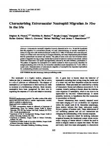
jections were frequently roughly perpendicular iB). Knob-like uropods
mm.
phalloidin
the
lasted 15-25 mm and contained 5-10 cells. 39 series of moving images were acquired by confocal microscopy through a single optical series typically lasted 10-15 mm and contained
leading edge. Extension from the leading edge
chamber. for 5
After
the were
through
lower
to characterize
migrating cells were clearly polarized, with a leading edge and tail similar to those seen in 2-D (Fig. 1B). Cells migrating in a 3-D matrix appeared more cylindrical than those migrating on a 2-D surface, which often have a broad, flattened
across
plated
unattached
paraformaldehde
(wlv)
were
were
and
of 10 nM
migra-
polvethlene
during
Neutrophils buffer
during
124-well
and
matrix
neutrophils represented 15 ± 3% of cells in any given volume frame. After 3-D rendering, unpolarized cells appeared spherical, with some ruffling and protrusion ofpseudopods at the surface (Fig. lA) In contrast, actively
rinse
scanning
stained
pores
)Poretics
filters
experiments.
then
PBS-S
cells
in PBS.
rinsed
inserts
blocked
Dickinsonj
also
3-.tm
at 37#{176}Cin incubation
In some
pore
Becton assays.
albumin for 5
with
0.8-tm
For immunofluorescence
were
inserts
amniotic
nonmignating
microscopy.
tion
within
by spinning-disk
acquired
face
FL-
in PBS-S.
and
was
mained adherent or migrate. These
1 h in
BODIPY
(Sigma)
bs a 1-h rinse
were
proteinase.
rinsed
or rhodamine-phalloidin
followed
the
incubated
antibody
two
The 1 h in PBS-S
and
were
of secondary
mi-
rhodamine/SE) for
aim
erage series In addition, laser scanning plane. These
amnions
paraformaldehde.
permeablized
Primary
Probes)
with
(w/x)
lO%
above).
in PBS-S.
was
within to migrate
then
saponin.
by addition
phallicidin
focal
tg/mL
been
proteins allowed
1 h in 2% were
described
followed
in PBS.
for
amnion
antibody
were
had
fixed
contained
with
cells
amnions
then
cells inhibitor
cytoskeletal
first
was
antibody
(Chemicon).
grating
morphology
of neutrophils migrating in a 3-D amnionic to a chemoattnactant gradient of fMLP. these experiments, a total of 64 series
anti-a-actinin
antibody
isothiocvanate-goat
Our
doextracel-
antibody
anti-rabbit
Neutrophil
cytoplasmic integrin
secondary
secondary
polyclonal International.
anti-2
anti-rabbit
anti-rabbit
rabbit
Chemicon
anti-a5
Chemicon),
IL).
studies:
monoclonal
fluorescein-goat
fluorescein
To localize
the
polyclonal
(mAb1962;
Rockford.
.
in these (Ab1930;
mouse
fluorescein-donkey
ical
used
domain
Chemicon),
Sigma).
icon),
were
cytoplasmic
Temecula,
,
main
staining
migration of the
Neutrophil
D).
over longer chemoattractant
migration
Several
upward for at least the predominantly
within
times was gradient
cells
showed
pant of the horizontal
tnajectories observation movement,
downward in the direcas evidenced by the
a three-dimensional
matrix
191
D -
2
‘s_
C G)
a_-a.’-.
.
E
#{149}
I
.!
#{149}#{149}#{149}..;4-..’
a
‘
4
-
1,
S
S
x Fig.
1
(40
nM
.
Three-dimensional fMLP)
was
volume-rendering
in the
spherical,
with
a trailing
uropod.
Pseudopods
migration
through
amniotic
basement in minutes.
membrane, toward Panel D indicates
bered primarily
192
cells.
extension
lower
The in the
Journal
starting x-y
of
plane,
Leukocyte
chamber,
and
retraction are
of calcein-labeled which of short
continuously
membrane
primarily
was
neutrophils
downward
pseudopods.
(B)
extended
from
occurs
in a plane
the source ofchemoattractant. the location of the centroids
location with
of the net
Biology
cells,
displacement
Volume
in the
either
at t
61,
migrating
view
shown
=
February
within
human
(A)
Non-migrating
here.
Migrating
cells
leading
edge,
whereas
to the
basement
the
parallel
display
Numbered arrows in panel (circles) at 1 -mm intervals
downward
displacement
0 or upon in the
1997
entering
z-axis.
Bars:
the (A,
amnion,
a polarized none
are
extended
membrane.
(C,
by D)
10
en not
face.
The
polarize
with from
Net
is indicated
B) 5 tm,
do
morphology
C indicate the locations of eight cells from the field,
viewed cells
the
migration
an
active
uropod. occurs
chemoattractant
and
remain leading
(C,
roughly edge
and
D) Neutrophil
perpendicular
to the
ofindividual cells; times are indicated sequence shown in (C), including numan tm.
open
circle.
Note
that
motion
occurs
accumulation membrane [29,
over
31].
much of the
of cells into the amnion near the 20- to 60-mm incubation
However,
greater than chemoattractant
Fig.
1D).
the
speed
the
net velocity (by roughly
As discussed
related
to the
of individual
below,
orientation
the basement time (Fig. iD) neutrophils
was
vector in the direction one order of magnitude;
the
horizontal
of matrix
movement
was
fibnils.
Subcellular localization of proteins found in adhesion plaques On
2-D
to the
surfaces, substrate
neutrophil [9, 39],
leading
and
they
edges
contain
focal adhesion complexes in other cell distribution ofthese proteins (e.g. F-actin, teins,
and
integrmns)
could
indicate
adhere proteins
types actin
which
tightly found
[9, 40]. binding
neutnophil
in The pro-
struc-
tunes, including leading edges, pseudopods, and uropods, are involved in cell adhesion and motility in our 3-D system. To study the distribution of these proteins, cells were fixed during
fMLP-stimulated
migration
through
the
matrix
and
stained for proteins connecting the ECM to the cytoskeleton. In addition to a subcortical distribution (Fig. 2A), F-actin was enriched in three major structures: leading edges, pseudopods,
and
pods
were
uropods
(Fig.
identified
pseudopods that could
were identified be distinguished
of F-actin the uropod
staining stained
grating
through
a-Actinin edge and
2, A-D).
Leading
monphologically
as protrusions from leading
is consistent for F-actin amnion
(i.e.,
co-localized pseudopods,
edges
and
in well-polarized from edges.
the cell body The pattern
with that in 2-D except more intensely in cells Fig.
2,
B and
with F-actin but it was not
unocells;
that mi-
D).
in both the found in the
leading uropod
(Fig. 2, B and C). Immunostaining for the cytoplasmic domain of the integrin a5 revealed a distribution similar to that of a-actinin; a co-localized with F-actin in the leading edges and pseudopods of migrating cells, but was not found in the ilar
unopods
(Fig.
distribution
2D).
to that
Integrmn of a5
a5
(data
not
was
found
in a sim-
Fig.
2.
and
shown).
F-actin,
a-actinin,
pseudopods,
man
amnion
but
neutrophils can in the matrix
utilize
physical
(red) ing
To investigate the dynamic role of neutrophil structures in 3-D cell motility, moving images of neutrophils obtained by spinning disk confocal microscopy were examined closely for examples ofpseudopod extension during migration. While migrating
nent
ci-actinin
(green) rows)
to extend
with
respect
and
rowhead).
is
in the
cell
Bars:
5 .tm.
body
(A),
pseudopods (B, with but
by indirect
complex
from
uropods.
(D)
is excluded
im-
in leading
arrows),
adhesion
in the from
leading the
hu-
F-actin
is enriched
C, thin
C) The
F-actin
edges
within
microscopy.
and (A,
excluded
co-localized
viewed
confocal
distribution
D arrowheads). (green)
is found
gradient,
scanning
arrows),
and constriction rings structures. Anchored pseudopods as lateral protrusions
(Fig. 3). In addition to these anchored pseudopods, neutrophils occasionally were observed to extend a pseudopod that continued to expand as the cell transferred its mass into the growing structure (Fig. 4, A and B). The connection between the pseudopod and the original cell body formed a constriction ring that remained stationary relative to the matrix (Fig. 4, A and B). This process resembled a balloon being squeezed thnough a hole and expanding on the
remained forward
other
currence
side.
(B,
fMLP
in leading migrating
and
trail-
compo-
a
Integrin
edge
trailing
(thick uropod
ar(ar-
through
seen
the matrix, neutrophils were frequently lateral pseudopods that remained stationary to the matrix as the cell body migrated past
in a cortical
(A, B, D, thick uropods
laser
co-localize
of neutrophils
to an
and
is found
edges
integrins
uropods,
in response
munofluorescence
Migrating structures
and
not
As discussed
below,
these
anchored
anchored
et at.
Neutrophil
closely
were defined from polarized,
associated
with
pseudopods
were
of footholds
was
migration
within
seen
in eight
greatly
a three-dimensional
moved These
different
disk confocal microscopy, imaging of the matrix. probably
matrix
in 3-D moving images migrating cells that
fixed with respect to the matrix as the cell past them in three consecutive volume frames.
through the use of spinning did not permit simultaneous
pseudopods
Mandevitte
were
cells which The oc-
underestimated
matrix
193
Fig.
3.
using
Neutrophils
spinning
relative
to the
due our
use
footholds
disk
confocal
matrix
as
in the
the
cell
from
and
Constriction rings were nections between the main remained fixed with respect as exhibiting a constriction
During
fMLP-stimulated
surface-rendered,
migrates
to the angle of viewing imaging system.
appeared
matrix.
microscopy.
the
the
and left
to the
temporal
right
(>
by 3-D focal
reconstruction
transferred
of cells
microscopy.
currence
As for
from
by use
the
of constriction
of
defined as narrow annular concell body and a pseudopod that to the matrix. A cell was scored ring only if the mass of the cell
to be continuously
the cell
of spinning
scoring
rings
was
body period times
disk
of footholds,
con-
the
underestimated
oc-
To determine rings
opposed media
whether
were
anchored
in fact
to positionally [41]), simultaneous
was performed
pseudopods
associated fixed
and
constnic-
a5
Staina comin both
and density, could represent
that olution
of light
oriented When
parallel viewed
images
lowing
ing),
were
maximal
and
ascertain ing through
microscopy.
The
to the basement by laser scanning obtained time
some
to
as well as a diffuse interstitial a hydrated gel of fibnils below
from
resolution
adjustment
whether constriction openings in the
ofthe cells and that constriction
matrix rings
in the
ECM,
through
which
D). Constriction
rings
of fibrils
a single
optical
and minimal of the focal plane rings matrix.
the
staining the nes-
membrane. confocal microscopy,
cell
were
(Fig.
10 times
imaging method. In addition, the opening could also stretch
pseudopods
matrix,
into
the
pores
(footholds)
through
4,
by this
but
large
enough
membranes.
pores
were
those
on filters
(Fig. face
(n
B);
surface =
pores
38% (n
Biology
Volume
61.
February
1997
more 5).
containing efficiently
In addition,
were
versus
the
than mor-
highly
9%
polarized
on
on the smooth
sun-
94).
exhibited
along contact
The fibnils in the amniotic parallel to the basement
is lim-
membranes
(Fig.
of cells 107)
=
Neutrophils migrate matrix fibrils Neutrophils
a small
in the absence on adsorbed
migrating on these porous surfaces was more that of cells migrating on the smooth surface
A and
the porous
on
significantly
fibnils). oriented
Leukocyte
by stain-
to accommodate
plated
to migrate without
ofcells than 6,
Cells
able
ical, deformation The observation
of
as seen
pseudopod. Motility assays were performed of any added proteins either in the medium
amnion
Journal
structures
conditions in sized holes matrix struc-
F-actin (data not shown). Cells were the 3-.tm pores. 2 integrmn stained
through
194
these
Polycanbonate membranes were used to directly test the hypothesis that neutrophils could productively use anchored pseudopods to facilitate migration. Cell migration assays in
phology polarized
expansion the matrix
rings
since
appropriate geometric presence of appropriately sufficient density ofthese
nion present an elastic barrier to neutnophil migration that can be circumvented by mechanical, as opposed to chemconstriction
5 .tm.
cell surface diffusely, whereas F-actin, a-actinin, and integrin localized to the expanding portion of the cell, distal to the constriction ring (Fig. 4, F and G). In general, F-actin tended to localize near cell-substratum contacts.
to the
to accommodate the cell (Fig. 4E). This deformation of the matrix directly demonstrates the exertion of mechanical force by the cell on the ECM. Therefore, some regions ofthe am-
of the matrix. offootholds and
imaged stationary
to crawl
(al-
by squeezimaging
migrating
of the
were
remains
mov-
optical plane showed by narrow openings was
Bar:
and
response to a uniform application of 10 nM fMLP (chemokinetic stimulation) were performed on polycarbonate membnanes with or without O.8-.tm pores, too small for the cell
photobleachwas necessary
observed
in minutes.
architecture
neutrophils
is extended
were
section
were caused Simultaneous
through a single were produced
C and
dual cell/matrix of the cell through
majority
is indicated
by the
calcein-labeled (arrow)
albumin-coated filters with 0.8- and 3-rim pore diameters, respectively. Although cells could not crawl through the O.8-.tm pores, they did migrate across them, inserting lat-
the
microscopy.
caliber
amnion,
tunes to be utilized by cells during the observation period and sufficient rigidity of the matrix against which neutnophils can exert locomotive force). To model footholds and constriction rings in a more simplified system, we employed
(as
confocal
structures
ited
Time
the
pseudopod
can only exist under the amnion (i.e., the on gaps in the matrix,
pseudopods projecting into the imaging of the cells and matrix
scanning
matrix
field.
A lateral
ing for l2 integnn and able to squeeze through
ing of the amnion with rhodamine/SE demonstrated plex network of interconnecting fibrils that varied
ing
by laser
with
through
face.
enal
in these
studies.
tion
en
of the
resolution
the pseudopod during the entire observation 5 mm). Constriction rings were observed seven
into
migration
viewed
(i.e.,
cell
and
distort
guidance
attachment
to and membrane membrane,
during
migration
migration
along
are generally perpendicu-
Fig.
4.
phils.
Expansion
viewed
fling
disk
en
confocal
labeled
constriction
appear
microscopy.
the
matrix
(red).
the
matrix
as the
coated
distal
exhibiting
microscopy.
neutrophils
confocal
of pseudopods face.
PET
The cell
the
lan to the migrating
Image
(red(.
the
panel
contains
is migrating
across
the
hole
simulating acquired
by
line-scan
both
stain
gradient neutrophils
during
distal
that
using
of chemoattractant. revealed that
appears (E)
plane
laser
to the
scanning
constriction
Images
by
and
i.e.
ring
leading
Dynamic images of migration occurred pri-
edge.
to the
attractant.
pulled
80_90%
to
migrate
The dynamic vicinity
imaging
neutrophils were along visible fibers. leading edge is a highly of
extensions in a groping
of the associated active
matrix with
structure.
the cells contacted fashion. This kind
many
ofgroping
showed and and fibers
while
(G)
Same
as
(A.
B. C.
E) Time
for
of a few
tended through
this
it;
difficult
and
pull
F.
pulling
and
but
is indicated
by spin-
(arrows).
Calceinscanning
a hole
(arrowheads)
in
elastic
deformation
of
pore
in an albumin-
of migration).
Indirect
is distributed
through-
stained
for
in minutes.
for neutnophils been suggested
actin
(green)
Bars:
and
5 tm.
migrating on to contribute
details in the environment [20]. wrapped around a fiber and distortion
is readily
to appreciate
(green)
neutro-
imaged by laser
a 3-tm
direction
112 integrin
reported it has
micrometers
but
through
panel
matrix
Note
arrow,
were
obtained
through
microscopy.
filter;
and
plane
migrating
detection ofstructural Frequently, the lamellipods
that
in still
of fibers
apparent images.
oven
distances
in moving This type
images of grab
behavior was seen continuously during migration all (n > 50) neutnophils imaged by laser scanconfocal microscopy, often with several fibers being
of virtually
in the behavior
tlande,ilte
edge).
in the
optical
migrating
of the
has been previously grooved surfaces,
of calcein-labeled cells
openings
(green)
neutrophil
(leading
B) Examples
a single confocal
surfaces
microscopy.
the
manly parallel to the basement membrane (Fig. iC), but with net downward migration toward the source of chemoSimultaneous
scanning ofa
bottom
constriction confocal
ring.
from cell
laser
(A.
Volume-rendered
through
are
BODIPY-SM-labeled
top
force. amnion.
of pseudopods
(F) Cross-section
(arrowheads. distal
human
red.
obtained
transmigration.
locomotive
through
by expansion
neutrophil.
localizes
to generate
migration
caused
optical
ring
to the
be used
amnion
a single
(arrows(
(red(
are
a single
a constriction
integrin
which
rings
may during
rhodamine-labeled
each
expands
rings (arrows)
D( Constriction and
cell
of a
cell.
Q-actinin
rings
and
membrane.
immunofluorescence out
(C. green.
to constriction
ning
et at.
Neutrophil
migration
within
a three-dimensional
matrix
195
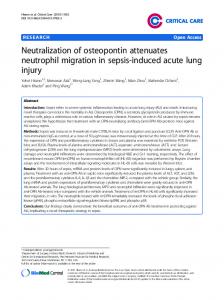
100
80
60
0
40
20
Fn Coated
Pores
No
Pores
Control
Fig.
5.
Neutrophils
ing
pores.
Cells
or
without
dishes
0.8-.tm
as a control.
counted
as motile.
each
condition
and
migrate were
were
more
plated
on
pores
or
on
Cells
able
to migrate
were
averaged
cells.
to be
significantly
on
membranes
data
than
7 tm
three
shown
different
filters
with
coverslip-bottom
more over
The
contain-
membrane
fibronectin-coated
Data
n > 150
found
efficiently polycarbonate
in 200
s were
experiments are
mean
from
each
and values
other
for
SE
±
by
t-test
(P < 0.05).
Fig.
6.
Cells
pores
have
face.
Cells
without and
pulled clung
toward the cell simultaneously. to the uropod ofthe migrating
ping back to its original In fields of heavy cell constant the
position density,
The matrix frequently neutrophil, finally snapafter the cell pulled the amniotic matrix
away. was in
Migration cell was
tortion
tended currently
to continue migrating,
by the grasping
Periodically, this tended pendicular parallel havior
along which
pseudopods
the fiber exhibited
than
did
a cell changed its direction to occur along an adjacent to the current fiber, rather
fiber (Fig. was observed
the
majority
the
observation
7, A and > 20 times
of cells
Germany) City,
did
parallel
fibers.
of migration, fiber that ran than switching
not change
and pento a
fibers
ized
morphology Bar:
fibers behind
tnacellular amnion, rhodamine shown).
In addition,
cells
were
matrix, and matrix density
noted
cells (data
to accumulate
in-
vesicles containing rhodamine, used to label the over the time course of migration even though the could not be detected in the media (data not These observations implied that neutnophils were
digesting the matrix intnacellularly.
during
migration,
either
extracellularly
on
196
Journal
of
Leukocyte
Biology
Volume
61.
a cooled
CCD
though
no
with
with
fMLP
proteins
(Leitz
(Dage-MTI
0.8-tm
sur(A)
or
fixed
immunofluorescence
Microscope camera
with
filters
stimulated Digital
0.8-.tm
a smooth
pores were
Wetzlar. Inc..
exhibited
present
Micha polar-
on
the
mem-
tim.
or generation the migrating
matrix. into
Fig.
We expected to observe pericellular proteolysis during neutrophil migration through the matrix because neutrophils express a number of pnoteinases, some of which are bound to the cell surface [42]. Confocal sections of cells within the matrix frequently revealed a slight penicellular halo of
not shown).
then Diavert
on membranes even
10
were
a Leitz
on
membrane
phalloidin.
with
containing those
However, time-lapse moving images evidence of active fiber degradation,
On
February
1997
the
pre-existing
weight
Extracellular proteolysis is not required for migration through amnion
than the surrounding found in areas oflower
Cells
than
of free cells.
contrary, areas
pnoteinase 8,
B and
did such
fiber ends, or cleaning cells
were
of low matrix
thermore, neutrophils were able the presence ofa high-concentration
during
time.
lower fluorescence were commonly
IN).
Cells
rhodamine with
membranes
polycarbonate
pores.
equipped
on
morphology
on
obtained
igan
migrate
on which less dis-
B). This fiber-switching bein 12 different cells, although
(n > 50)
plated
with
were
to
polarized
0.8-jim
stained
brane.
motion.
were
(B)
images
induced
a more
inhibitors
clearing of the often
density
any of
of tunnels penicellulan
seen (Fig.
to migrate cocktail
(see
not reveal as severing
to migrate 8A).
Fun-
into a matrix of low-molecular-
Materials
and
in
Methods;
C).
DISCUSSION Neutnophils
and
other
motile
animal
a complex, nescence
3-D matrix in vivo. microscopy to directly
behavior We
of neutnophils crawling found that many aspects
sue.
in a 3-D
matrix
are
functionally
cells
We have observe
migrate
through
used confocal the dynamic
fluomotile
through a biological tisof neutrophil migration similar
to those
on
a flat
surface but there are significant differences. The 3-D matnix can impede migration by presenting a physical barrier, such as the basement membrane [31], but neutnophils are also apparently capable of using the physical structure of the matrix to promote locomotion. The orientation of ma-
tnix fibers
was found
to be a major
range direction of cell tnix can be reversibly A central
theme
movement. deformed
to the
proposal
3-D matrix is different from that cells are able to use the architecture dition to specific ligand-neceptor force. niotic
determinant
Our direct observations matrix used in these
of the
that
motility
through
with
respect
amar-
unique pseudo-
relative to the substrate during inserted into footholds in the that contain a high density of
provide a variegated surface into which be extended. Movement of the anchored
rearward
a
a 2-D surface is that of a 3-D system in adinteractions to generate
support these ideas. The studies had a heterogeneous
pods that remain stationary cell translocation are indeed matrix. Areas of the matrix
ma-
on
chitectune in which a variety of motile cell behaviors to 3-D systems could be observed. We found that
fibnils could
short-
In some regions, the during cell migration.
to the
cell
body
will
pseudopods pseudopod propel
the
cell
forward with respect to the matrix, not unlike a climber using crevices to scale a wall. We also noted the expansion of pseudopods distal to constniction
which these
use
would
the
rigidity
too wide, tion non caliber
caused by narrow the cells migrated rings for locomotion,
rings,
through to utilize
be
dependent
of the provides a potential narrow
and
cannot migrate through, the opening, using the is somewhat
elastic,
to accommodate
of the
the
matrix
opening
of the
cell
can
is
to cell transmigraConversely, if the is rigid,
then
the
although it could migrate hole as a foothold. But if the
the
and
opening
distort
the
The
matrix
enough
observation
rings in amnion through a matrix
cell
across matrix of foot-
supports by use
the idea of simple
forces.
The idea that used to generate
these force
by pseudopods
is supported
choned
size caliber
an obstacle of traction.
transmigration.
holds and constriction that cells could migrate mechanical
the
If the
neither source
it
is too
on
matrix.
openings in the matrix (Fig. 4). If cells are able we speculate that such
two
footholds and for locomotion
constriction through
the
rings are amnion
additional findings. First, these ancontain integrins, F-actin, and a-actinin
(Fig. 2), a molecule that links integnins to the cytoskeleton [43]. Therefore, the lateral pseudopods contain appropniate machinery to support traction. Second, cells were observed Fig. otic through optical
7. Neutrophils matrix. Images
exhibit contact of calcein-labeled
rhodamine-labeled plane by use of laser
is indicated leading
in seconds. edges
while
fibril tended
s). The
cell
(arrowhead. to the
does was
not detach migrating
began fiber
migrating (arrowhead,
amnion scanning
Pseudopods uropods
Cell to the left of the of thin fibrils (arrow, 150
switched 300 upper at the upward
fibrils s).
left
The along
10
fibrils within amni(green) migrating
(red) were acquired confocal microscopy, were
exhibited
panel was 0 s) and
migrating switched
continuously
little
change
a second cell in the a diagonal
time
parallel fibril
to the left (arrowhead,
to the right after switching onto 70 s). In the last panel. the cell
(A)
horizontal panel 0 s),
exbut
of the panel 0 s), and
a second, horizontal again changes fibers The cell that entered along a fibril that is panel (arrow, 400 s).
tm.
Mandevitle
the
matrix
during
both
retrograde
and while squeezing such deformation could
motion
through only be
holes medi-
ated
from
to the sheet (arrowhead,
right of the (arrowhead.
to deform
of lateral pseudopods in the matrix, and
a single the time
extended
to a different
lower fibril
to head downward. (70 s) was migrating in focus in the last
from and
in morphology.
to the left, to a vertical
tail. (B) The neutrophil along the vertical fiber
(arrowhead, 400 s) and begins the field from the lower right visible with the slight change Bars:
guidance along neutrophils
et at.
by transmission of force from the cell to the matrix. Because amnion provides chemical as well as mechanical interactions, we could not separate these to demon-
strate a role for mechanical interactions in facilitating migration. However, the studies with synthetic membranes cleanly indicate that both lateral pseudopods inserted into matrix footholds and squeezing through constriction rings can allow forward migration in the absence of specific adhesion proteins. The enhanced migration on polycarbonate membranes containing 0.8-.tm pores (Fig. 5) is particulanly informative because the enhanced migration could only be attributed in the substrate,
Neutrophil
migration
to mechanical (not the pores providing
within
chemical) footholds
a three-dimensional
differences into which
matrix
197
Fig.
8.
induced (roscopy.
A cocktail
of proteinase
to migrate
into
(A)
the- pa1.
The
migrating
in minutes.
the cells presence
architecture. amniotic
ment
membrane
chemoattractant ance appears
Journal
bottom
right
Bars:
could insert of pores also
along
does
of the
through 10
panel
Leukocyte
neutrophil in the
migrates
presence
downward,
a pre-existing
hole
demonstrating
the
anchored the cells
in the
pseudopods. to assume
the amniotic both in terms
Much of the motion fibnils that were oriented and perpendicular to gradient. Thus, in our to override chemotactic
of
prevent (red)
migration distorting
matrix
distribution
into
amniotic
of proteinase the
(arrows).
matrix.
inhibitors matrix
Images
of neutrophils
and
as
it moves;
were
taken
after
30-mm
Calcein-labeled imaged
laser
cell
enters
another from
neutrophils
using
a single
focal
migration
through
cells
progressively
the
at the
Time
matrix
were
confocal
field
plane. the
(green)
scanning
mitop
of
is indicated
in the
presence
.tm.
lateral allowed
(Fig. 6). to its elasticity, structure,
not
amnions
C) Stereo-projections
inhibitors.
gated shape In addition a heterogeneous
198
at the
to the
(B and
of l)re)ttinase
and
cell
inhibitors
rhodamine-labeled
Biology
The an elon-
matrix also has of composition
of neutrophils was parallel to the basethe direction of the system contact guidguidance on a local
Volume
61,
February
1997
scale.
However,
with direction
tnix
the ing dicate impart
since
time, net migration of the chemoattractant
chemoattractant a localized
binds haptotactic
still
occurs gradient.
to the matrix, gradient. Our
that the influence of the a directional preference
move
into
the
ma-
downward in the It is possible that thereby genenatobservations in-
chemoattractant when cells
are
may be to switching
from one fibnil to another. We observed that, during fibril switching, cells pnefenentially migrate along fibnils exhibiting the least deformation
(i.e. the greatest
tension/rigidity).
Although
tify this behavior, it appeared ing the tension in the network along
the
fibnil
may
most
stable
fibers.
be
related
to
we did
as though of fibers, This
recent
the cells favoring
not quan-
ACKNOWLEDGMENTS
were testmigration
This
work
preference
for the
stiffer
grants
observations
with
beads
Medical
was
supported
GM34770 Scientist
Training
in an optical trap as they bind to integrins [441. When the strength of the trap was increased, the resistance of the beads to displacements increased, possibly due to in-
(J.
creased linking
and/or crosscomponents
tions, Drs. Richik Ghosh croscopy, and Dr. Satyajit
through along
labeling.
polymerization of actin of surface receptors to the
of adhesion complexes
complexes. would favor
high-tension
matrix
in contact cell
with
Increased extension
fibrils,
stable
migration
[45,
adhesion migration
and
and
surfaces
filaments cytoskeleton
stabilization can
such rigid,
(Fig.
needed cilitate
8A),
through which
the
direction
and
one
of
proteinases
are
required
amniotic
[31].
In
if
4. 5.
tissues, idea may
neutrophil
between
Neutrophil
tems.
However,
gradients matrix ofthe can
are additionally matrix, which override
cell
with
forces
2-D
and
similar migrating
10.
differ-
3-D
sun-
1 1.
in both syson a flat
chemoattractant or neutrophils migrating
12.
haptotactic through
guidance
architecture
on a local of the
matrix
scale
a physiological
3-D
matrix
(Fig.
provides
that can be used to generate to 3-D (Figs. 3 and 4). We propose
through
9.
at the
a
13.
1).
14.
the
I 5.
traction that cells
generate
16.
trac-
through
interactions,
mined
a combination of chemical and mechanical the relative contribution of which is deterby the ECM and the adhesive state of the cells. Our
help to define tion of cells in 3-D matrices. techniques may be directed observations
behaviors has been
1 7. 18.
questions unique to the migraFuture studies using similar toward the quantitation of 3-D
during migration through biological matrices done for cells crawling on 2-D surfaces [41,
47]. The methods we have described to the study of other 3-D migratory cell
on
footholds
unique
migrating tion
the
8.
influenced by the physical structure provides contact guidance (Fig. 7) that
chemotactic
Furthermore,
migration
on
7.
specialized regions basement membrane
polarization appears while neutrophils
rely primarily for orientation,
surface
6.
out in the absence of sepnoteases. It is likely that
studies, the cells accumulated basement membrane and did not penetrate it. In conclusion, we have found both similarities and faces.
3.
faof
of cells through not support this
breaching such as the
2.
enlarge
prevent that pro-
carried serum
1.
in the matrix could active degradation
in our
ences
grant
and
5-T32GM07367
Kim and members of Dr. Samfor help with amnion preparaand Ken Dunn for help with miMayor for help with fluorescent
REFERENCES
ma-
inhibitors accepted
for
matrix,
fact.
in the
mechanically
openings not observe
the migration our results do
be that our studies were rum and lack activatable of the
could
openings
nor did a battery of proteinase in the matrix. It is generally facilitate reason
teinases
pre-existing
they
(Fig. 4). Although migration, we did
the matrix migration
Program
of Health
(M. A. L.),
46J.
to migrate
tnix
thank Sonia laboratory
Institutes
T32AG00189
of pseudopods
change
The extracellulan matrix can provide directional cues to cells by acting as a physical barrier to migration. Cells were preferentially found in areas of lower matrix density, and tended
T. H. M.). The authors uel Silverstein’s
by National
(F. R. M.),
should systems
19.
as 46, 20.
be applicable such as tumor
21.
invasion.
.liandeiilte
et at.
Devreotes. P. N.. Zigmond, S. H. (1988) Chemotaxis in eukariotic cells: a focus on leukocytes and Dictyoszelium. Annu. Rev. Cell Bail. 4, 649-686. Stossel, T. P. (1994) The machinery of blood cell movements. Blood 84, 367-379. Lee. J.. Ishihara. A.. Jacobson, K. (1993) How do cells move along surfaces? Trends Cell Bail. 3. 366-370. Hynes. R. 0. (1992) Integrins: versatility. modulation, and signaling in cell adhesion. Cell 69. 11-25. Stossel. T. P. (1993) On the crawling of animal cells. Science 260, 1086-1094. Berton, G.. Laudanna, C., Sorio, C.. Rossi, F. (1992) Generation of signals activating neutrophil functions by leukocyte integrins: LFA-l and gpiSO/95. but not CR3. are able to stimulate the respiratory burst of human neutrophils. .1 Cell Biol. 116, 1007-1017. Jaconi. M. E.. Theler, J. M.. Schlegel, W.. Appel, R. D.. Wright. S. D.. Lew. P. D. (1991) Multiple elevations of cytosolic free Ca2 in human neutrophils: initiation by adherence receptors of the integrin family. J. Cell Biol. 112. 1249-1257. Hendey. B.. Klee. C. B.. Maxfield. F. R. (1992) Inhibition of neutrophil chemokinesis on vitronectin by inhibitors of calcineurin. Science 258. 296-299. Lawson. M. A.. Maxfield, F. R. (1995) Ca2and calcineurin-dependent recycling of an integrin to the front of migrating neutrophils. Nature 377. 75-79. Maxfield. F. R. (1993) Regulation of leukocyte locomotion by Ca2 . Trends Cell Biol. 3. 386-391. Oloole. T. E.. Katagii-i. Y.. Faull. R. J.. Peter, K.. Tamura. R.. Quaranta. V. Loftus. J. C.. Shattil. S. J.. Ginsburg. M. H. (1994) Integrin cytoplasmic domains mediate inside-out signal transduction. J. Cell Biol. 124. 1047-1059. Condeelis. J. (1993) Life at the leading edge: The formation ofcell protrusions. Annu. Ret. Cell Biol. 9, 411-444. Sibille. Y.. Lwebuga-Mukasa. J. S.. Polomski. L.. Merrill. W. W., Ingbar, D. H.. Gee. J. B. L. (1986) An in vitro model for polymorphonuclearleukocyte-induced injury to an extracellular matrix. Am. Rev. Respir. Di.s. 134. 134-140. Brown. A. F. (1982) Neutrophil granuloctes: adhesion and locomotion on collagen substrata and in coHagen matrices. .L Cell Sci. 58. 455-467. Hurst. 1.. Wilton. J. (1990) Polymorphonuclear leukocyte phagocytosis of Capnocvtophaga ochracea in three-dimensional plasma clots. J. Cell Sci. 95. 487-498. Haston, W. S.. Shields, J. M., Wilkinson, P. C. (1982) Lymphocyte locomotion and attachment on two-dimensional surfaces and in threedimensional matrices. j Cell Biol. 92, 747-752. Brown. A. (l984( Neutrophil and monocyte behaviour in three-dimensional collagen matrices. Scanning Electron Microsc. 2, 747-754. Schmalstieg. F. C.. Rudloff. H. E., Hillman. G. R., Anderson, D. C. (1986) Two-dimensional and three-dimensional movement of human polymorphonuclear leukocvtes: two fundamentally different mechanisms of locomotion. J. Leukoc. Biol. 40. 677-691. Lanir. N.. Ciano. P. 5.. Van de Water. L.. McDonagh. J.. Dvorak. A. M.. Dvorak. H. F. (1988) Macrophage migration in fibrin gel matrices. II. Effects of clotting factor XIII. fibronectin. and glycolaminoglycan content on cell migration. J. Immunol. 140, 2340-2349. \Iathes. T.. Gruler. H. (1988) Analysis ofcell locomotion: Contact guidance of human polsmorphonuclear leukocytes. Eur. Bioplws. J. 15, 343-357. Schmidt. J. A.. von Recum. A. F. (1992) Macrophage response to microtextured silicone. Biomateriafs 13. 1059-1069.
Neutrophil
migration
within
a three-dimensional
matrix
199
22.
23.
24. 25. 26. 27.
28.
29.
30.
31. 32.
33.
34.
35.
200
W#{243}jciak.Stothard, B., Curtis, A. S. G., Monaghan, W., McGrath, M.. Sommer, I., Wilkinson, C. D. W. (1995) Role of the cytoskeleton in the reaction of fibroblasts to multiple grooved substrata. Cell Motil. Cytoskel. 31, 147-158. Guido, S.. Tranquillo, R. (1993) A methodology for the systematic and quantitative study of cell contact guidance in oriented collagen gels: Correlation of fibroblast orientation and gel birefringence. J. Cell Sci. 105, 317-331. Wilkinson, P. (1987) Leucocyte locomotion: Behavioural mechanisms for accumulation. J. Cell Sci. 8 (Suppl.), 103-119. Travis, J. (1988) Structure, function, and control of neutrophil proteinase. Am. J. Med 84, 37-42. Ciano P. 5., Colvin, R. R, Dvorak, A. M., McDonagh, J.. Dvorak, H. F. (1986) Macrophage migration in fibrin gel matrices. Lab. Invest. 54, 62-70. Parkhurst, M. R., Saltzman, W. M. (1992) Quantification of human neutrophil motility in three-dimensional collagen gels. Effect of collagen concentration. Biophys. J 61, 306-315. Parkhurst, M. R., Saltzman, W. M. (1994) Leukocytes migrate through three-dimensional gels of midcycle cervical mucus. Cell. IrnmunoL 156, 77-94. Furie, M. B., Naprstek, B. L., Silverstein, S. C. (1987) Migration of neutrophils across monolayers of cultured microvascular endotheial cells. An in vitro model of leukocyte extravasation. J. Cell Sci. 88, 161-175. Aplin, J. D., Campbell, S., Allen, 1. D. (1985) The extracellular matrix of human amniotic epitheium: Ultrastructure, composition, and deposition. I Cell Sci. 79, 119-136. Azzarelli, B., Lafuze, J. (1987) Amniotic basement membrane: a barrier to neutrophil invasion. Am. J. Obstet. Gynecol. 156, 1130-1136. Furie, M., Cramer, E., Naprstek, B., Silverstein, S. (1984) Cultured endothelial cell monolayers that restrict the transendotheial passage of macromolecules and electrical current. I. Cell BaiL 98, 1033-1041. Loike, J. D., El Khoury, J., Can, L., Richards, C. P., Rascoff, H., Mandeville, J. T. H., Maxfield. F. R., Silverstein, S. C. (1995) Fibrin regulates neutrophil migration in response to interleukin 8, leukotriene B4, tumor necrosis factor, and formyl-methionyl-leucyl-phenylalanine. J. Ex,a Med. 181, 17631772. Mandeville. J. T. H., Ghosh, R. N., Maxfield, F. R. (1995) Intracellular calcium levels correlate with speed and persistent forward motion in migrating neutrophils. Biophys. J. 68, 1207-1217. Kremer, J. M. H., Esker, M. W. J. v. d., Pathmamanoharan, C., Wiersema,
Journal
of
Leukocyte
Biology
Volume
61,
February
1997
36.
37.
38.
39.
40. 41
42.
43.
44. 45.
46.
47.
.
P. H. (1977) Vesicles of variable diameter prepared by a modified injection method. Biochem. 16, 3932-3935. Marks, P. W., Maxfield, F. R. (1990) Transient increases in cytosolic free calcium appear to be required for the migration of adherent human neutrophils. J. Cell Biot. 110, 43-52. Marks. P. W., Hendey, R, Maxfield, F. R. (1991) Attachment to fibronectin or vitronectin makes human neutrophil migration sensitive to alterations in cytosolic free calcium concentration. J. Cell BaiL 112, 149-158. Wells, K. S., Sandison, D. R., Stricker, J., Webb, W. W. (1990) Quantitative fluorescence imaging with laser scanning confocal microscopy. In Handbook ofBiological Confocal Microscopy (J. B. Pawley, ed.) New York: PIenum, 27-39. Sullivan, J. A., Mandell, G. L. (1983) Motility of human polymorphonuclear neutrophils: microscopic analysis of substrate adhesion and distribulion of F-actin. Cell Motil. 3, 31-46. Bereiter-Hahn, J., Fox. C. H., Thorell, R (1979) Quantitative reflection contraM microscopy of living cells. ,.t Cell Bail. 82. 767-779. Shutt, D. C., Weasels, D., Wagenknecht, K.. Chandrasekhar. A.. Hitt. A. L., Luna, E. J., Soll, D. R. (1995) Ponticulin plays a role in the positional stabilization of pseudopods. J Cell Biol. 131, 1495-1506. Owen, C. A., Campbell, M. A., Sannes, P. L., Boukedes, S. S., Campbell, E. J. (1995) Cell surface-bound elastase and cathepsin G on human neutrophils: a novel, non-oxidative mechanism by which neutrophils focus and preserve catalytic activity of serine proteinases. I. Cell Biol. 131, 775789. Otey, C. A., Vasquez. G. a. Burridge. K.. Erickson. B W. (1993) Mapping of the alpha-actinin binding site within the beta-i integrmn cytoplasmic domain. j Bail. Chem. 268, 21193-21197. Choquet, D., Felsenfeld, D. P., Sheetz. M. P. Extracellular matrix rigidity causes strengthening of integrin-cytoskeletal linkages. Cell, in press. Ueda, M., Ogihara. S. (1994) Microtubules are required in amoeba chemotaxis for preferential stabilization of appropriate pseudopods. J. Cell Sci. 107, 2071-2079. Wessels, D., Vawter-Hugart, H., Murray, J.. Soll. D. R. (1994) Threedimensional dynamics of pseudopod formation and the regulation of cell turning during the motility cycle of Dictyostelium. Cell Motil. Cytoskel. 27. 1-12. Murray, J., Vawter-Hugart, H., Voss, E., Soll, D. R. (1992) Threedimensional motility cycle in leukocytes. Cell Motil. Cywskel. 22. 211-223.