32nd Annual International Conference of the IEEE EMBS Buenos Aires, Argentina, August 31 - September 4, 2010
Dynamic Neural Network Detection of Tremor and Dyskinesia from Wearable Sensor Data Bryan T. Cole, Serge H. Roy, Carlo J. De Luca, Life Fellow, IEEE, and S. Hamid Nawab, Senior Member, IEEE
Abstract—We present a dynamic neural network (DNN) solution for detecting time-varying occurrences of tremor and dyskinesia at 1 s resolution from time series data acquired from surface electromyographic (sEMG) sensors and tri-axial accelerometers worn by patients with Parkinson's disease (PD). The networks were trained and tested on separate datasets, each containing approximately equal proportions of tremor, dyskinesia, and disorder-free data from 8 PD and 4 control subjects performing unscripted and unconstrained activities in an apartment-like environment. During DNN testing, tremor was detected with a sensitivity of 93% and a specificity of 95%, while dyskinesia was detected with a sensitivity of 91% and a specificity of 93%. Similar sensitivity and specificity levels were obtained when DNN testing was carried out on subjects who were not included in DNN training.
I. INTRODUCTION
A
DVANCES in machine learning algorithms [1] and fast evolving wearable sensor technology have created the potential for developing systems that may someday allow clinicians to remotely and unobtrusively detect functional activities and/or movement disorders of sensor-wearing patients in their natural environments of daily living. This type of detection would offer a more accurate and reliable alternative to self-report diaries that are currently used in clinical practice for medical management of patients with motor disorders. For example, the best agreements between self-report diaries of motor disorders and expert assessments have been reported [2] to range only between 0.49 and 0.78 using a kappa statistic. We have been developing algorithms to address the specific problem of detecting movement disorders associated with Parkinson‟s disease (PD), such as tremor [3] and dyskinesia [4]. The algorithms are applied to data from wearable miniaturized sensors that are attached to parts of Manuscript received April 1, 2010. This work was supported in part by the Public Health Service/National Institute of Health (PHS/NIH) under Grant 1 R01 EB007163-04 and under Supplementary Grant 3 R01 EB007163-03S1 from the National Institute of Biomedical Imaging and Bioengineering (NIBIB). B. T. Cole is with Dept. of Electrical and Computer Engineering (ECE), Boston University, Boston, MA 02215 USA (phone: 617-353-3559; email:
[email protected]). S. H. Roy is with Neuro-Muscular Research Center (NMRC) at Boston University (e-mail:
[email protected]). C. J. De Luca is with Biomedical Engineering (BME), ECE, and NMRC at Boston University (e-mail:
[email protected]). S. H. Nawab is with ECE, BME, and NMRC at Boston University (email:
[email protected]).
978-1-4244-4124-2/10/$25.00 ©2010 IEEE
DYNAMIC NEURAL NETWORK Input Nodes
Hidden Nodes Output Nodes
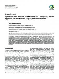
Fig. 1. Conceptual diagram of our tremor and dyskinesia system is illustrated. The sensors attached to various parts of the body of the subject wirelessly transmit ACC and sEMG signals to a dynamic neural network system to detect movement disorders.
the body as illustrated in Figure 1. Each customized sensor acquires and wirelessly transmits three channels of tri-axial accelerometer (ACC) data and one channel of surface electromyographic (sEMG) data, all sampled at 1 KHz (with appropriate anti-aliasing filtering). In our laboratory, we have acquired an extensive database of such sEMG/ACC data for a period of 4 hours per subject while they carried out unscripted and unconstrained activities in an apartment-like environment. Each subject was videotaped during the experiments and the resulting videotapes were annotated on a per second basis by individuals trained in identifying PD motor signs (tremor, dyskinesia, and others). These annotations were then used to guide the development of a process for detecting the presence of dyskinesia and/or tremor through the analysis of ACC and sEMG data. In order to understand the challenges of our tremor and dyskinesia detection problem, consider the raw 12-second ACC and sEMG signals in Figure 2 collected from three different subjects. Figure 2(a) shows the signals from a subject who exhibits neither tremor nor dyskinesia; there is little fluctuation in the ACC signals except during a short period of voluntary movement beginning around the 6 second mark; the sEMG signal also shows very little activity except for a period of force generation in the associated muscle during the voluntary movement. Figure 2(b) shows the signals from a subject who exhibits tremor except during a short period (shaded in the figure) between the 6 and 10 second marks. Being able to differentiate between the presence and absence of a disorder for such short episodes motivates us to carry out the overall detection process on a per second basis. However, on such a small time scale it is
6062
II. PREVIOUS WORK
sEMG
ACC
(a)
sEMG
ACC
(b)
sEMG
ACC
(c)
0
2
4
6
[s]
8
10
12
Fig. 2. Sensor signals from the combined sEMG/ACC sensor attached to the dominant arm as illustrated in top picture. The signals in panel (a) are from a control subject with no dyskinesia or tremor. The signals in panel (b) are from a PD subject with tremor. The signals in panel (c) are from a PD subject with dyskinesia. The shaded regions in panels (b) and (c) indicate intervals where the corresponding movement disorders are absent.
challenging to detect the periodicity features of tremor for the following two reasons: 1. Extra fluctuations (typically non-periodic) in the ACC signal during voluntary movements. 2. Extra components (typically non-periodic) in the sEMG signal during force exertions that may occur during voluntary movements. In Figure 2(b) one can also see one of the reasons why we have employed both ACC and sEMG sensor data. Note that a voluntary wrist extension movement (between the 2 second and 4 second marks) is followed by an interval where the periodicity of tremor is hard to detect in the ACC signals but easy to detect in the sEMG signal. Similarly, in regions (not shown in Figure 2) where the muscle is exerting force but there is no limb movement, the periodicity of tremor can be easily extracted from the ACC signal but not from the sEMG signal. Finally, Figure 2(c) shows the ACC and sEMG signals from a subject who exhibits dyskinesia except during the interval that is shaded (2 to 6 s). We observe that the dyskinesia regions are marked by relatively large “spikelike” erratic fluctuations in the ACC signals as opposed to the more gradual fluctuations of voluntary movements.
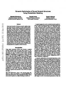
In previous work by our research group [5], we have used static neural networks and a neuro-fuzzy inference framework to detect instances of a set of 11 scripted activities of daily living (e.g. sit-to-stand, tooth-brushing, shirt-buttoning) performed by sensor-wearing stroke patients in a constrained laboratory environment. Both sEMG and ACC sensor data was collected from each patient. The resulting system performed the detections with 95% mean sensitivity and 99.7% mean specificity and it had a misclassification rate of below 10% on an additional set of 10 scripted activities of daily living that were selected for their similarity to the first set. This research established the efficacy of a combined sEMG-ACC approach for the classification of patient activity, but it was limited in the sense that each activity of interest had to be performed in isolation during well-delineated time segments. Clearly, the isolated-activity limitation is too restrictive for our current investigation where the activities of interest (tremor and dyskinesia) occur in the midst of other unscripted activities being carried out by the PD subjects. In previous work by Salarian et al. [6], the detection of tremor was considered on a per-second basis using tri-axial gyroscope signals from subjects performing a scripted sequence of activities such as tooth-brushing while standing and eating while sitting. Their algorithm yielded 99.5% sensitivity on tremor-only data and 94.2% specificity on tremor-free data. This algorithm, however, was not designed to discriminate between tremor and dyskinesia and was therefore not tested on datasets containing instances of dyskinesia. The detection of dyskinesia from ACC sensors worn by PD patients carrying out unscripted and unconstrained activities was considered by Keijsers et al. [7] using static neural network processing. They reported accurate detection of dyskinesia on a minute by minute basis. However, they did not consider the problem of discriminating dyskinesia from tremor, in this or other publications. III. METHODS We used the dynamic (as opposed to static) neural network approach [8] to design machine learning algorithms for detecting time-varying tremor and dyskinesia. While a static neural network (SNN) is constrained to learn timeindependent weights to apply to the features of the underlying data, a DNN can learn time-dependent weights to apply to the features of the underlying data. This allows DNNs to be trained to learn how features of tremor or dyskinesia change over time rather than being restricted to learning from static snapshots of their features at particular times. The DNN we have designed for detecting tremor is a multi-layered neural network with a hidden layer of 4 nodes (See Figure 3). The hidden nodes and the output node use
6063
u1
i1
FIR
FIR
FIR
FIR
h1
h2
h3
h4
FIR
FIR
FIR
FIR
i4
i7
Fig. 3. Dynamic Neural Network used for detecting tremor. There are 7 input nodes, 4 hidden nodes, and one output node. Each FIR filter has 5 weights.
the weights of a 5-point FIR filter to be applied to timedelayed and time-advanced versions of their respective input data. The seven input nodes consist of various features extracted from 2-second windowed sections of the sEMG and ACC sensor signals:
Energy of ACC signal after it is lowpass filtered with a cutoff frequency of 2.5 Hz. Energy of ACC signal after it is highpass filtered with a cutoff frequency of 2.5 Hz. Lag of first peak (not at origin) in autocorrelation of highpass ACC signal. Ratio of height of first peak (not at origin) to height of peak at origin in autocorrelation of highpass ACC signal. Energy of sEMG signal. Lag of first peak (not at origin) in autocorrelation of sEMG signal, provided significant peaks also exist at integer multiples of that lag. Ratio of height of first peak (not at origin) to height of peak at origin in autocorrelation of sEMG signal, provided significant peaks also exist at integer multiples of the first peak‟s lag.
The DNN we have designed for detecting dyskinesia is a multi-layered neural network with a hidden layer of 2 nodes (See Figure 4). The hidden nodes and the output node use the weights of a 5-point FIR filter to be applied to timedelayed and time-advanced versions of their respective input data. The input nodes represent the same four features that pertain to the ACC signals in the case of the tremor net. After designing the dyskinesia and tremor DNNs, we conducted two experiments to determine how well these DNNs perform on ACC and sEMG data collected from 8 different PD subjects and 4 control subjects. The data we used in these experiments was collected from a hybrid sEMG/ACC sensor that was attached near the wrist extensor muscle of the dominant arm (see top of Figure 2) of each subject as he/she carried out unscripted and unconstrained activities.
Experiment 1: The purpose of this experiment was to test how well our DNNs perform when they are trained on one set of data and tested on a different set of data from the same set of subjects. Two datasets (A and B) were created. Each dataset contained 10 minutes of sensor data from 4 PD subjects exhibiting tremor and voluntary movement in the dominant arm, 10 minutes of sensor data from 4 PD subjects exhibiting dyskinesia and voluntary movement in the dominant arm, and 10 minutes of sensor data from 4 control subjects exhibiting no tremor or dyskinesia while carrying out voluntary movements in the dominant arm. The data were selected to represent a variety of situations from each subject‟s 4-hour unscripted, freeform experiment. The video data, the sEMG data, and the ACC data within sets A and B were visually inspected to set on a per second basis the “truth” of whether the subject was exhibiting tremor and/or dyskinesia in the dominant arm. One tremor DNN (referred to as Tr-DNN-A) was trained on set A and one tremor DNN (referred to as Tr-DNN-B) was trained on set B. Similarly, one dyskinesia net (Dy-DNN-A) was trained on set A and the other dyskinesia net (Dy-DNN-B) was trained on set B. The trained DNNs were tested on the dataset that was excluded from its training and the detection results were evaluated for sensitivity and specificity. u1
FIR
FIR
i1
FIR
FIR
h1
h2
FIR
FIR
i2
FIR
FIR
i3
FIR
FIR
i4
Fig. 4. Dynamic Neural Network used for detecting dyskinesia. There are 4 input nodes, 2 hidden nodes, and one output node. Each FIR filter has 5 weights.
Experiment 2: The purpose of this experiment was to test how the trained DNNs from Experiment 1 perform on data from subjects who had not been included in the training of the DNNs. Two subjects were selected for this purpose and we created a dataset (C) containing five minutes of tremor data and five minutes of dyskinesia data from each of the two selected subjects. The trained DNNs from Experiment 1 were then applied to dataset C and evaluated for sensitivity and specificity. IV. RESULTS Experiment 1 Results: The sensitivity and specificity results of Experiment 1 are summarized in Table I. Clearly, sensitivities and specificities are above 90% in every case. As an addendum, we also repeated Experiment 1 with the
6064
number of FIR weights per hidden/output node of the DNN reduced to 1. This turns the DNN into an SNN. The performance of the network degraded, particularly in tremor regions with movement such as the one illustrated in Figure 5. The SNN mistakenly declares the region around the 10 second mark as containing no evidence for tremor. However, the DNN does not make that error. Experiment 2 Results: The sensitivity and specificity results of applying the four trained neural networks of Experiment 1 to dataset C are shown in Table II. Clearly, the sensitivities and specificities are of the same order as in Table I, even though the test data for Table II was from subjects who were not used for training the DNNs.
TABLE I EXPERIMENT 1 RESULTS Network
In this paper, we have presented a DNN solution for detecting two motor signs (tremor and dyskinesia) of Parkinson‟s disease using sEMG and ACC data from wireless miniaturized sensors that can be conveniently worn by PD patients. The DNN solution was found to have high specificity levels and high sensitivity levels on a dataset that included data from subjects with tremor, data from subjects with dyskinesia, and data from subjects with no motor disorders. These high levels of sensitivity and specificity were also observed when the test data was from subjects who were not included in the training of the DNNs. In our ongoing research, we are also developing and evaluating DNN solutions for detecting other PD motor signs (such as bradykinesia and akinesia) from sEMG and ACC wearablesensor data. In future research, we will be combining these DNN solutions within a larger artificial intelligence framework to carry out not only the detection but also the quantitative assessment of PD motor signs.
Network
Spec
ACKNOWLEDGMENT We would like to thank Santosh Ganesan and Shey-Sheen Chang for their assistance in carrying out the DNN experiments reported in this paper. We would also like to thank L. Don Gilmore for the custom implementation of the miniaturized sensors and for his assistance in carrying out the data collection from PD subjects.
[1] [2]
-0.2 30
[3]
[V]
Sens
Tr-DNN-A 87.2% 92.0% Tr-DNN-B 91.7% 93.0% Dy-DNN-A 93.4% 94.9% Dy-DNN-B 95.7% 93.6% The four DNNs of Experiment 1 were applied to dataset C which contains ten minutes of tremor data and ten minutes of dyskinesia data from subjects who did not have any of their data included in the training of the DNNs.
0
[mV]
ACC-Y
Spec
REFERENCES
0.2
sEMG
Sens
TABLE II EXPERIMENT 2 RESULTS
V. CONCLUSIONS
SNN DNN
Test Data
Tr-DNN-A B 92.4% 96.7% Tr-DNN-B A 92.9% 93.7% Dy-DNN-A B 93.5% 91.2% Dy-DNN-B A 90.3% 95.1% Tr-DNN-A and Tr-DNN-B represent the tremor DNNs that were respectively trained on dataset A and dataset B. Dy-DNN-A and DyDNN-B represent dyskinesia DNNs that were respectively trained on dataset A and dataset B. Each dataset contains 10 minutes of tremor data, 10 minutes of dyskinesia data, and 10 minutes of data without either disorder. There is no common data between dataset A and dataset B.
[4]
0 [5]
-30 Tremor
Tremor
Tremor [6]
Tremor
8
9
No Tremor
10 [s]
Tremor
11
12
Fig. 5. Top two panels show an ACC and an sEMG signal recorded from a sensor at the wrist extensor while the subject was flexing the wrist. In contrast to the DNN, the SNN makes an error by declaring “no tremor” at the 10 s mark.
[7]
[8]
6065
R. O. Duda, P. E. Hart, D. G. Stork, Pattern Classification. New York: John Wiley & Sons, 2001. J. Reimer, M. Grabowski, O. Lindvall, P. Hagell, „„Use and interpretation of on/off diaries in Parkinson‟s disease,” J. Neurol. Neurosurg. Psychiatry, vol. 75, pp. 396-400, 2004. R. J. Elble, „„Tremor: clinical features, pathophysiology, and treatment,‟‟ Neurol. Clin., vol. 27, no. 3, pp. 679-695, Aug. 2009. J. Jankovic, „„Motor fluctuations and dyskinesias in Parkinson‟s disease: clinical manifestations,‟‟ Movement Disorders, vol. 20, suppl. 11, pp. S11-S16, 2005. S. H. Roy, M. S. Chang, S. S. Chang, J. Moore, G. De Luca, S. H. Nawab, and C. J. De Luca, “A combined sEMG and accelerometer system for monitoring functional activity in stroke,” IEEE Trans. Neural Syst. Rehabil. Eng., vol. 17, no. 6, pp. 585-594, Dec. 2009. A. Salarian, H. Russmann, C. Wider, P. R. Burkhard, F. J. G. Vingerhoets, K. Aminian, „„Quantification of tremor and bradykinesia in Parkinson‟s disease using a novel ambulatory monitoring system,‟‟ IEEE Trans. Biomed. Eng., vol. 54, no. 2, pp. 313-322, Feb. 2007. N. L. Keijsers, M. W. Horstink, S. C. Gielen, „„Automatic assessment of levodopa-induced dyskinesias in daily life by neural networks,‟‟ Movement Disorders, vol. 18, no. 1, pp. 70-80, 2003. E. Wan, „„Discrete time neural networks,‟‟ Journal of A pplied Intelligence, vol. 3, pp. 91-105, 1993.