Send Orders for Reprints to
[email protected] Current Computer-Aided Drug Design, 2017, 13, 000-000
1
RESEARCH ARTICLE
Dynamic Simulation, Docking and DFT Studies Applied to a Set of Anti-Acetylcholinesterase Inhibitors in the enzyme β-Secretase (BACE-1): An Important Therapeutic Target in Alzheimer’s Disease Edeildo F. Silva-Júnior1, Paulo H. Barcellos França2, Lucindo J. Quintans-Júnior3, Francisco J. B. Mendonça-Junior4, Luciana Scotti5, Marcus T. Scotti5, Thiago M. de Aquino1 and João X. de Araújo-Júnior1,* 1
Laboratory of Medicinal Chemistry, Federal University of Alagoas, Nursing and Pharmacy School, Maceió-AL, Brazil; Chemistry and Biotechnology Institute, Federal University of Alagoas, Maceio-AL, Brazil; 3University of Sergipe, Postgraduate Program in Health Science, Aracaju-SE, Brazil; 4State University of Paraiba, Laboratory of Synthesis and Drug Delivery, João Pessoa-PB, Brazil; 5Federal University of Paraíba, João Pessoa-PB, Brazil 2
Abstract: Background: Alzheimer's disease (AD) affects mainly elderly people over 60 years of age. Currently, there are more than 35 million people with this disease worldwide. The enzyme β-secretase is involved in the processing of the amyloid precursor protein and plays a key role in the physiopathology of AD. The action of some acetylcholinesterase inhibitors (AChEI) as β-secretase inhibitors has been reported. ARTICLE HISTORY Received: February 26, 2016 Revised: February 27, 2017 Accepted: February 27, 2017 DOI: 10.2174/1573409913666170406150905
Objective: The aim of this study was to highlight the modes of the binding of acetylcholinesterase ligands onto the active site of the β-secretase enzyme. Method: Molecular dynamics and docking were used in order to identify pivotal interactions that favor the inhibitory activity and provide a rational basis for planning novel β-secretase inhibitors. Additionally, density functional theory (DFT) was used to provide accurate energy values for the complexes. A mechanistic study of the amide hydrolysis was also performed at the M06/6-31G(d) basis set. Results: Of the 100 AChE inhibitors, 10 were able to interact with Asp32 and/or Asp228 residues from the enzyme BACE-1, suggesting that these could act as multi-target compounds. These inhibitors were selected for DFT studies in order to provide more accurate energy values. Interestingly, the range of energy values (-27.01 to -8.64 kJ mol-1) obtained was in agreement with the anti-AChE activity. The results obtained in the mechanistic study of compound 93 using DFT are in agreement with theoretical studies described in the literature. Conclusion: The results reported in this study will advance our understanding of the influence of the distinct chemical structures of inhibitors at the active site and aid the development of new virtual screening protocols to design novel AChE multi-target inhibitors.
Keywords: β-secretase, molecular docking, dynamic simulations, DFT, Alzheimer’s disease, acetylcholinesterase. 1. INTRODUCTION Alzheimer's disease (AD), discovered by the German neuropathologist Alois Alzheimer in 1907, is a neurodegenerative disease of the brain that usually affects people over 60 years of age [1-8]. Less often, it also affects younger people (around 40 years of age) with a more dramatic and aggressive effect and the severe impact on the quality of life *Address correspondence to this author at the Nursing and Pharmacy School, Federal University of Alagoas, P.O. Box: 57072-900, Maceió, Brazil; Tel: +55-82-99901-9991; E-mail:
[email protected].
1573-4099/17 $58.00+.00
occurs when the patient is fully productive. Additionally, it is a type of progressive and irreversible dementia, whose initial clinical manifestation comprises memory loss and cognitive decline [1, 6, 7, 9]. In the World Alzheimer Report, Alzheimer's Disease International reported that in 2015, there were 47 million people with AD, and that number is expected to exceed 75 million in 2030 and 131 million in 2050 [10-12]. Furthermore, according to the World Alzheimer Report 2016, the total estimated worldwide cost of dementia is US$ 818 billion, and it will become a trillion dollar disease by 2018 [10]. © 2017 Bentham Science Publishers
2
Current Computer-Aided Drug Design, 2017, Vol. 13, No. 3
The most frequently prescribed anti-AD drugs act by increasing acetylcholine (ACh) levels in the brain via inhibition of the enzyme acetylcholinesterase (AChE) and/or butyrylcholinesterase (BuChE) [13]. AChE is present at neuromuscular junctions and cholinergic brain synapses, which hydrolyze this neurotransmitter and yield choline and the acetyl group and terminate the relaying signal [12]. The enzyme β-secretase (also known as β-APP or BACE1) is one of the two proteases responsible for processing the amyloid precursor protein (APP) to give 40/42 residues of βamyloid peptide (Aβ), the primary constituent of the amyloid plaques observed in the brains of Alzheimer's patients [2, 6, 9, 10, 14-18]. The cleavage of APP appears to be the limiting step in the production of Aβ in mice with the complete absence of Aβ, without significant side effects. Hence, BACE-1 appears to be an attractive therapeutic target for the treatment of AD [14, 15]. BACE-1 (β-amyloid precursor protein cleaving enzyme, β-secretase) has been characterized as the first known example of a membrane-associated pepsin-like aspartyl protease. The crystal structure of this soluble domain shows a high degree of similarity with the tertiary structures of other aspartyl proteases in some fungi and mammals. This enzyme has two catalytic residues (dyad) related to inhibition, Asp32 and Asp228 (or Asp93 and Asp289 according UniProt isoform, respectively), and peptidomimetics described in the literature are effective inhibitors of this enzyme [14, 19]. Additionally, studies with BACE-1 have shown that the proton transfer between the Asp dyad through a water molecule plays a critical role in the mechanism of the catalytic activity [19, 20]. Despite extensive crystallographic studies, the protonation states of the active site amino acid residues in the presence of the inhibitors are as yet obscure [19]. This is related to the absence of hydrogen coordinates, which prevents the identification of proton donors and acceptors [20]. According to Xu et al. [15], interaction with Asp32 is essential for the inhibitory activity towards this enzyme. However, other authors report that the Asp93 and Asp289 residues are responsible for the recognition of substrates, both positioned at the site of the peptide bond hydrolysis. The inhibition of BACE-1 is considered as the most important mechanism for the treatment of AD. The possibility of interrupting the Notch pathway through the use of γsecretase inhibitors is not a very attractive approach [15]. The inhibition of γ-secretase may cause some mechanismbased adverse effects, such as interference in T-lymphocyte production [2]. In computational chemistry applied to medicinal chemistry, molecular dynamic (MD) simulations have been used to provide logical explanations and rationally predict the enzyme character (e.g., thermal stability, solvent tolerance, and ligand binding) to elucidate the physical basis of the structure of macromolecules in the order of nanoseconds (ns) [21]. Molecular docking studies are the most common virtual screening approach and play an important role in the identification of hits or the optimization of hit compounds [22]. In addition, several studies based on molecular mechanics or quantum mechanics approaches to enzyme-catalyzed reac-
Silva-Júnior et al.
tions have contributed to evaluating the role of the catalytic residues and to clarifying the reaction mechanisms [23, 24]. Based on compounds reported by Huang et al. [25] and Peng et al. [26] and due to the high affinity of AChEI compounds and the non-selectivity of some examples from this class, in this study, we used molecular dynamics, docking, and DFT studies to investigate the possibility for the activity of AChEI molecules toward the BACE-1 enzyme, which would suggest that the higher effectiveness of some antiAChE compounds is a multi-target mechanism. 2. MATERIALS AND METHOD 2.1. Dataset Building A total of 120 compounds were used to build a dataset of molecules, containing 100 compounds that are active toward the enzyme AChE (selected as a positive control for the theoretical procedures), and 20 compounds which are not active toward AChE (selected as the negative control and for validation of the methods), determined in other studies through experimental tests (see supplementary material, Table 1). 2.2. Dynamic Simulations The initial coordinates for building the molecular model were taken from the X-ray crystal structure of β-secretase (BACE-1) from Homo sapiens with PDB entry 1M4H at a resolution of 2.1 Å. Hydrogen atoms were added to the crystal structure of BACE-1 using the Gromacs program (https:// http://www.gromacs.org/). The pKa values of the structure of PDB entry 1M4H were calculated for physiological conditions. Counterions (Na+ and Cl-) were placed at optimal electrostatic positions around the system to obtain electroneutrality. The whole system was placed in a cubic box of water molecules (1000 Å3). The protein and water molecules were described by the OPLS force field, implemented in the Gromacs program. The final system contains a total of 256,514 atoms and was relaxed for 10 ns by means of quantum mechanics/molecular mechanics molecular dynamics using the simple point charge (SPC) model [12]. Energy minimization of the prepared system was carried out up to a maximum of 1000 steps using the steepest descent method or until a gradient threshold (-25.0 kJ mol-1) was reached, followed by 10 ps of temperature stabilization at 300 K [12,20]. Additionally, 10,000 snapshots were extracted during the 10,000 ps production run using the NPT ensemble with a time step of 2 femtoseconds (fs) with no restraints [12,20]. The root-meansquare deviation (RMSD) of each collected snapshot in comparison to the crystal structure was obtained using the Grace package v. 4.1.2 (http://plasmagate.weizmann.ac.il/Grace/doc/Users Guide. Html#ss1.1). The extracted coordinates obtained from the last ps snapshot were used for the docking studies [20]. To validate the BACE-1 enzyme 3D structure (post-dynamics), the Ramachandran plots was employed using Maestro software, version 11.0.15 (https://www.schrodinger.com/ freemaestro). 2.3. Docking Studies In the molecular docking study the ligand structures were drawn in the SMILES format and they were converted to
Dynamic Simulation, Docking and DFT Studies Applied
three-dimensional MOL2 files using Corina (Molecular Networks, http://www.molecular-networks.com/). The Austin model (AM1) was employed as the energy minimization method in ArgusLab v. 4.0.1 (http://www.arguslab.com/ arguslab.com/Argus Lab.html). All final structures were converted to *.pdbqt format in AutoDock Tools v. 4.2.5 (http://autodock.scripps. edu) [27]. All ligand structures were considered at pH 7.4 (physiological conditions). All ligands, ions and solvent molecules that were present in the original structures downloaded from the Protein Data Bank (PDB) were manually removed and hydrogen atoms were added using the AutoDock Tools v. 4.2.5 (http://www.autodock. scripps.edu/downloads). A blind-docking search was employed based on the possible existence of an allosteric site in this enzyme. A grid box with centered coordinates at x = 0.06, y = 0.096, and z = 0.289 Å was established in AutoDock Tools v. 4.2.5 [27]. The grid was used to evaluate the steric boundary of the protein and van der Waals and/or hydrogen bond interactions between the ligand and the protein. Finally, the molecule was then converted to the *.pdbqt extension [27]. The ligand and receptor preparations were carried out in the AutoDock 4.2.5 program. After the binding site definition and ligand preparation, the molecular docking of 120 compounds was performed in AutoDock Vina (http://vina.scripps.edu/). All calculations were carried out considering the binding site as rigid and the ligand as flexible [28]. 2.4. Density Functional Theory (DFT) Calculations After the docking calculations, the density functional theory (DFT) calculations were performed using quantum mechanics (QM) models from the Spartan'14 program to determine the corrected binding energies for the BACE1/ligand complexes. We then investigated the potential dual compounds by theoretical methods. Initially, the correct structures of the drugs and their target enzyme are required. The optimized geometries of compounds able to interact with the catalytic dyad were taken from docking analysis. In addition, the coordinates of the BACE-1 enzyme were taken from the crystal structure (PDB ID: 1M4H), and the ligand and water molecules were removed [28]. The binding energies of the enzyme/ligand complexes were calculated applying the M06/6-31G(d) basis set. The M06 method was carried out applying the global hybrid functional, which is the top performer within the 6 functionals of the main group, thermochemistry, kinetics and noncovalent interactions. Moreover, frequency calculations were performed to confirm the nature of the stationary point at the same level [28]. QM binding energies were obtained applying the following formula in which the binding energies (Ebinding) were calculated as the difference between the energy of the complex (Eenzyme-ligand) and the sum of the enzyme (Eenzyme) and drug (Eligand) energies considered separately [28]. Ebinding = (Eenzyme/ligand - (Eenzyme + Eligand)) Several theoretical calculations and X-ray and neutron diffraction studies have shown that one of the Asp residues remains protonated and the other unprotonated during the
Current Computer-Aided Drug Design, 2017, Vol. 13, No. 3
3
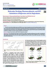
catalytic cycle [29-32]. Thus, a mechanistic study was performed for the compound that was able to interact with the two residues of the Asp dyad simultaneously. In these calculations, the electrostatic and steric effects of the protein environment surrounding the active site were completely ignored [29]. The final energies of the optimized structures were improved by including the single point energy from the 631G(d) basis set unscaled zero-point energy (ZPE) and thermal corrections (at 298.15 K and 1 atm) estimated at the same level of theory [29], using the Spartan'14 program. The potential energy surface (PES) diagram was generated for each reaction step. 4. RESULTS AND DISCUSSIONS 4.1 Dynamic Simulations MD simulation represents an alternative way to gain molecular-scale insights into the interactions between solvents and enzymes that to date have not been exploited. Additionally, MD provides a wealth of information on cation-anion and protein-ion interactions [33]. In general, in an aqueous medium, the surface area to volume ratio of the enzyme decreases and the hydrogen bond and ion-pair interactions increase [34]. The conformational stability of the BACE-1 enzyme was investigated during the MD simulation. It was observed that the structure relaxes to a root-mean-square deviation (RMSD) of around 2.45 Å after 8.7 ns of MD simulation (Fig. 1). The low RMSD value indicates that the enzyme exhibits little conformational deviation from its starting structure.
Fig. (1). Molecular dynamics trajectory plots correlating RMSD deviation from the initial BACE-1 enzyme Cα-atoms as a function of time.
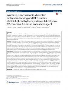
Additionally, the 3D structure of the BACE-1 enzyme generated by MD simulations was validated. The validation of the backbone and amino acid side chain conformations for this model was performed by generating and analyzing the corresponding Ramachandran plots, which represent the distributions of the amino acid dihedral angles ϕ and ψ (Fig. 2). The Ramachandran plots were used for the validation of the
4
Current Computer-Aided Drug Design, 2017, Vol. 13, No. 3
Silva-Júnior et al.
Fig. (2). Ramachandran plots showing the distribution of the phi (ϕ ) and psi (ψ) dihedral angles (in degrees) of the amino acid side chains in the model of the enzyme BACE-1. Glycine is plotted as triangles, proline as squares, and all other residues as circles. The red regions are the favored regions, the yellow regions are allowed regions, and the white regions are disallowed regions.
BACE-1 enzyme. For the BACE-1 enzyme generated by the MD simulations, 95.4% of the residues are located in the most favored regions (red) and 2.68% are located in allowed regions (yellow). Only 1.91% of the amino acid residues were found in the disallowed region (white). The plots in the top- and bottomleft quadrants indicate that the model contains both β-sheet and α-helix structures. Moreover, the plots in the top- and bottom-right quadrants represent a loop structure. These results verify that the model is highly plausible. Lastly, the structure generated by molecular dynamics for physiological conditions was used to evaluate the antiBACE-1 potential of the set of anti-acetylcholinesterase molecules by molecular docking. 4.2. Docking Studies According to Jain et al. [35], for BACE-1 inhibitors, a group that acts as an H-bond donor is needed for H-bonding interactions with the catalytic aspartate dyad, that is, Asp32 and Asp228. Furthermore, large hydrophobic pockets (S1, S3 and S2') are key regions and they require hydrophobic groups to fill them. The S1 hydrophobic cleft is formed by the side chains of Tyr71, Phe108, Trp115, Ile118, and Leu30. The S1 and S3 sites are also largely hydrophobic pockets formed by side chains of Leu30, Tyr71, Trp115, and Ile110, as well as a main chain of Gln12, Gly230, Thr232, and Ser35, while S2' is comprised of Ile126, Trp76, Val69, Arg128, and Trp198 amino acid residues [35]. For the docking studies, 100 anti-AChE compounds (75% of the dataset, see supplementary material) were ana-
lyzed to determine their inhibitory capacity toward the enzyme BACE-1. In all cases, the anti-AChE compounds are capable of interacting with the amino acid residues at the S1, S2', and S3 sites. However, interactions were observed predominantly with amino acids at S1 and Asp32. The most frequent amino acid residues observed were Asp32, Gly34, Tyr71, Phe108, Ile110, Trp115, Glu230, and Thr232 (Fig. 3a). In general, electrostatic interactions or hydrogen bonds with Asp32 were observed for 10 compounds (see DFT section). Only compound 93 was able to interact with the Asp32-Asp228 dyad. Results obtained with pharmacophore models developed by Al-Nadaf et al [36] suggest that hydrogen-bonding and electrostatic interactions occur with the hydroxyl and carboxylate side chains of Ser35, Asp32, and Asp228. A total of 20 anti-AChE compounds (see supplementary material) which are not active toward BACE-1 were used as a negative control (15% of the dataset). In general, all compounds interact close to sites S1 and S3, but none of them is able to interact with Asp32 and Asp228 residues (catalytic dyad). In addition, they were able to predominantly interact with residues Arg54, Tyr60, Gln55, Arg61 and 64 hydrophobically (Fig. 3b). Furthermore, these compounds are able to interact with Trp277, Tyr320, and His362 via hydrogen bonding. In the BACE-1/compound complexes (for the negative control), no interactions were observed via hydrogen bonding. Cluster analysis of the negative control revealed that these complexes are able to interact with the enzyme in a distinct region in relation to the catalytic dyad (Fig. 3b), explaining their inactivity in experimental trials.
Dynamic Simulation, Docking and DFT Studies Applied
Current Computer-Aided Drug Design, 2017, Vol. 13, No. 3
5
Fig. (3). Dataset of complexes between BACE-1 and inactive compounds. (a) AChEI cluster at catalytic site and (b) inactive AChEI cluster. The colors for the molecular surfaces were obtained with the rainbow chains module in the AutoDock Tools software.
4.3. DFT Calculations Since docking calculations are not able to provide the correct binding energies, the QM models were used to obtain these values [28]. For this purpose, only the enzyme/ligand complexes involved in interactions with Asp32 and/or Asp228 residues were selected and optimized at the M06/631G(d) level of theory to calculate the binding energies (Fig. 4). Furthermore, the corrected relative energies were determined applying the zero point energy (ZPE) [28]. In general, the binding energy results generated by DFT show excellent correlation with the IC50 values obtained from the literature. Compounds 10, 11 and 13 showed high stability (Ebinding) in the complex with the enzyme BACE-1, Ebinding = -20.46, -18.14, and -27.01 kJ mol-1, respectively. For compound 10, can be observed that there are hydrophobic interactions between the oxo-heterocycle ring with the Thr329; pyridine with the Tyr71 and the terminal phenyl group with the Asp32 residue. In addition, for compound 10 hydrogen bonds were observed between the heterocycle and the nitrogen in the Arg128 residue, with distances of 1.72
and 2.37 Å. The oxo-heterocycle ring at compound 11 interacts with the Tyr71 and Asp228 residues; and the terminal morpholine group with the Tyr 71, via hydrophobic contacts. However, a hydrogen bond was observed between NH2 in Arg128 and the morpholine ring from this compound, with a distance of 1.6 Å. Regarding compound 13, hydrophobic interactions were observed with residues Gly30, Tyr71, and Thr72. However, the iminothioazole ring forms a hydrogen bond with the Asp32 residue (around 2.44 Å). In addition, compounds 32, 35, 38, and 93 show relatively good stability values, Ebinding = -12.38, -12.96, -12.65, and -12.17 kJ mol-1. Compound 32 interacts with the Tyr71, via π-π stacking, through the tricycle system. In addition, there is hydrophobic contacts between the methoxyl group interacts with the Thr231 residue. Finally, a hydrogen bond was observed between the heterocycle and COO- in Asp32, with a distance of 1.94 Å. For compound 35, hydrogen bonds with Asp32 and Thr232 residues were observed, with distances of 1.9 and 2.8 Å, respectively. In addition, hydrophobic interaction with the Tyr71 residue was verified. For compound 38, there is a π-π stacking interaction between the tricycle system and the
6
Current Computer-Aided Drug Design, 2017, Vol. 13, No. 3
Silva-Júnior et al.
Fig. (4). Calculated relative binding energies (Ebinding) of the AChEI/BACE-1 complexes at M06/6-31G(d) level of theory. The noncritical hydrogen atoms are omitted from the figure for clarity.
Tyr71 residue. However, hydrogen bonds were observed with the Asp32 and Gly230 residues, with distances of 1.7 and 2.5 Å, respectively. Compound 93 only performs hydrogen bonding at the BACE-1 active site. These interactions occur with the Asp32, Asp228 and Thr231 residues, with distances of 1.6, 1.7 and 2.8 Å, respectively. Compound 29 shows intermediate stability (Ebinding = -11.46 kJ mol-1) in comparison with the other compounds. In addition, this compound exhibits hydrophobic interaction with the Tyr198 residue, and interaction with Asp32 and Gly34 via hydrogen bonds, with distances of 1.94 and 2.05 Å, respectively. Compounds 30 and 31 showed poor stability (Ebinding = -8.9 and -8.64 kJ mol-1) when in a complex with the active site of the BACE-1 enzyme. Compound 30 shows interactions with the Gly34, Gln73 (hydrophobic) and Asp32 residues (via hydrogen bonding - 1.9 Å). However, compound 31 shows the same interactions as compound 30, but a hydrogen bond is present (1.79 Å). The stability information generated by M06/6-31G(d) DFT calculations suggests that the amino acid residues which confer notable stability to the complexes are Asp32, Tyr71 and Gly230. On comparing compounds 10, 11 and 13 (better stability) it can be noted that the interaction with Asp32 is more energetically stable than Asp228 (observed for compound 11 complex). In addition, the complexes with intermediate stability (compounds 32, 35, 38 and 93) interact with Aps32 and Tyr71 residues,
reinforcing our hypothesis. However, compound 38 exhibits interactions with the same amino acids observed for the most stable complexes (compounds 10, 11 and 13), showing intermediate stability. This finding can be explained by the additional interaction with the Thr232 residue, suggesting that this reduces the stability of the final complex. For the less stable complexes (compounds 29, 30 and 31) it was observed that they only interact with the Asp32 residue, which explains their low Ebinding values. It is known that the protonation state preference of BACE-1 can easily change for different inhibitors, since the ligand has a strong impact on the protonation state of the dyad. This is mostly due to the hydrogen atom(s) of the ligand. For instance, if the hydrogen of the OH- interacting group containing the ligand is facing Asp32, the dyad is mono-protonated (deprotonated Asp32 and protonated Asp228) [19, 27, 29]. In general, the Asp32 residue acts as an acid and donates a proton to the ligand. However, Asp228 acts as a base and pulls a proton from the catalytic water and there is a nucleophilic attack of the OH- of the water molecule on the electrophilic group [19], generating a gem-diol intermediate. Lastly, amino hydrolysis occurs [29]. Based on the docking results, quantum mechanics (QM) was employed
Dynamic Simulation, Docking and DFT Studies Applied
Current Computer-Aided Drug Design, 2017, Vol. 13, No. 3
7
Fig. (5). Hydrolysis mechanism proposed for compound 93 catalyzed by the enzyme BACE-1.
to investigate the mechanism associated with the hydrolysis of compound 93 (Fig. 5). Regarding Fig. (5), in the optimized reactant (SI) for compound 93, the activation of the catalytic water (H2O3) by the deprotonated Asp228 (O3H···O4 = 1.4 Å) generates the hydroxyl ion (-O3H-) nucleophile that simultaneously attacks the carbonyl carbon (C=O1) of the amide group. This process is accompanied by the concomitant delivery of a proton from the Asp32 to the O1 atom of the C=O1 group (O1···H-O2 = 1.5 Å). Additionally, both Asp32 and Asp228 residues are involved in a network of hydrogen bonds in SI. In the first transitional state (TSI), all of the critical bond distances suggest that this process is concerted (O1-H = 0.9, O2-H = 1.6, O3-H = 1.6, and O4-H = 0.9 Å). For this step the computed energetic barrier was 23.38 kcal mol-1. This step leads to the formation of the gem-diol intermediate (SII), which is 17.02 kcal mol-1 endothermic for SI. This intermediate is stabilized by a network of hydrogen bonds formed by Asp32 (O1 -
H···O2 = 1.6 Å) and Asp228 (O3-H···O4 = 1.5 Å). Cleavage of the amide bond occurs in SII, Asp228 donates a proton to the N4-H group of the C-N4 bond and Asp32 concomitantly accepts the O2-H proton. This step leads to cleavage of the amide bond and produces the separated carboxyl (CO1O3H) and amine (-N4H2) terminals. Furthermore, in the second optimized transitional state (TSII), all of the key distances show that this process is synchronous (O1-H = 1.0, O2-H = 1.4, C-N4 = 1.5, N4-H = 1.6, and O4-H = 1.0 Å). For TSII, the barrier for this process is 39.28 kcal mol-1 from the SI reactant and 22.26 kcal mol-1 from SII. Lastly, the formation of SIII is -5.6 kcal mol-1 exothermic for SI and 11.42 kcal mol-1 for SII. These results are very similar to those obtained by Barman & Prabhakar [29]. The potential energy surface (PES) diagram for these reaction steps is shown in Fig. (6). According to the theoretical protocols used in this study, compounds 10, 11, 13, 29, 30, 31, 32, 35, 38, and 93 can be
8
Current Computer-Aided Drug Design, 2017, Vol. 13, No. 3
Silva-Júnior et al.
Fig. (6). Potential energy diagram for the cleavage of the amide bond of compound 93 catalyzed by the enzyme BACE-1.
considered as potential dual inhibitors (anti-AD). However, data in the literature concerning activity toward BACE-1 is only available for compound 93. This AChEI presents an IC50 value of 46.64 µM relation to the enzyme BACE-1. In experimental studies, this compound showed potent activity toward the enzymes AChE and BACE-1. Furthermore, receptor-binding studies on this compound and BACE-1 showed several essential interactions with the enzymes, involving the Aps32 residue [25]. CONCLUSION In this theoretical study, protonation states of the critical Asp dyad (Asp32 and Asp228) of BACE-1 were predicted for physiological conditions. It was observed that the enzyme reaches the relaxation state at around 8.7 ns. Moreover, it was verified that the Ramachandran plots are in agreement with a favorable and allowed structure for this enzyme. Of the 100 AChE inhibitors, 10 were able to interact with Asp32 and/or Asp228 residues from the enzyme BACE-1 (via electrostatic interaction and H-bonding), suggesting that these could act as multi-target compounds (dual activity). These inhibitors were selected for DFT studies in order to provide more accurate energy values. Interestingly, the range of energy values (-27.01 to -8.64 kJ mol-1 ) obtained was in agreement with the anti-AChE activity. The results obtained in the mechanistic study of compound 93 using DFT are in agreement with theoretical studies described in the literature. Additionally, this compound was able to inhibit the enzyme BACE-1 in experimental assays (IC50 = 46.64 µM). The amide bond cleavage is exothermic and leads to the amine and carboxyl acid fragments. The results reported in this study will advance our understanding of the influence of the distinct chemical structures of inhibitors at the active site and aid the development of new virtual screening protocols to design novel AChE multi-target inhibitors. CONFLICT OF INTEREST The authors confirm that this article content has no conflicts of interest.
ACKNOWLEDGEMENTS The authors acknowledge CAPES and CNPq for financial support. SUPPLEMENTARY MATERIAL Supplementary material is available on the publisher’s web site along with the published article. REFERENCES [1] [2] [3]
[4]
[5] [6]
[7] [8]
[9]
[10]
Smith, M.D.A.C. Doença de Alzheimer. Rev. Bras. Psiquiatr., 1999, 21, 3-7. Rzeszotarski, W.I. Alzheimer’s Disease: Search for Therapeutics. In Burger’s Medicinal Chemistry; Abraham, D.J., Ed.; Wileyinterscience: Viriginia, 2003; pp. 243-261. Scotti, L.; Lima, E.; da Silva, M.; Ishiki, H.; Oliveira Lima, I.; Oliveira Pereira, F.; Mendonça Junior, F.; Scotti, M. Docking and PLS studies on a set of thiophenes RNA polymerase inhibitors against Staphylococcus aureus. Curr. Top. Med. Chem., 2013, 14, 64-80. Varadaraju, K.R.; Kumar, J.R.; Mallesha, L.; Muruli, A.; Mohana, K.N.S.; Mukunda, C.K.; Sharanaiah, U. Virtual screening and biological evaluation of piperazine derivatives as human acetylcholinesterase inhibitors. Int. J. Alzheimers. Dis., 2013, 2013, 653962. Younes, S.H.; Hasan, A.A.; Mohammad, M.A. Molecular docking studies of Ginkgo biloba against acetylcholinesterase enzyme. Int. J. Pharm. Sci. Rev. Res., 2013, 21, 321-324. Huang, H.-J.; Chen, H.-Y.; Lee, C.-C.; Chen, C.Y.-C. Computational design of apolipoprotein e4 inhibitors for Alzheimer’s disease therapy from traditional Chinese medicine. BioMed Res. Int., 2014, 2014, 1-13. Schwarz, S.; Lucas, S.D.; Sommerwerk, S.; Csuk, R. Amino derivatives of glycyrrhetinic acid as potential inhibitors of cholinesterases. Bioorg. Med. Chem., 2014, 22, 3370-3378. Seniya, C.; Khan, G.J.; Uchadia, K. Identification of potential herbal inhibitor of acetylcholinesterase associated Alzheimer’s disorders using molecular docking and molecular dynamics simulation. Biochem. Res. Int., 2014, 2014, 1-7. Zuo, Z.; Luo, X.; Zhu, W.; Shen, J.; Shen, X.; Jiang, H.; Chen, K. Molecular docking and 3D-QSAR studies on the binding mechanism of statine-based peptidomimetics with Β-secretase. Bioorg. Med. Chem., 2005, 13, 2121-2131. Prince, M.; Comas-Herrera, A.; Knapp, M.; Guerchet, M.; Karagiannidou, M. World Alzheimer Report 2016. Improving healthcare for people living with dementia - coverage, quality and
Dynamic Simulation, Docking and DFT Studies Applied
[11]
[12]
[13]
[14]
[15]
[16]
[17]
[18]
[19]
[20] [21]
[22]
[23]
costs now and in the future. Alzheimer's Disease International (ADI), London, 2016. Ambure, P.; Kar, S.; Roy, K. Pharmacophore mapping-based virtual screening followed by molecular docking studies in search of potential acetylcholinesterase inhibitors as Anti-Alzheimer’s agents. BioSystems, 2014, 116, 10-20. Dhanjal, J.K.; Sharma, S.; Grover, A.; Das, A. Use of ligand-based pharmacophore modeling and docking approach to find novel acetylcholinesterase inhibitors for treating Alzheimer. Biomed. Pharmacother., 2015, 71, 146-152. Chand, K.; Alsoghier, H.M.; Chaves, S.; Santos, M.A. Tacrine(hydroxybenzoyl-Pyridone) hybrids as potential multifunctional anti-Alzheimer’s agents: AChE inhibition, antioxidant activity and metal chelating capacity. J. Inorg. Biochem., 2016, 163, 266-277. Holloway, M.K.; McGaughey, G.B.; Coburn, C.; Stachel, S.J.; Jones, K.G.; Stanton, E.L.; Gregro, A.R.; Lai, M.T.; Crouthamel, M.C.; Pietrak, B.L.; Munshi, S.K. Evaluating scoring functions for docking and designing Β-secretase inhibitors. Bioorganic Med. Chem. Lett., 2007, 17, 823-827. Xu, W.; Chen, G.; Zhu, W.; Zuo, Z. Molecular docking and structure-activity relationship studies on benzothiazole based nonpeptidic BACE-1 inhibitors. Bioorg. Med. Chem. Lett., 2010, 20, 6203-6207. Huang, D.; Liu, Y.; Shi, B.; Li, G.; Wang, G.; Liang, G. Comprehensive 3D-QSAR and binding mode of BACE-1 inhibitors using R-group search and molecular docking. J. Mol. Graph. Model., 2013, 45, 65-83. Palakurti, R.; Sriram, D.; Yogeeswari, P.; Vadrevu, R. Multiple Epharmacophore modeling combined with High-Throughput Virtual Screening and Docking to identify potential inhibitors of Βsecretase(BACE1). Mol. Inform., 2013, 32, 385-398. Zhang, T.; Xu, W.; Mu, Y.; Derreumaux, P. Atomic and dynamic insights into the beneficial effect of the 1,4-Naphthoquinon-2-YlL-Tryptophan inhibitor on Alzheimer’s AB1-42 dimer in terms of aggregation and toxicity. ACS Chem. Neurosci., 2014, 5, 148-159. Kocak, A.; Erol, I.; Yildiz, M.; Can, H. Computational insights into the protonation states of Catalytic Dyad in BACE1 acyl guanidine based inhibitor complex. J. Mol. Graph. Model., 2016, 70, 226235. Sabbah, D.A.; Zhong, H.A. Modeling the protonation states of ΒSecretase binding pocket by molecular dynamics simulations and docking studies. J. Mol. Graph. Model., 2016, 68, 206-215. Cao, H.; Wang, M.; Nie, K.; Zhang, X.; Lei, M.; Deng, L.; Wang, F.; Tan, T. Β-Cyclodextrin as an additive to improve the thermostability of Yarrowia lipolytica lipase 2: Experimental and Simulation Insights. J. Taiwan Inst. Chem. Eng., 2016, 0, 1-7. Silva, M.M.; Savariz, F.C.; Silva, E.F.; De Aquino, T.M.; Sarragiotto, M.H.; Santos, J.C.C.; Figueiredo, I.M. Interaction of Β-carbolines with DNA: Spectroscopic studies, correlation with biological activity and molecular docking. J. Braz. Chem. Soc., 2016, 27, 1558-1568. De Oliveira, E.B.; Humeau, C.; Maia, E.R.; Chebil, L.; Ronat, E.; Monard, G.; Ruiz-Lopez, M.F.; Ghoul, M.; Engasser, J.M. An approach based on Density Functional Theory (DFT) calculations to assess the Candida Antarctica Lipase B selectivity in rutin,
Current Computer-Aided Drug Design, 2017, Vol. 13, No. 3
[24]
[25]
[26]
[27]
[28]
[29]
[30]
[31]
[32] [33]
[34]
[35]
[36]
9
isoquercitrin and quercetin acetylation. J. Mol. Catal. B Enzym., 2010, 66, 325-331. Li, L.; Jiang, Y.; Zhang, H.; Feng, W.; Chen, B.; Tan, T. Theoretical and experimental studies on activity of Yarrowia lipolytica lipase in methanol/water mixtures. J. Phys. Chem. B, 2014, 118, 1976-1983. Huang, W.; Tang, L.; Shi, Y.; Huang, S.; Xu, L.; Sheng, R.; Wu, P.; Li, J.; Zhou, N.; Hu, Y. Searching for the multi-target-directed ligands against Alzheimer’s disease: Discovery of quinoxalinebased hybrid compounds with AChE, H 3R and BACE 1 inhibitory activities. Bioorg. Med. Chem., 2011, 19, 7158-7167. Peng, D.Y.; Sun, Q.; Zhu, X.L.; Lin, H.Y.; Chen, Q.; Yu, N.X.; Yang, W.C.; Yang, G.F. Design, synthesis, and bioevaluation of benzamides: Novel acetylcholinesterase inhibitors with multifunctions on Butylcholinesterase, AB aggregation, and B-secretase. Bioorg. Med. Chem., 2012, 20, 6739-6750. Barman, A.; Prabhakar, R. Protonation states of the catalytic Dyad of Β-secretase (BACE1) in the presence of chemically diverse inhibitors: A molecular docking study. J. Chem. Inf. Model., 2012, 52, 1275-1287. Khosravan, A.; Marani, S.; Sadegh, M.; Googheri, S. The effects of fluorine substitution on the chemical properties and inhibitory capacity of Donepezil anti-Alzheimer drug; density functional theory and molecular docking calculations. J. Mol. Graph. Model., 2016, 71, 124-134. Barman, A.; Prabhakar, R. Elucidating the catalytic mechanism of Β-Secretase (BACE1): A Quantum Mechanics/Molecular Mechanics (QM/MM) approach. J. Mol. Graph. Model., 2013, 40, 1-9. Coates, L.; Erskine, P.T.; Mall, S.; Gill, R.; Wood, S.P.; Myles, D.; Cooper, J.B. X-Ray, neutron and NMR studies of the catalytic mechanism of aspartic proteinases. Eur. Biophys. J., 2006, 35, 559566. Coates, L.; Tuan, H.H.; Tomanicek, S.; Kovalevsky, A.; Mustyakimov, M.; Erskine, P.; Cooper, J. The catalytic mechanism of an aspartic proteinase explored with neutron and X-Ray diffraction. J. Am. Chem. Soc., 2008, 130, 7235-7237. Rajamani, R.; Reynolds, C.H. Modeling the protonation states of the catalytic aspartates in Β-secretase. J. Med. Chem., 2004, 47, 5159-5166. Burney, P.R.; Pfaendtner, J. Structural and dynamic features of Candida rugosa lipase 1 in water, octane, toluene, and ionic liquids BMIM-PF6 and BMIM-NO3. J. Phys. Chem. B, 2013, 117, 26622670. Foresti, M.L.; Ferreira, M.L. Computational approach to solventfree synthesis of ethyl oleate using Candida rugosa and Candida antarctica B lipases. I. Interfacial activation and substrate (ethanol, oleic Acid) adsorption. Biomacromolecules, 2004, 5, 2366-2375. Jain, P.; Wadhwa, P.K.; Rohilla, S.; Jadhav, H.R. Rational design, synthesis and in vitro evaluation of allylidene hydrazinecarboximidamide derivatives as BACE-1 inhibitors. Bioorg. Med. Chem. Lett., 2016, 26, 33-37. Al-Nadaf, A.; Sheikha, G.A.; Taha, M.O. Elaborate ligand-based pharmacophore exploration and QSAR analysis guide the synthesis of novel pyridinium-based potent Β-secretase inhibitory leads. Bioorg. Med. Chem., 2010, 18, 3088-3115.