Aug 22, 1994 - (Kershaw,. Cunningham and Kenwright. 1993). Variable cyclic move- ments have been measured on the frame of the Orthofix fixator after.
DYNAMISATION J. B. RICHARDSON.
From
OF TIBIAL T. N. GARDNER,
Glenfield
General
Fixator.
In
the
early
J. R. W. HARDY,
Hospital
We studied the effect fractures in six patients
and
stages,
peak
fixator
Royal
cyclic
EVANS,
column
tibial Axial
movement
at
of movement correlated fracture stiffness. After at six
weeks,
progressive
closure of the gap averaged 1.3 mm (0.1 to 3.5). Cyclic movement is produced by early weight-bearing with the fixator column locked. Progressive closure occurs
after
associated effects should
Received
and
Joint
Surg
23 Ma
the
a reduction
of dynamisation be defined
movement J Bone
unlocking
with
column, in cyclic
[Br]
is
often The
on movement at the fracture site separately, in terms of cyclic
of progressive 1994:
and movements.
closure.
l995:77-B:4l2-6. Accepted
22 August
J.-H.
Jnfirmar
0.75 mm (0.19 to 1.02) on and 0.86 mm (0.21 to 1.25)
on the lateral side. The amount with the applied load and the the
Leicester
M.
of ‘dynamisation’ on treated by the Dynamic
two to four weeks averaged the medial side of the bone
unlocking
FRACTURES KUIPER,
J. KENWRIGHT
England
Some
movement
at a fracture
healing
by callus
fixators
can
ment and Kenwright best
formation
be
modified
is believed
pattern
to control
of movement
movements improvement
the
for healing
Dynamic
Axial
at different
Fixator
(Orthofix at four
SRL, to six
the
determination
fixator
of these
PATIENTS We
Richardson, MD. FRCS, Professor of Orthopaedics Kuiper, MSc. PhD, Biomechanical Engineer Robert Jones and Agnes Hunt Orthopaedic Hospital. SY1O 7AG, UK.
column.
Oswestry.
LE3
412
to Mr J. R. W. Hardy. of Bone
and
Joint
fixator cyclic
of
Verona,
weeks
Italy),
and
gave
an
The
9QP,
Surgery
Shrop-
UK.
stiffness
Windmill
and
normal pattern of or the true effect
development
of new
at the fracture site now allows accurate
METHODS
six consecutive
patients
with
tibial
diaphyseal
was
measured when
(Richardson
at each fracture
by all
from 22 to 57 years (Table I). with six I 30 mm bone screws
the anteromedial aspect of the tibia. the fixator column was unlocked.
removed
degree
(PohI
treated by an Orthofix Dynamic Axial Fixator movement at the fracture gap. They were
inserted into after fracture, were
weeks
movements.
men and their ages ranged The fixator had been applied
T. N. Gardner. BSc, DPhiI, Senior Research Scientist M. Evans. Biomechanical Engineer J. Kenwright, MA, MD, FRCS, Professor of Orthopaedics Nuffield Orthopaedic Centre NHS Trust. University of Oxford. Road, Headington. Oxford OX3 7LD. UK.
©1995 British Editorial Society 0301 -620X/95/3961 $2.00
AND
studied
fractures measuring
be sent
of
average healing time of 15.5 weeks for closed tibial fractures (De Bastiani et al 1984). The ability of these frames to allow axial movement has been questioned. In the Dynabrace early cyclic movements at the fracture site were
techniques to measure displacement (Evans et al 1994; Gardner et al 1994)
should
stages
weeks from injury gave a 20% rates (Kenwright et al 1991). For
is recommended
of unlocking
Correspondence
1984; the
small, of the order of 0.2 mm at two weeks (Kershaw, Cunningham and Kenwright 1993). Variable cyclic movements have been measured on the frame of the Orthofix
1994
J. R. W. Hardy, FRCS, Lecturer in Orthopaedics Glenfield General Hospital. Groby Road, Leicester
of move-
and Brivio to determine
fixator after unlocking at four to six Williams 1989; Ralston et al 1990). There have been few reports of the movement in externally fixated fractures,
J. B. J.-H. The shire
External
amount
maturation. that, using the Dynabrace UK). the application
within two in healing
unlocking
to induce
et al 1970).
force (De Bastiani, Aldegheri et al 1991) and it is important
callus formation and We have shown (Richards, Cambridge.
the
site
(Lindholm
two-weekly
stiffness
et al 1994)
and
Six weeks Fracture
visit.
had
Frames 15 Nm!
reached
no further
splint
was
applied. We measured with simultaneous movement Oxford
at
movement recording the
fracture
Micromovement
in two ways. During walking, of weight-bearing, the relative site Transducer
THE
JOURNAL
was
measured (OMT;
OF BONE
AND
using Gardner JOINT
the et al
SURGERY
DYNAMISATION
Table
I. Details
of the
six patients
studied.
They
were
all
Peak
cyclic movement
Age (yr)
AO class of fracture*
I 2 3 4
22 23 33
42B2.22 42B3.3 42B2.3
0 I
57
42B1.2
0
1.13
5
28
42A3.3
0
6
23
42A2.3
0
0.85 1.25
Case
Muller
*
wound
Left leg
male
Healing time (wk)
(mm)
t
0.92 I .03
26 16 10.8 13 17 24.1
1.04
Medial
The
OMT
is fixed
centre
0.025#{176}in all three
of the
these
movements
termed
cyclic.
to the fixator
pins
and
between the sets of three rotational components
and
fracture,
with planes
were
fully
pins
edges
the
bone
an accuracy and
axes.
of 0.025
In each
recoverable
gait
on either
fragments,
side
of the
migration
or
and henceforth
radiographs were made
in each
and
This
deviation movement
termed
taking
are
into
account
the
an
fracture.
closure
measured from Measurements plane
the
fracture
using a Vernier by one observer
average
of
of
taken
calliper at three
of
the
these measurements non-recoverable and
was
gap
was
gauge. points
results. was was
The
0.2 mm. therefore
maximum
cyclic
axial,
and the maximum ground Figure 2. The maximum at different
medial reaction ground
and forces reaction
lateral
movements
are represented force is seen
in to
Five
varied.
cyclic smaller
of unlocking patients
movements movements
(Figs 2b to 20. was associated cyclic
VOL.
77-B.
the fixator
(cases
column
2 to 6) showed
at six weeks relatively
of up to 1 mm in the early of about 0.3 mm during In these five patients, with an immediate
movement.
after another fracture.
showing movements
The
sixth
the position of the fixator relative to the fracture gap.
patient
(case
1 ) showed
and
of
medial,
small
axial
and
movements
of
0.2 mm during the early stage, larger movements (0.7 mm) during the intermediate stage and small movements (0.2 mm) during the late stage of healing (Fig. 2a). Unlocking the fixator increased the cyclic movement, and this decreased Before
only later the fixator
on the lateral 12% greater
stiffness was rising. the average movement
side of the bone (away from the fixator) was than that on the medial side. Unlocking
this ratio: 2% larger
Progressive
when fracture was unlocked,
average movements on the medial than those on the lateral side.
movements.
Unrecoverable
movement
seen in all patients after unlocking the fixator, of 1.3 mm (0.1 to 3.5; Table II). Transverse not migrate as far as the oblique or fractures.
side was
with a mean fractures did comminuted
pendent weeks.
weight-bearing
for
the
whole
group
was
17.8
rates.
Cyclic movements. Recoverable movement was seen in all patients at two weeks after injury with a mean of 0.74 mm (0.20 to 0.98). The amount of movement correlated with the applied load (Fig. 3), reducing as the fracture stiffness increased. The effect
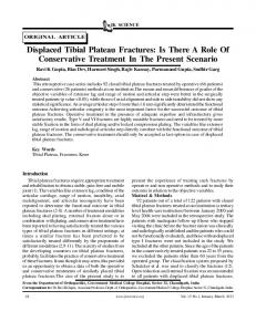
Diagram
I
All patients showed steady progress in fracture healing, although the rate of healing revealed by stiffness measurements varied widely (Fig. 4). The average time to inde-
RESULTS
rise
Fig.
changed became
progressive.
The
movements
mm cycle
and bending movements (Fig. I ), and of the change in length of the soft
effects of both axial providing a measure Relative
change
into three of motion at
lateral
of
length
transforms
From each set of measurements, we determined the maximum axial component of the relative motion at the centre of the fracture. From the bone geometry as measured on radiographs, we transformed the relative movements at the fracture centre into length changes between the oppos-
standard
Lateral
(1976)
the
tissues
length
Axial
and Anderson
relative movement translational and
ing
413
FRACTURES
et al (1990)
t Gustilo
1994).
of
Type
OF TIBIAL
The two
No. 3. MAY
weeks,
1995
reduction despite
unlocking moderate
of movement increased
period.
later
large with periods
the fixator reduction in was loading
greater of the
DISCUSSION Our previous studies have indicated that cyclic applied by a pump soon after fracture correlate
movements with faster
healing rates as shown by measurements of fracture ness (Goodship and Kenwright 1985; Kenwright 199 1 ). We have now shown that cyclic movements
occur
during walking on an Orthofix unilateral a locked column, giving axial movements
amplitude of 0.74 mm, applied at that time (Fig. of early
cyclic
movement
usually 3). This than
stiffet al do
fixator with with a mean
in proportion to the load is a larger average amount the 0.2 mm
Dynabrace fixator (Kershaw et al 1993). We found a wide variation in the
reported
amount
for the of
cyclic
414
J. B. RICHARDSON,
T. N. GARDNER,
J. R. W. HARDY.
ET
AL
Case
Case
2
1000 3
3
800
0
z
(DID
-
0
600
0.6
400
0.4
200
0.2..
-
V
!
CD
.3
...0
0.2
0
5 Time
10 since
15
fracture
c)
0
20
5
10 since
Time
(weeks)
15
fracture
Fig.
2a
Fig.
2b
Case
3
Case
4
20
(weeks)
Unlocking
1000
1000
+
800
O.8
600
600
0.6
!
0.4
400 200
5
10
15
Time since fracture
1000
Fig.
2c
Case
5
200 0
20
MH1 0.4
400 200
1000#{149}
.
3g.
U
fracture
(weeks)
20
15
Unlocking
800
0.8
600
0.6 0.4
200
20
3
3g.
0.2.. 0
0
Fig.
since
15
400
0.2
10
10
CaseS
3
0.6
since fracture
5 Time
600
5
0 0
0.8
Time
3g.
0.2
Fig. 2d
Unlocking
0
0.4
400
(weeks)
800
!
0.6
0
t
0
.
!
3.
0.2 0
0.8
800
0
(weeks)
5
10
Time since
2e
15
fracture Fig.
-0 20
(weeks)
2f
Axial
-0-----
Medial
-.--
Lateral
-0--
Load
showing of the fracture
Charts
cyclic pattern
movements are marked
and weight-bearing M on the medial
forces side.
for each
patient.
THE
The
JOURNAL
diagrams
OF BONE
AND JOINT
SURGERY
DYNAMISATION
OF TIBIAL
415
FRACTURES Table II. unlocking
600 Case
500
gressive
3
Fracture gap measurements the fixator column, and
closure
after unlocking Fracture
Case
After unlocking (mm)
Progressive movement (mm) 0.4 3.0 0.2 0.8 0.1 3.5
C,
300 CD
Case
C)
5
....;
t
:
Case
200
6
0-
0.4
0.0
3.8
0.8
3
1.2 1.9 1.5 3.7
1.0 1.1 1.4 0.2
I
I
0
0.2
0.4 Axial
0.6
0.8
displacement
1
(mm)
In all five
Load versus
movement weight-bearing.
at two
to four
unstable
weeks
for each
case
during
walking
movement
is similar
actual
that
15 V
reported
Case
2
.-.--.
Case
3
-G---
Case
4
able on mechanical grounds. screws act as a spring, and
6
bearing.
The inverse relationship movement and progressive
Cl) Cl)
a) C
CI)
Case5
A
Case
-.--&.----
10
5
15
20
25
has
been
‘Healed’
transfer of rising
fracture
4 for each
the
case.
load
due
to the variations loads applied by
and
by
was
measurements
larger
than
that
resorption
measured at the
bone
the
The reopen
locked fixator and bone the fracture gap after it
application fixator
fracture,
of allows
thus
by the fixator.
screws,
problems
and
this
which we found between cyclic closure in this study is predict-
the
the
taken
bone
screw (0.20 to 0.98 mm), largely geometry, tissue properties
for
may
reported
load the
and stops the spring-like any gap probably allows
through
axial
stiffness
closed Unlocking
permanently closure of
Weeks Fig.
be
fixator
by Kay, Ross and Powell shortening of 1 mm. The
of bone
-0--
E
z
may because
the
averaging 1 .3 mm II). This amount of
ends, but since we record only relative movements, method of measurement is probably satisfactory.
Casel
U
movement in fracture
to
displacement
radiographically,
Pattern
unlocking
movement gap (Table
made on the body of the fixator (1989): 50% of patients showed
20
0
fractures,
associated with progressive with closure of the fracture
Fig. 3
with
I 2
4 5 6
100
gap
Before unlocking (mm)
a, .
and after of pro-
J/Case4
400
.
before amount
the
weightto
close
action. Permanent more direct loadreducing
This
by gap
must
explain
reduce
the
by De Bastiani
low
the
share
of
the load
on
incidence
of
et al (1984).
The term ‘dynamisation’ has become associated with both cyclic movements (Paley et al 1990; Calhoun et al 1 992) and with progressive, non-recoverable displacement
the patients. In the only patient showing small early movements (Fig. 2a), there was no visible fracture gap at the lateral cortex. The cyclical subperiosteal movement at the lateral cortex was 12% larger than that at the medial cortex, as a result of
(Melendez and Colon 1989; Egger caused some to expect increased
bending
failed to obtain cyclic movement after unlocking a fixator: they expected dynamisation to increase movement. We suggest that movement at a fracture site should always be
explain lateral
in the plane
of the bone
the common observation callus in patients treated
screws.
This
finding
may
of greater amounts of in unilateral external
fixation. Unlocking the reduction in cyclic
fixator was movement,
generally associated either immediately
with a or within
two weeks. The reduction in cyclic movements in the later weeks of healing may be explained by rising fracture stiffness but does not explain the immediate fall at the time of unlocking (Fig. six weeks increase and we consider of the fracture VOL.
77-B,
4). In only the cyclic
one patient movement
did unlocking (case 1 Fig.
this to be due to compressive site at the time of reduction
No. 3, MAY
1995
,
prestressing and fixation.
at 2a),
‘dynamisation’ gressive concern
defined The
by
unlocking,
while
movement or gap closure. of Pohl and Williams (1989)
as either cyclic unpredictability
fractures
et al 1993), and cyclic movement
may
both cyclic series. The
partly
has after
expect
pro-
This may explain the when they sometimes
or progressive. of healing times
be explained
movement patients
others
this
for individual
by the wide
and progressive with early cyclic
tibial
variation
in
closure in our small movements of more
than 0.5 mm usually healed faster, as measured objectively by fracture stiffness (Fig. 4). One patient (case 6), however, with large early cyclic movements of I .25 mm and later a progressive movement of 3.5 mm had slow healing. In this case
the initial
fracture
gap
of 3.7 mm
probably
contributed
416
J. B. RICHARDSON,
to the
delay.
Relatively
act as a stimulus while increased more
appropriate
large
movements
to the growth of callus stability and compression stimuli
in the later
tion. Much larger studies optimum size, frequency, which are likely to influence We have shown that obtained in some fractures the fixator six weeks
cyclic
column usually
T. N. GARDNER,
may
in the early of callus
phase
of callus
well
weeks, may be matura-
will be required to define the and direction of movements fracture healing. early cyclic by allowing
locked. Unlocking allows progressive
movement weight-bearing
will
be with
the column at four to movement which may
be beneficial, but may also cause a reduction in cyclic movements. For research purposes it is important to consider the effect of ‘dynamisation’ in terms of both cyclic also
movement The
authors
and
authors
like
closure
at the fracture
site.
our patients for their co-operation in this for financial support. Although none of the authors have received or will receive benefits for personal or professional use from a commercial party related directly or indirectly to the subject of this article, benefits have been or will be received but are directed solely to a research fund, foundation, educational institution, or other non-profit institution with which one or more of the
study,
would
progressive
J. R. W. HARDY.
ET
Al
Gardner TN, Evans M, Simpson AHRW, Turner-Smith AR. Threedimensional movement at externally fixated tihial fractures and osteotomies during normal patient function. C/in Biomecli I 994:9:5 1 -9. Goodship upon [Br/
AE, Kenwright J. The the healing of experimental l985:67-B:650-5.
Gustilo RB, Anderson iT. thousand and twenty-five and prospective analysis. Kay
PR, Ross an external
Kenwright
ERS, fixator
of
tihial
Surg [Br]
Kershaw weight
induced
fractures.
micromovement
J Bone
Joint
Siti
Prevention of infection in the treatment of one open fractures of long hones: retrospective J Bone Joint [A,n/ 1976:58-A:453-8.
Powell ES. monitoring
Development and system. J Bio,ned
i, Richardson
and tibial fractures: Joint
influence
iB, Cunningham a controlled randomised 1991:73-B:654-9.
Ci, Cunningham JL, Kenwright bearing and fracture movement.
clinical application Eig 1989:11:240-4.
of
JL, et al. Axial movenient trial of treatment. J Bone J. Tibial C/in Ort/iop
external fixation. 1993:293:28-36.
Lindholm RV, Lindholm TS, Toikkanen S, Leino R. Effect of forced inter-fragmental movements on the healing of tihial fractures in rats. Ada Orthop Scand I 970:40:72 1-8.
to thank
and the Wishbone
Trust
is associated.
Melendez Orthofix
EM, Colon C. Treatment of open fixator. C/in Orthop 1989:241:224-30.
Muller ME, classification
Nazarian S. offractures
Koch
P,
(if long
fractures
with
the
Schatzker J. The toinprehensive 1ones. Berlin. etc: Springer-Verlag.
1990. Paley D, Fleming B, Catagni M, Kristiansen evaluation of external fixators used in limb
REFERENCES
tihial
T, Pope lengthening.
M.
Mechanical C/in Orthop
1990:250:50-7.
Calhoun JH, Li F, Ledhetter fixator for fracture fixation.
De Bastiani a dynamic Egger
G, Aldegheri axial
fixator.
BR, Gill CA. C/in Orthop
Biomechanics 1992:280:15-22.
R, Brivio LR. The J Bone Joint Surg
EL, Gottsauner-WoIf F, Palmer axial dynamization on bone healing.
Evans M, Gardner TN, Hardy The effect of ‘dynamisation’ D.A.F. fixator. J Bone Joint
J,
of the Ilizarov
treatment of fractures [Br] 1984:66-B:538-45.
Am HT, Chao EY. Effects J Trauma 1993:34:185-92.
PohI with
AP,
Orthofix
Williams
fixator.
DR. Proc
The dynamic 131/i hit Conf
axial loading properties Minnesota. etc: I 989:63.
Ralston of
JRW, Richardson JB, Ten Voorde WB. on fracture site movement with the orthofix Surg [Br] 1994:76-B Supp II & 111:88.
JL, Brown TD, Nepola JV, Williams DR, Marsh iL. ical analysis of the factors affecting dynamization of the Dynamic Axial Fixator. J Orthop Trauma 1990:4:449-57.
Richardson JB, Cunningham iL, Goodship wright J. Measuring stiffness can define Bone Joint Surg [Br] 1994:76-B:389-94.
THE
JOURNAL
AE, healing
OF BONE
O’Connor of tibial
AND JOINT
of
the
MechanOrthofix
BT, Kenfractures. J
SURGERY