Experimental Neurology 283 (2016) 246–254
Contents lists available at ScienceDirect
Experimental Neurology journal homepage: www.elsevier.com/locate/yexnr
Review Article
Dysferlin function in skeletal muscle: Possible pathological mechanisms and therapeutical targets in dysferlinopathies Ana M. Cárdenas a,⁎, Arlek M. González-Jamett a,b, Luis A. Cea b, Jorge A. Bevilacqua b, Pablo Caviedes c a
Centro Interdisciplinario de Neurociencia de Valparaíso, Facultad de Ciencias, Universidad de Valparaíso, Valparaíso, Chile Programa de Anatomía y Biología del Desarrollo, ICBM, Facultad de Medicina, Departamento de Neurología y Neurocirugía, Hospital Clínico Universidad de Chile, Universidad de Chile, Santiago, Chile c Programa de Farmacología Molecular y Clinica, ICBM, Facultad de Medicina, Universidad de Chile, Santiago, Chile b
a r t i c l e
i n f o
Article history: Received 27 April 2016 Received in revised form 22 June 2016 Accepted 23 June 2016 Available online 25 June 2016 Keywords: Muscular dystrophies Dysferlin Dysferlinopathies Membrane repair Vesicle trafficking Connexin hemichannels Inflammatory processes
a b s t r a c t Mutations in the dysferlin gene are linked to a group of muscular dystrophies known as dysferlinopathies. These myopathies are characterized by progressive atrophy. Studies in muscle tissue from dysferlinopathy patients or dysferlin-deficient mice point out its importance in membrane repair. However, expression of dysferlin homologous proteins that restore sarcolemma repair function in dysferlinopathy animal models fail to arrest muscle wasting, therefore suggesting that dysferlin plays other critical roles in muscle function. In the present review, we discuss dysferlin functions in the skeletal muscle, as well as pathological mechanisms related to dysferlin mutations. Particular focus is presented related the effect of dysferlin on cell membrane related function, which affect its repair, vesicle trafficking, as well as Ca2+ homeostasis. Such mechanisms could provide accessible targets for pharmacological therapies. © 2016 Elsevier Inc. All rights reserved.
Contents
1. Introduction . . . . . . . . . . . . . . . . . . . . . . . . . . . 2. Clinical background . . . . . . . . . . . . . . . . . . . . . . . . 3. Dysferlin splice variants . . . . . . . . . . . . . . . . . . . . . . 4. The ferlin family, a unique group of proteins with multiple C2 domains. 5. The different dysferlin domains display independent functions . . . . 6. Role of dysferlin in the membrane repair machinery . . . . . . . . . 7. Other roles of dysferlin in skeletal muscle . . . . . . . . . . . . . . 8. Expression and function of dysferlin in other tissues . . . . . . . . . 9. Pathological mechanisms underlying dysferlinopathy . . . . . . . . . 10. Role of connexin hemichannels in dysferlinopathies . . . . . . . . . 11. Potential therapies for dysferlinopathies . . . . . . . . . . . . . . 12. Conclusions, future directions . . . . . . . . . . . . . . . . . . . Acknowledgements . . . . . . . . . . . . . . . . . . . . . . . . . . References. . . . . . . . . . . . . . . . . . . . . . . . . . . . . . .
Abbreviations: LGMD, limb girdle muscular dystrophy; CK, creatine kinase levels; MG53, mitsugumin 53; Cx, connexins; TRPV2, transient receptor potential vanilloid type 2; STB, syncytiotrophoblasts. ⁎ Corresponding author at: Centro Interdisciplinario de Neurociencia de Valparaíso, Facultad de Ciencias, Universidad de Valparaíso, Gran Bretaña 1111, Playa Ancha, Valparaíso 2360102, Chile. E-mail address:
[email protected] (A.M. Cárdenas).
http://dx.doi.org/10.1016/j.expneurol.2016.06.026 0014-4886/© 2016 Elsevier Inc. All rights reserved.
. . . . . . . . . . . . . .
. . . . . . . . . . . . . .
. . . . . . . . . . . . . .
. . . . . . . . . . . . . .
. . . . . . . . . . . . . .
. . . . . . . . . . . . . .
. . . . . . . . . . . . . .
. . . . . . . . . . . . . .
. . . . . . . . . . . . . .
. . . . . . . . . . . . . .
. . . . . . . . . . . . . .
. . . . . . . . . . . . . .
. . . . . . . . . . . . . .
. . . . . . . . . . . . . .
. . . . . . . . . . . . . .
. . . . . . . . . . . . . .
. . . . . . . . . . . . . .
. . . . . . . . . . . . . .
. . . . . . . . . . . . . .
. . . . . . . . . . . . . .
. . . . . . . . . . . . . .
. . . . . . . . . . . . . .
. . . . . . . . . . . . . .
. . . . . . . . . . . . . .
. . . . . . . . . . . . . .
. . . . . . . . . . . . . .
. . . . . . . . . . . . . .
. . . . . . . . . . . . . .
. . . . . . . . . . . . . .
. . . . . . . . . . . . . .
. . . . . . . . . . . . . .
. . . . . . . . . . . . . .
. . . . . . . . . . . . . .
. . . . . . . . . . . . . .
. . . . . . . . . . . . . .
246 247 247 247 248 249 250 250 250 251 251 252 252 252
1. Introduction The hereditary myopathies comprise a large family of degenerative muscular disorders genetically determined by over 350 different mutations in distinct genes, for which novel causing mutations and genes are identified each year (Kaplan and Hamroun, 2014). The pathological
A.M. Cárdenas et al. / Experimental Neurology 283 (2016) 246–254
mechanisms underlying these ailments are largely unknown, and therapy mostly lies within the realm of tertiary care. Among these, muscular dystrophies represent a sizable amount of such disorders, with a worldwide distribution and an estimated incidence of 1/2000 live births. Although the genetic background is clear for most muscle dystrophies, their pathophysiology remains elusive. Indeed, mutations that render truncated or dysfunctional proteins that are critical for normal muscle integrity and function is a common feature of these disorders, but the underlying mechanisms by which such mutations result in the compromise of the skeletal muscle remain unclear. Moreover, in many cases the lack of knowledge of the function of the protein in question is the key issue. Among the causes of such myopathies are those linked to mutations in the dysferlin gene (DYSF), a large-sized gene spanning over 150 kb of genomic DNA that is located on chromosome 2p13 (Bashir et al., 1998; Liu et al., 1998; Aoki et al., 2001) and that produces a 237 kDa single-pass transmembrane protein (Anderson et al., 1999; Matsuda et al., 1999). Reduction or absence of dysferlin, resulting from autosomal recessive mutations in the gene that encodes for DYSF [MIM# 603009, GenBank NM_003494.2] comprises a number of different muscular dystrophy phenotypes known as dysferlinopathies. The term dysferlinopathy was introduced after setting forth that Miyoshi's myopathy (MM) and limb girdle muscular dystrophy (LGMD) type 2B, the two most common dysferlinopathy phenotypes, corresponded to allelic disorders (Bashir et al., 1998; Liu et al., 1998; Illarioshkin et al., 2000). At present, more than 2000 polymorphic variants and over 260 diseasecausing mutations associated with different dysferlinopathy phenotypes have been reported in the Leiden Muscular Dystrophy database (www.dmd.nl/dysf_seqvar.html), with the majority being point mutations and small insertions/deletions. Yet, many other non-pathogenic variants have also been included (Aoki et al., 2001). The exact incidence and prevalence of dysferlinopathy worldwide are unknown, but it has been estimated that it represents up to 30% of progressive recessive muscular dystrophies in the Middle East and India (Urtizberea et al., 2008). In the present review, we discuss current evidence of the role of dysferlin in critical membrane related events, particularly in membrane repair and vesicle trafficking. We also discuss possible pathological mechanisms involved in dysferlinopathy, including the de novo expression of non-selective Ca2+ permeable channels, which might contribute to the inflammatory process associated to dysferlinopathy, and could provide readily accessible targets for pharmacological therapies. 2. Clinical background Clinically, dysferlinopathy most commonly begins between the second and third decades in a previously asymptomatic patient. At onset, most patients complain of lower limb weakness, difficulties upon running or climbing stairs, sometimes aggravated by pain. These symptoms are usually accompanied by marked increase in plasma creatine kinase levels (CK) (Galassi et al., 1987; Barohn et al., 1991, 1998; Linssen et al., 1997; Rosales et al., 2010). Three main clinical phenotypes and some other variants of dysferlinopathy have been described (Miyoshi et al., 1967, 1986; Liu et al., 1998; Bashir et al., 1998; Illa et al., 2001; Nguyen et al., 2005, 2007; Laval and Bushby, 2004; Okahashi et al., 2008; Paradas et al., 2009; Klinge et al., 2008). There is also a marked inter- and intrafamiliar phenotypical variability (Miyoshi et al., 1986; Illarioshkin et al., 1996; Ueyama et al., 2002; Guglieri et al., 2008; Rosales et al., 2010, Woudt et al., 2016). However systematic muscle magnetic resonance imaging and functional assessment of patients with these different phenotypes do not show substantial differences in terms of distribution and severity of muscle compromise (Paradas et al., 2010; Díaz et al., 2016). After the first years of evolution, the disease mainly affects lower limbs, and later muscle weakness progresses involving paravertebral and proximal upper girdle muscles, to finally affect forearm flexor muscles. Head and neck muscles are not or very
247
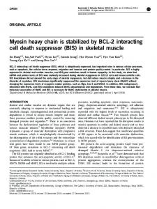
lately affected. Independent ambulation is lost on average after 10 years of disease course (Linssen et al., 1997; Woudt et al., 2016; Díaz et al., 2016). Although cardiac involvement is absent in most patients (Nguyen et al., 2007; Klinge et al., 2010a; Takahashi et al., 2013, Woudt et al., 2016), some recent evidences indicate that subclinical involvement is present in several patients with dysferlin deficiency (Wenzel et al., 2006; Choi et al., 2009; Rosales et al., 2010). So far, no central nervous system involvement has been described in dysferlin deficient patients, and respiratory involvement is mild, and presents itself in later stages of the disease (Takahashi et al., 2013; Woudt et al., 2016). Dysferlin protein analysis is essential to confirm diagnosis, as the clinical features tend to overlap with other genetic disorders (i.e. mutations on calpain and caveolin-3 genes). Further, molecular analysis of the DYSF gene is most desirable in order to elucidate specific mutations that may relate to the disease (Bushby, 1999; Krahn et al., 2009). Clinical differential diagnosis of dysferlinopathy is mainly with other types of LGMD (Barohn et al., 1998; Bushby, 1999; Urtizberea et al., 2008; Guglieri et al., 2008; Fanin et al., 2009), as all LGMD patients may show a similar clinical picture. These include increased CK levels, weakness and wasting restricted to the limb musculature and relative sparing of heart and bulbar muscles (depending on the genetic subtype). This is particularly evident in LGMD type 2B and proximodistal dysferlinopathy; however, the combination of muscle atrophy of the posterior leg compartment and marked increase of CK levels in an adolescent or young adult is very suggestive of dysferlinopathy. To differentiate LGMD subtypes, immunohistochemistry and western blot are necessary in order to detect specific protein deficits and subsequently perform genetic analysis (Nguyen et al., 2007; Krahn et al., 2009; Fanin et al., 2009, Rosales et al., 2010). Genetic diagnosis after identification of the protein deficit is necessary to verify that this specific deficit is the cause of the dystrophy, and not secondary to mutations in other related genes (Krahn et al., 2009; Fanin et al., 2009, Rosales et al., 2010). 3. Dysferlin splice variants The dysferlin gene is susceptible to suffer alternative splicing. In 2004, Salani and collaborators demonstrated that human primary myogenic cells express a dysferlin mRNA that lacks exon 17 and whose expression inversely correlates with muscle differentiation. This variant is later completely replaced by full-length dysferlin in adult skeletal muscle (Salani et al., 2004). Two years later, a novel human dysferlin transcript named DYSF_v1 was identified, which differs from the canonical dysferlin transcript in the sequence of the first exon (Pramono et al., 2006). The authors predicted that exon 1 of this new human dysferlin transcript shares 85% homology with the corresponding exon of mouse dysferlin and 89% with that of rat dysferlin. As in the canonical dysferlin, exon 1 of DYSF_v1 encodes for the first Ca2+-binding domain (C2A), but, as discussed below, they differ in their Ca2+-affinity (Fuson et al., 2014). Other dysferlin human transcript variants, produced by inclusions in exons 5a and 40a, were also reported (Pramono et al., 2009). All these variants could translate into different dysferlin isoforms, which as discussed later, appear to differ in their tissue distribution, sensibility to Ca2+, and interaction with other proteins. 4. The ferlin family, a unique group of proteins with multiple C2 domains Dysferlin belongs to the ferlin family, a group of single-pass transmembrane proteins that possess a short C-terminal extracellular domain and multiple (five to seven) tandem cytosolic C2 domains. These proteins also contain variable numbers of Fer and DysF domains. Fig. 1 shows how these different domains are organized in the ferlin family members dysferlin, myoferlin and otoferlin. Myoferlin is a mammalian ferlin highly expressed in developing skeletal muscle (Davis et al., 2000), where it is known to regulate myoblast fusion and muscle regeneration (Doherty et al., 2005). Myoferlin is also involved in membrane
248
A.M. Cárdenas et al. / Experimental Neurology 283 (2016) 246–254
Fig. 1. Comparison of dysferlin structure with those of otoferlin and myoferlin. Ferlins have a short C-terminal transmembrane domain (with 15–20 extracellular residues), five to seven tandem C2 domains and a variable number of Dysf and Fer domains (Adapted from Kobayashi et al., 2012).
repair during accelerated proliferation of tumor cells (Leung et al., 2013). Otoferlin is another mammalian ferlin mainly expressed in the inner ear, where it regulates SNARE-mediated exocytosis (Johnson and Chapman, 2010). Mutations in otoferlin result in non-syndromic deafness (Pangršič et al., 2012). These mammalian ferlins reportedly exhibit specific subcellular location that appears to correlate with their functions (Redpath et al., 2016). For example, dysferlin and myoferlin are abundantly expressed in the plasma membrane, according with their functions in membrane repair and muscle regeneration, whereas otoferlin mainly localizes in intracellular compartments, where it plays a role in exocytosis (Redpath et al., 2016). In addition to dysferlin, myoferlin and otoferlin, other three ferlin proteins (Fer1L4, Fer1L5 and Fer1L6) have been described in mammals; although their functions remain unknown (Lek et al., 2012). Ferlin proteins are also expressed in Caenorhabditis elegans (Fer-1) and drosophila (misfire). Fer-1 regulates Ca2 +-mediated membrane fusion during spermatogenesis (Washington and Ward, 2006). Misfire is required for Ca2+-dependent breakdown of the sperm plasma membrane during fertilization (Smith and Wakimoto, 2007). While the functions of Fer and DysF domains remain unknown, most actions of ferlins appear to be mediated by their C2 domains (Lek et al., 2012). C2 domains are lipid-binding modules of about 130 residues that are organized in an eight-β-strand structure (Corbalan-Garcia and Gómez-Fernández, 2014). Many C2 domains, as those present in synaptotagmin and some protein kinase C isoforms, have the ability to bind Ca2 + and regulate Ca2 +-dependent events (Corbalan-Garcia and Gómez-Fernández, 2014). In 2009, Therrien et al. (2009) reported that the seven C2 domains of dysferlin are able to bind acidic phospholipids, but only the C2A domain associates to phosphatidylserine and phosphoinositides in a Ca2+-dependent manner. On the other hand, a more recent work shows that the seven dysferlin C2 domains exhibit variable affinity for Ca2 +, being the C2A and C2C the most sensitive, with KD values of 3.7 and 1.5 μM, respectively (Abdullah et al., 2014). Furthermore, the seven dysferlin C2 domains bind anionic lipids, and this association is enhanced by Ca2+ in concentrations between 1 and 10 μM (Abdullah et al., 2014). In a recent study, Fuson et al. (2014) crystallized the dysferlin C2A domain and its splice variant C2Av1, and analyzed their affinity for Ca2+.They observed that the canonical C2A domain has two classes of
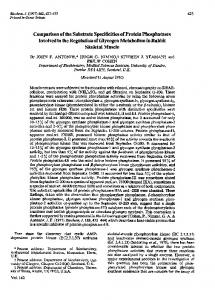
Ca2 + binding sites. One of them has a high affinity for Ca2 + (KD ~60 nM), while the other has a lower affinity (KD ~53 μM), suggesting that the first site is occupied by Ca2 + at resting conditions, whereas the second binds Ca2+ after the plasma membrane is disrupted. The affinity for Ca2+ of both types of sites significantly increases in the presence of anionic phospholipids. On the other hand, the C2Av1 domain, whose expression in human skeletal muscle is around 23% (Pramono et al., 2006), does not bind Ca2+ in the absence of lipids, but in the presence of the latter, a high-affinity Ca2+ binding site is exposed. These authors also indicate that both C2A domains display highly flexible structures, a property that would allow them to undergo a variety of conformational states to interact with other proteins in resting or elevated Ca2 + conditions. Dysferlin partners that bind the C2A domain are Mitsugumin 53 (Matsuda et al., 2012) and AHNAK (Huang et al., 2007). Mitsugumin 53 (MG53) is a protein that belongs to the tripartite motif (TRIM 72) family involved in membrane repair in different tissues (Cai et al., 2009; Duann et al., 2015), where it facilitates the recruitment of dysferlin and vesicles to repair sites (Cai et al., 2009; Matsuda et al., 2012). On the other hand, AHNAK is a 700-kDa scaffolding protein involved in the regulation of L-type Ca2+ channels, cytoskeletal organization and membrane repair (Davis et al., 2014). Further, the dysferlin C2A domain, as well as the C2B domain, also interacts with α-tubulin (Azakir et al., 2010). 5. The different dysferlin domains display independent functions Regarding C2 domains of dysferlin, it seems that they have independent functions. For instance, it has been reported that the dysferlin C2A domain mediates the fusion of lysosomes with the plasma membrane (Han et al., 2012), and that it is also required for MG53-dependent accumulation of dysferlin at damage sites (Matsuda et al., 2012). The Cterminal region of dysferlin, which contains the C2F and C2G domains, also appears to mediate membrane repair. This dysferlin fragment, which shares structural resemblance with synaptotagmin, a protein with a transmembrane region and two C2 domains (Bai and Chapman, 2004), was called “minidysferlin”. The expression of minidysferlin in dysferlin-deficient mice restored the membrane repair activity of skeletal muscle fibers (Krahn et al., 2010). However, it did not correct the dystrophic phenotype (Lostal et al., 2012), suggesting that other
A.M. Cárdenas et al. / Experimental Neurology 283 (2016) 246–254
dysferlin domains, which are missing in minidysferlin, are involved in other critical functions of dysferlin in skeletal muscle. Minidysferlin seems to be originated by calpain-dependent cleavage of dysferlin during skeletal muscle membrane injury (Lek et al., 2013). In this regard, Redpath et al. (2014) proposed that the separation of this dysferlin fragment from the rest of the protein could be a regulatory checkpoint for expediting dysferlin-mediated vesicle fusion during membrane repair. As reported by the authors, calpain cleaves dysferlin in a motif encoded in exon 40a. Transcripts including exon 40a are poorly expressed in skeletal muscle (Pramono et al., 2009), but expressed at higher levels in other tissues such as placenta, lung, liver, kidney and pancreas (Redpath et al., 2014). Furthermore, activation of calpain cleaves dysferlin and releases minidysferlin in different types of cells, including myocytes, astrocytes and endothelial cells, among others (Redpath et al., 2014). Therefore, this dysferlin fragment could play a role not only in the skeletal muscle, but also in other tissues. On the other hand, the C2B-FerI-C2C motif regulates dysferlin expression at the plasma membrane, as well as its endocytic rate (Evesson et al., 2010). The endocytosis of dysferlin, which is also regulated by caveolin-3 (Hernández-Deviez et al., 2008), is apparently a crucial step for the membrane repair process (Idone et al., 2008; McDade et al., 2014). Indeed, a recent report showed that membrane damage promotes the formation of endocytic dysferlin-containing vesicles which are recruited to lesion sites, where they form a large dysferlinrich vesicle that may act as a membrane patch that reseals the wounded sarcolemma (McDade et al., 2014). In summary, the distinct domains of the dysferlin protein seem to contribute to sarcolemma repair, probably by regulating different steps of the resealing process. Furthermore, some dysferlin domains appear to be unnecessary for the repair process (N-terminal region including C2A to C2E domains), yet indispensable for other critical functions. A schematic representation summarizing these findings is shown in Fig. 2. 6. Role of dysferlin in the membrane repair machinery Being one of the most malleable tissues of our body, the skeletal muscle is constantly adapting to functional demands by changing its protein expression, mass and contractile properties. In spite of this plasticity, sometimes the stress resulting from muscle mechanical work exceeds its own adaptation, producing microlesions in the plasma membrane (Tidball, 2011). These membrane disruptions cause a massive entry of Ca2+ to the cell, with the consequent recruitment of vesicles and other membranous organelles to the injury sites. The recruited vesicles fuse with one another and with the surrounding plasma membrane, thus forming a patch that reseals the membrane lesion (McNeil, 2002). A critical component of the sarcolemma repair machinery is dysferlin (Anderson et al., 1999). Muscle biopsies from dysferlinopathy patients show absence or markedly reduced sarcolemmal expression of dysferlin, an excess of immature fibers, evidence of dystrophy and inflammatory cell infiltration (Chiu et al., 2009; Gallardo et al., 2011; Woudt et al., 2016; Yin et al., 2015). Ultrastructural analysis of dysferlinopathy muscle fibers shows
249
disruption of the plasma membrane and its replacement by layers of small vesicles (Selcen et al., 2001; Cenacchi et al., 2005). Loss of membrane integrity and accumulation of subsarcolemmal vesicles have also been observed in muscle fibers from dysferlin-deficient mice (Bansal et al., 2003; Hornsey et al., 2013). Those findings stress the importance of this protein in membrane repair, where a mechanism that involves vesicle aggregation and/or fusion at membrane damage sites is at hand. In fact, both dysferlinopathy patients and dysferlindeficient mice myocytes exhibit defective membrane resealing (Bansal et al., 2003; Lostal et al., 2012; Philippi et al., 2012). Meanwhile, in myocytes that normally express dysferlin, vesicles containing the protein are recruited to resealing sites, where they fuse to each other, and also with other membranous organelles, forming a patch that reseals the membrane lesion (McNeil, 2002, 2009; McDade et al., 2014; McDade and Michele, 2014). The exact mechanism of membrane repair is still unclear, and many steps involved in this process are unknown. However, reportedly it comprises the participation of dysferlin and its partners. One of them is MG53, which accumulates, before dysferlin at injury sites in an oxidation-dependent, but Ca2 +-independent manner (Cai et al., 2009); next MG53 recruits, in a Ca2+-dependent manner, vesicles containing dysferlin (Matsuda et al., 2012; Duann et al., 2015). Then, dysferlin appears to promote vesicle aggregation and fusion by a mechanism that depends on its association with annexins (McNeil, 2009). Annexins are proteins that bind membranes containing acidic phospholipids, in a Ca2 +-dependent manner (Gerke et al., 2005; Marg et al., 2012). In particular, annexins A1 and A2 are implicated in vesicle trafficking, membrane fusion, endocytosis, actin cytoskeleton remodeling and membrane repair (Gerke et al., 2005; McNeil et al., 2006; Hitchcock et al., 2014). Most of these actions are related to their ability to create membrane microdomains that facilitate the interactions of membrane proteins with cytosolic partners, including actin filaments (Hayes et al., 2004; Hitchcock et al., 2014). This property of annexins of forming specialized membrane microdomains, -particularly annexin A2 which forms a heterotetrameric complex with its partner S100A10-, explains their ability to promote vesicle aggregation (Jones et al., 1994; De la Fuente and Parra, 1995) and their recruitment to exocytotic sites (Chasserot-Golaz et al., 2005). Indeed, annexin A2 provides the lipid environment required for vesicle docking and SNAREmediated membrane fusion (Chasserot-Golaz et al., 2005; Umbrecht-Jenck et al., 2010; Gabel et al., 2015). Most of the work carried out to study the interaction of dysferlin with annexins has been conceived in a membrane repair context. Lennon et al. (2003) showed that dysferlin co-precipitates and colocalizes at the sarcolemma with annexins A1 and A2. Using fluorescence lifetime imaging microscopy, they observed that in non-injured myocytes, both annexins interact with dysferlin in a Ca2+-dependent form. However, after sarcolemma injury, the association of annexin A1 with dysferlin is disrupted, while annexin A2 and dysferlin remain associated. They also observed that the distribution of these annexins is significantly modified in dysferlin-deficient muscle. On the other hand, annexin A6, which has also been involved in membrane repair
Fig. 2. Functions and protein interactions of the distinct dysferlin regions. The scheme summarizes some of the roles described for different Dysferlin domains.
250
A.M. Cárdenas et al. / Experimental Neurology 283 (2016) 246–254
(Swaggart et al., 2014), does not interact with dysferlin in cultured myotubes (Lennon et al., 2003). The temporal recruitment of dysferlin and annexins to injury sites has also been studied in vivo in zebrafish myofibers. During sarcolemma repair, dysferlin-enriched membranes are rapidly accumulated at lesion sites, where they form a membrane patch. Annexin A6 also arrives rapidly, whereas annexin A2 is recruited later. Finally, annexin A1 covers the lesion. Interestingly, the arrival of annexins A1 and A2, but not of annexin A6, to resealing sites depends on dysferlin (Roostalu and Strähle, 2012). Thus, this latter work also points out a role of dysferlin in mediating the recruitment of annexins A1 and A2 to repair sites. A recent report of Codding and collaborators highlights the possibility that dysferlin itself acts as an effector of the SNARE-mediated membrane fusion (Codding et al., 2016). Indeed, dysferlin seems to accelerate the Ca2+-dependent assembly of the SNARE-complex via a direct interaction with syntaxin 4 and SNAP-23, suggesting its participation during the fusion between the resealing-vesicles and wounded plasma membrane (Codding et al., 2016).
7. Other roles of dysferlin in skeletal muscle Besides its localization in the sarcolemma, dysferlin is richly expressed in T-tubule membranes (Klinge et al., 2010b; Kerr et al., 2013), where it colocalizes with the dihydropyridine receptor, caveolin-3, MG53, annexin A1 and AHNAK (Ampong et al., 2005; Huang et al., 2007; Waddell et al., 2011), and contributes to the maintenance of the T-tubule system (Kerr et al., 2013; Demonbreun et al., 2014). In fact, dysferlin-deficient muscle exhibit abnormal T-tubule morphology (Klinge et al., 2010b) and an enhanced sensitivity to damage by glycerol exposure (Demonbreun et al., 2014) or osmotic shock (Kerr et al., 2013). Since the dihydropyridine receptor antagonist diltiazem protected dysferlin-null fibers from damage induced by osmotic shock, Kerr et al. (2013) proposed that dysferlin contributes to keep Ca2 + homeostasis during mechanical stress. On the other hand, Demonbreun et al. (2014) proposed that dysferlin also contributes to the biogenesis and remodeling of the T-tubule system. Since T-tubule formation requires an advanced system of vesicle trafficking and membrane fusion (Di Maio et al., 2007), it is probable that the mechanism by which dysferlin contributes to T-tubule biogenesis and remodeling involves those cellular processes. Dysferlin also appears to participate in other cellular events that depend on vesicle trafficking and membrane fusion in myoblasts. In this regard, dysferlin-deficient myoblasts display impaired secretion of the cytokine MCP-1 (Chiu et al., 2009) and a reduced recycling rate of transferrin and insulin-like growth factor (IGF) receptors (Demonbreun et al., 2011). The association of dysferlin, via its C2A and C2B domains, with α-tubulin and microtubules in the skeletal muscle (Azakir et al., 2010), also supports the idea that dysferlin is involved in vesicle trafficking. Analysis of protein-protein interaction networks has become a useful tool for predicting protein functions. In 2013, Blandin and collaborators published a large-scale study addressed to establish a proteomic map of protein-protein interactions centered on different proteins involved in LGMD-dystrophies (Blandin et al., 2013). Using experimental and informatics approaches, these authors reported a strong interconnectivity between all the LGMD proteins studied (Blandin et al., 2013). Regarding dysferlin, a number of novel partners appeared including the cardiomyopathy-associated protein 5, the intermediatefilament protein desmin, the actin-binding proteins filamin-C and nebulin, the kinesin family member 1B, optineurin, the SAM domain and HD domain-containing protein 1, the Alstrom syndrome1 protein and APPL1 among others (Blandin et al., 2013). These data suggest new roles of dysferlin in gene regulation and muscle cytoskeletal remodeling.
8. Expression and function of dysferlin in other tissues Besides skeletal muscle, dysferlin is expressed in other tissues including brain, heart, liver and lungs (Redpath et al., 2016). High expression of dysferlin has been also reported in peripheral blood monocytes (Ho et al., 2002). The expression of dysferlin in this latter tissue reportedly correlates with dysferlin levels in skeletal muscle in both healthy and dysferlinopathy patients (Gallardo et al., 2011). Interestingly, dysferlin-deficient monocytes exhibit increased motility, pointing to a role of dysferlin in the formation of focal adhesions and cellular interactions (de Morrée et al., 2013). In 2007, Vandré and collaborators demonstrated the expression of high levels of dysferlin in trophoblasts of human placental microvillous (Vandré et al., 2007). As differentiating-muscle cells, they form syncytial structures and become a large cell layer of syncytiotrophoblasts (STB) on whose apical surface nutrient transport occurs (Jansson and Powell, 2006). Sprout-like structures containing heterochromatic nuclei and cellular debris emerge from the STB apical-membrane and are thrown into the maternal circulation (Ikle, 1964). Interestingly, a greater density of syncytial sprouts and STB-derived cellular debris has been associated with preeclampsia (Goswami et al., 2006; Heazell et al., 2007), suggesting that a destabilization of the STB-apical membrane is related with this pregnancy disorder. In this regard, dysferlin expression is reportedly reduced in human pre-eclamptic placentas (Lang et al., 2009), and its presence in STB appears to contribute to repair a damaged placental membrane (Robinson et al., 2008). Thus dysferlin also seems to play a role in the repair and stability of STB apical-membrane in placental tissue.
9. Pathological mechanisms underlying dysferlinopathy Most of the studies directed to elucidate the pathomechanism underlying dysferlinopathy have focused on the role of dysferlin in membrane repair. As aforementioned, defects in membrane resealing are observed in the sarcolemma of dysferlin-null-mice muscle fibers (Bansal et al., 2003) and in myocytes of dysferlinopathy patients (Philippi et al., 2012). Dysferlin-deficient skin fibroblasts also exhibit impaired plasma membrane resealing (Matsuda et al., 2015), indicating that dysferlin is a critical actor in membrane repair not only in skeletal muscle, but also in other types of cells where it is expressed. Despite this, several evidences demonstrate that the reversion of the defective plasma membrane repair is not enough to revert or improve the dystrophic phenotype in dysferlin-deficient tissues. In this regard, Krahn et al. (2010) reported a large homozygous deletion that leads to the production of a “minidysferlin”. The patient bearing this mutation presented a late-onset and moderate dysferlinopathy. Interestingly, the expression of minidysferlin in dysferlin-deficient mice restored membrane repair activity in skeletal muscle fibers (Krahn et al., 2010), yet it failed to correct the characteristic dysferlinopathy muscle histology (Lostal et al., 2012). The overexpression of myoferlin in dysferlin deficient animals also rescues the sarcolemma resealing without correcting the dystrophic phenotype (Lostal et al., 2012). Therefore, the pathomechanism of dysferlinopathy cannot be explained solely by a defective plasma membrane repair. Proper muscle integrity and function requires active intracellular membrane trafficking (Towler et al., 2004). As aforementioned, dysferlin is required for the biogenesis and maintenance of the Ttubule system (Klinge et al., 2010b; Kerr et al., 2013; Demonbreun et al., 2014), as well as for the recycling of plasma membrane protein (Demonbreun et al., 2011) and secretion of cytokines (Chiu et al., 2009). Furthermore, dysferlin interacts with proteins involved in vesicle trafficking, such as the actin-binding protein alpha-actinin, annexin A2, alpha-tubulin and microtubules (Assadi et al., 2008; Azakir et al., 2010). Then, it is likely that a defective intracellular trafficking also contributes to the pathological mechanism in dysferlinopathy.
A.M. Cárdenas et al. / Experimental Neurology 283 (2016) 246–254
Dysferlinopathy, as well as other muscle dystrophies, are present with an inflammatory process that could contribute to disease progression. In fact, components of the inflammasome pathway are upregulated and activated in muscle of dysferlinopathy patients and mice (Rawat et al., 2010, Mariano et al., 2013). Dysferlin-deficient muscles also exhibit increased protein oxidative damage (Terrill et al., 2013), activation of the ubiquitin-proteasomal pathway (Azakir et al., 2012; Fanin et al., 2014; Rajakumar et al., 2014), up-regulation of the complement system factors (Han et al., 2010) and macrophage infiltration (Roche et al., 2015). Furthermore, monocytes from dysferlinopathy patients and dysferlin-deficient mice exhibit increased phagocytic activity that might also contribute to the inflammatory response (Nagaraju et al., 2008). However, anti-inflammatory agents, such as the glucocorticoid deflazacort or Celastrol, an anti-inflammatory drug with antioxidant properties, are not sufficient to improve the dysfunction in dysferlin-deficient muscles (Walter et al., 2013; Dillingham et al., 2015). It has been proposed that the defective plasma membrane repair caused by dysferlin deficiency contribute importantly to the dysferlinopathy phenotype. Indeed, a loss of the plasma membrane integrity leads to disruption of intracellular Ca+2 and redox homeostasis, and release of ATP and inflammatory mediators (Han, 2011). However, as we discuss below, the de novo expression of non-selective Ca2+ permeable channels, such as connexin based channels, might also contribute to the inflammatory process associated to dysferlinopathy. 10. Role of connexin hemichannels in dysferlinopathies Connexins (Cx) are a group of plasma membrane proteins, which assemble in homomeric or heteromeric hexameric structures to allow the passage of small molecules like Ca2 +, ATP, glucose and ascorbate, among others (Li et al., 2001; Kang et al., 2008; Schalper et al., 2010; Hansen et al., 2014). These Cx structures, often known as hemichannels, allow the exchange of chemical signals between intracellular and extracellular compartments, or they can assemble in specialized intercellular connections called gap junctions (Saez et al., 2003). Cx hemichannels play key roles in autocrine and paracrine communication, but they are also reportedly associated to the pathogenesis of several inflammatory diseases, such as atherosclerosis and acute lung disease (Scheckenbach et al., 2011). They have also been implicated in neurodegenerative disorders such as Alzheimer, Parkinson and Huntington diseases (Eugenin et al., 2012). Recent reports indicate that Cx hemichannels are involved in pathological conditions that affect skeletal muscles (Cea et al., 2012; Cea et al., 2013). During myogenesis, Cx proteins (Cx39, Cx43 and Cx45) form gap junctions in myoblasts to coordinate myoblast fusion (Gorbe et al., 2007); but they are absent in normal adult skeletal muscles (Cea et al., 2012). Interestingly, de novo expression of Cx hemichannels is observed after chemical injury (Araya et al., 2005; Gorbe et al., 2005). This also occurs after denervation of skeletal muscle, where the presence of Cx hemichannels, along with other Ca2 + permeable channels, such as P2X7 receptors, and pannexin-1 and transient receptor potential vanilloid type 2 (TRPV2) channels, induces inflammatory signals. Such signals involve the nuclear factor κB activation and increase of proinflammatory cytokines, such as the tumor necrosis factor α and interleukin 1β (Cea et al., 2013). This inflammatory process is completely prevented in denervated skeletal muscle of Cx43 and Cx45 muscle deficient animals (Cea et al., 2013). Furthermore, the denervation-induced muscle atrophy was also partially, yet significantly prevented in Cx43 and Cx45 muscle deficient animals (Cea et al., 2012; Cea et al., 2013). Thus, these findings suggest that Cx hemichannels are involved in the inflammatory process of the skeletal muscles in response to injury or denervation. Cx hemichannels have also been involved in muscular diseases of genetic origin such as Duchenne muscular dystrophy, which is caused by mutations in the dystrophin gene. Indeed, in myofibers of an animal model of Duchenne muscular dystrophy (the mdx mice), these
251
hemichannels alter sarcolemma permeability, resulting in elevated intracellular Ca2+ levels, activation of NFκB, and expression of the inducible nitric oxide synthase and active caspase 3, and apoptosis-mediated death (Cea et al., 2016a). All of these features were totally prevented in Cx43 and Cx45 muscle deficient mdx mice (Cea et al., 2016a). Performance in the hanging test, which is reduced in mdx mice, was also partially prevented in Cx43 and Cx45 muscle deficient mdx mice. As observed with denervated myofibers, the Cx hemichannels were also present along with pannexin-1 and TRPV2 channels, and P2X7 receptors. It seems that Cx hemichannels act cooperatively with these other Ca2+ permeable channels, as the inhibition of any of these channels restores the altered sarcolemma permeability. In addition, Cx hemichannels appear to be upstream of the other Ca2 + permeable channels because the absence of Cx hemichannels prevented the expression of P2X7 receptors and TRPV2 channels (Cea et al., 2016a). Cx hemichannels have also been involved in the pathophysiology of dysferlinopathies. In this case, functional Cx hemichannels are expressed in mature human myocytes from patients harboring dysferlin mutations, where they are responsible of the elevated basal Ca2+ levels (Cea et al., 2016b). Down-regulation of dysferlin expression with small hairpin RNA also induces the expression of functional Cx hemichannels in mature myotubes (Cea et al., 2016b). Then, it seems plausible that the expression of Cx hemichannels constitutes a secondary mechanism triggered by the absence of dysferlin. This mechanism would explain at least partially why the reposition of the sarcolemma repairing function with minidysferlin or myoferlin is not enough to revert the muscle damage observed in dysferlinopathies (Lostal et al., 2012). 11. Potential therapies for dysferlinopathies There are currently no specific therapeutic alternatives available for patients bearing dysferlinopathy (Kobayashi et al., 2012). Efforts have been hindered by the lack of knowledge on the myriad of effects linked to dysferlin mutation. Although its role in membrane repair is clear, efforts to reinstate this function by transfecting functional variants of dysferlin genes in animal models have proved successful in this objective, yet they have failed to arrest progressive muscle wasting. This indicates that dysferlin possesses other functions in skeletal muscle that are yet unclear, and that restoration of all functions will require the presence of full length dysferlin. In this regard, recent studies by Escobar et al. (2016) have shown that stable, non-viral vector transfection of full length dysferlin DNA in dysferlin-deficient myoblasts, results in stable dysferlin expression and cell survival after engraftment in dysferlin-, immune-deficient mice. The grafted muscle cells exhibited normal regenerative capabilities in situ. Such ex vivo therapeutical approaches appear most attractive, yet developments along this field will necessitate greater study. Gene therapy approaches to dysferlinopathy appear indeed as the correct path to finding a cure, yet efforts along these lines have been hindered by limitations in the packaging capacities of adenoassociated viral vectors. In this regard, Sondergaard et al. (2015) presented an attractive alternative to deliver oversized genes such as that of dysferlin. This group developed a two vector system (named AAV.DYSF.DV), that packages the cDNA of the protein through the use of two discrete vectors defined by a 1 kb region of homology. Dysferlin-deficient mice and non-human primates (rhesus macaques) treated with this novel vector carrying a dysferlin cDNA, either by intramuscular or intravascular delivery, exhibited high levels of dysferlin expression in muscle. This strategy also restored membrane repair ability in dysferlin-deficient mice. Further, AAVrh.74.DYSF.DV treatment in macaques was found to stimulate dysferlin expression, with no apparent toxicity or systemic immune rejection, hence laying a foundation for preclinical safety studies. Although this study potentially overcomes outstanding limitations in gene therapy for dysferlinopathy, such as an efficient and safe full length dysferlin gene delivery, there is still much work to do to overcome efficacy and safety in the long term.
252
A.M. Cárdenas et al. / Experimental Neurology 283 (2016) 246–254
In the meantime, what alternatives could be suggested? Pharmacological approaches are certainly attractive, as they can yield potential therapies much faster, and have the added value of reversibility. Yet, results are still disappointing. Use of glucocorticoids has not yielded beneficial effects, in light of the use of such compounds in many patients due to initial misdiagnosis. Yet, controlled studies with deflazacort have clearly demonstrated no beneficial effects (Walter et al., 2013). Vitamin D supplementation has been proposed as a potential therapy, in light of studies showing increased dysferlin expression in carriers of one mutation in the dysferlin gene (De Luna et al., 2012), yet studies in actual patients are still lacking. Lately, the inflammatory response in muscles of dysferlinopathy patients has been linked to upregulated interleukin 1β, and in this regard, the inhibition of this cytokine or the use of interleukin 4 to counteract the M1-mediated immune response in dysferlin-deficient mouse muscle (Cohen et al., 2015). Another, open label study, showed improvements in the mobility of pelvic and shoulder girdles in two patients after treatment with rixutimab, which also correlated with a dramatic decrease in B-cell number (Lerario et al., 2010). Hence, specific targets within the immune system appear much more effective, compared to a generalized immune and inflammatory modulation sought with glucocorticoids. Also, promoting hypertrophy in dysferlinopathy has to be weighed carefully, as Lee et al. (2015) demonstrated that myostatin inhibition in mouse models of the disease, which induces hypertrophy, results in increased CK levels, suggesting increased muscle destruction. The latter evidence suggests that, initially, pharmacological treatment targeting specific aspects of the immune response, perhaps associated with controlled stimulation of muscle trophism, may be an interesting path in the quest for therapeutical alternatives for these patients. The final cure will necessarily involve gene therapy, and the recovery of full dysferlin expression, yet a lengthier road is anticipated in the achievement of this goal. 12. Conclusions, future directions In the recent years, great advances have been reached in the comprehension of the role played by dysferlin in the different tissues where it is expressed. The best understood function of dysferlin is its role in the repair machinery that reseals injured cell membranes. This role is particularly relevant in skeletal muscles where the repair and regeneration of wounded plasma membrane constitute critical phenomena to maintain muscle integrity and function. In fact, dysferlinopathy causing mutations seem to impair the sarcolemmal resealing processes in injured myofibers, strongly suggesting that defects in membrane repair underlie the pathological mechanisms that lead to muscular dystrophy. However, other processes that require dysferlin participation could be deregulated in dysferlin-deficient tissues and hence contribute to the pathophysiology of dysferlinopathy. The clarification of these mechanisms is pivotal in the quest for therapeutic targets in patients harboring mutations in the dysferlin gene. The novel suggested functions of dysferlin in vesicle trafficking and membrane remodeling events could open up interesting research fields in the search of therapies that soften the dystrophic phenotypes and slow down the progression of the disease. Finally, the persistent expression of connexin in several dystrophic conditions presents an interesting therapeutical target, readily accessible to address pharmacologically, and where lead compounds do exist (Cea et al., 2016b). Acknowledgements This work was supported by the grants ACT-1121 (PIA, CONICYT), FONDECYT 1151383 and 1160495, PAI/Conicyt Proyecto de Inserción en la Academia 79140023, Post-doctoral FONDECYT 3160311 and P09022-F from ICM-ECONOMIA, Chile. The Centro Interdisciplinario de Neurociencia de Valparaíso (CINV) is a Millennium Institute supported
by the Millennium Scientific Initiative of the Ministerio de Economía, Fomento y Turismo. References Abdullah, N., Padmanarayana, M., Marty, N.J., Johnson, C.P., 2014. Quantitation of the calcium and membrane binding properties of the C2 domains of dysferlin. Biophys. J. 106, 382–389. Ampong, B.N., Imamura, M., Matsumiya, T., Yoshida, M., Takeda, S., 2005. Intracellular localization of dysferlin and its association with the dihydropyridine receptor. Acta Myol. 24, 134–144. Anderson, L.V., Davison, K., Moss, J.A., Young, C., Cullen, M.J., Walsh, J., Johnson, M.A., Bashir, R., Britton, S., Keers, S., Argov, Z., Mahjneh, I., Fougerousse, F., Beckmann, J.S., Bushby, K.M., 1999. Dysferlin is a plasma membrane protein and is expressed early in human development. Hum. Mol. Genet. 8, 855–861. Aoki, M., Liu, J., Richard, I., Bashir, R., et al., 2001. Genomic organization of the dysferlin gene and novel mutations in Miyoshi myopathy. Neurology 57, 271–278. Araya, R., Eckardt, D., Maxeiner, S., Krüger, O., Theis, M., Willecke, K., Sáez, J.C., 2005. Expression of connexins during differentiation and regeneration of skeletal muscle: functional relevance of connexin43. J. Cell Sci. 118, 27–37. Assadi, M., Schindler, T., Muller, B., Porter, J., Ruegg, M., Langen, H., 2008. Identification of proteins interacting with dysferlin using the tandem affinity purification method. Open Cell Dev. Biol. J 1, 17–23. Azakir, B.A., Di Fulvio, S., Therrien, C., Sinnreich, M., 2010. Dysferlin interacts with tubulin and microtubules in mouse skeletal muscle. PLoS One 5, e10122. Azakir, B., Di Fulvio, S., Kinter, J., Sinnreich, M., 2012. Proteasomal inhibition restores biological function of mis-sense mutated dysferlin in patient-derived muscle cells. J. Biol. Chem. 287, 10344–10354. Bai, J., Chapman, E.R., 2004. The C2 domains of synaptotagmin–partners in exocytosis. Trends Biochem. Sci. 29, 143–151. Bansal, D., Miyake, K., Vogel, S.S., Groh, S., Chen, C.C., Williamson, R., McNeil, P.L., Campbell, K.P., 2003. Defective membrane repair in dysferlin-deficient muscular dystrophy. Nature 423, 168–172. Barohn, R.J., Miller, R.G., Griggs, R.C., 1991. Autosomal recessive distal dystrophy. Neurology 41, 1365–1370. Barohn, R.J., Amato, A.A., Griggs, R.C., 1998. Overview of distal myopathies: from the clinical to the molecular. Neuromuscul. Disord. 8, 309–316. Bashir, R., Britton, S., Strachan, T., et al., 1998. A gene related to Caenorhabditis elegans spermatogenesis factor fer-1 is mutated in limb-girdle muscular dystrophy type 2B. Nat. Genet. 20, 37–42. Blandin, G., Marchand, S., Charton, S., Danièle, N., Gicquel, E., Boucheteil, J., Bentaib, A., Barrault, L., Stockholm, D., Bartoli, M., Richard, I., 2013. A human skeletal muscle interactome centered on proteins involved in muscular dystrophies: LGMD interactome. Skelet. Muscle 3, 3. Bushby, K.M., 1999. Making sense of the limb-girdle muscular dystrophies. Brain 122, 1403–1420. Cai, C., Masumiya, H., Weisleder, N., Matsuda, N., Nishi, M., Hwang, M., Ko, J.K., Lin, P., Thornton, A., Zhao, X., Pan, Z., Komazaki, S., Brotto, M., Takeshima, H., Ma, J., 2009. MG53 nucleates assembly of cell membrane repair machinery. Nat. Cell Biol. 11, 56–64. Cea, L.A., Riquelme, M.A., Cisterna, B.A., Puebla, C., Vega, J.L., Rovegno, M., Sáez, J.C., 2012. Connexin- and pannexin-based channels in normal skeletal muscles and their possible role in muscle atrophy. J. Membr. Biol. 245, 423–436. Cea, L.A., Cisterna, B.A., Puebla, C., Frank, M., Figueroa, X.F., Cardozo, C., Willecke, K., Latorre, R., Sáez, J.C., 2013. De novo expression of connexin hemichannels in denervated fast skeletal muscles leads to atrophy. Proc. Natl. Acad. Sci. U. S. A. 110, 16229–16234. Cea, L.A., Puebla, C., Cisterna, B.A., et al., 2016a. Fast skeletal myofibers of mdx mouse, model of Duchenne muscular dystrophy, express connexin hemichannels that lead to apoptosis. Cell. Mol. Life Sci. 73, 2583–2599. Cea, L.A., Bevilacqua, J.A., Arriagada, C., Cárdenas, A.M., Bigot, A., Mouly, V., Sáez, J.C., Caviedes, P., 2016b. The absence of dysferlin induces the expression of functional connexin-based hemichannels in human myotubes. BMC Cell Biol. 17 (15) (Suppl 1). Cenacchi, G., Fanin, M., De Giorgi, L.B., Angelini, C., 2005. Ultrastructural changes in dysferlinopathy support defective membrane repair mechanism. J. Clin. Pathol. 58, 190–195. Chasserot-Golaz, S., Vitale, N., Umbrecht-Jenck, E., Knight, D., Gerke, V., Bader, M.F., 2005. Annexin 2 promotes the formation of lipid microdomains required for calciumregulated exocytosis of dense-core vesicles. Mol. Biol. Cell 16, 1108–1119. Chiu, Y.H., Hornsey, M.A., Klinge, L., Jørgensen, L.H., Laval, S.H., Charlton, R., Barresi, R., Straub, V., Lochmüller, H., Bushby, K., 2009. Attenuated muscle regeneration is a key factor in dysferlin-deficient muscular dystrophy. Hum. Mol. Genet. 18, 1976–1989. Choi, J.H., Park, Y.E., Kim, S.I., Kim, J.I., Lee, C.H., Park, K.H., Kim, D.S., 2009. Differential immunohistological features of inflammatory myopathies and dysferlinopathy. J. Korean Med. Sci. 24, 1015–1023. Codding, S., Marty, M., Abdullah, N., Johnson, C., 2016. Dysferlin binds snares and stimulates membrane fusion in a calcium sensitive manner. J. Biol. Chem. http://dx.doi. org/10.1074/jbc.M116.727016 jbc.M116.727016. Cohen, T.V., Many, G.M., Fleming, B.D., Gnocchi, V.F., Ghimbovschi, S., Mosser, D.M., Hoffman, E.P., Partridge, T., 2015. Upregulated IL-1β in dysferlin-deficient muscle attenuates regeneration by blunting the response to pro-inflammatory macrophages. Skelet. Muscle 5, 24. Corbalan-Garcia, S., Gómez-Fernández, J.C., 2014. Signaling through C2 domains: more than one lipid target. Biochim. Biophys. Acta 1838, 1536–1547.
A.M. Cárdenas et al. / Experimental Neurology 283 (2016) 246–254 Davis, D.B., Delmonte, A.J., Ly, C.T., McNally, E.M., 2000. Myoferlin, a candidate gene and potential modifier of muscular dystrophy. Hum. Mol. Genet. 9, 217–226. Davis, T.A., Loos, B., Engelbrecht, A.M., 2014. AHNAK: the giant jack of all trades. Cell. Signal. 26, 2683–2693. De la Fuente, M., Parra, A.V., 1995. Vesicle aggregation by annexin I: role of a secondary membrane binding site. Biochemistry 34, 10393–10399. De Luna, N., Díaz-Manera, J., Paradas, C., Iturriaga, C., Rojas-García, R., Araque, J., Genebriera, M., Gich, I., Illa, I., Gallardo, E., 2012. 1α,25(OH)2-vitamin D3 increases dysferlin expression in vitro and in a human clinical trial. Mol. Ther. 20, 1988–1997. de Morrée, A., Flix, B., Bagaric, I., Wang, J., et al., 2013. Dysferlin regulates cell adhesion in human monocytes. J. Biol. Chem. 288, 14147–14157. Demonbreun, A.R., Fahrenbach, J.P., Deveaux, K., Earley, J.U., Pytel, P., McNally, E.M., 2011. Impaired muscle growth and response to insulin like growth factor 1 in dysferlin mediated muscular dystrophy. Hum. Mol. Genet. 20, 779–789. Demonbreun, A.R., Rossi, A.E., Alvarez, M.G., Swanson, K.E., Deveaux, H.K., Earley, J.U., Hadhazy, M., Vohra, R., Walter, G.A., Pytel, P., McNally, E.M., 2014. Dysferlin and myoferlin regulate transverse tubule formation and glycerol sensitivity. Am. J. Pathol. 184, 248–259. Di Maio, A., Karko, K., Snopko, R.M., Mejía-Alvarez, R., Franzini-Armstrong, C., 2007. Ttubule formation in cardiacmyocytes: two possible mechanisms? J. Muscle Res. Cell Motil. 28, 231–241. Díaz, J., Woudt, L., Suazo, L., Garrido, C., Caviedes, P., Cárdenas, A.M., Castiglioni, C., Bevilacqua, J.A., 2016. Broadening the imaging phenotype of dysferlinopathy at different disease stages. Muscle Nerve http://dx.doi.org/10.1002/mus.25045. Dillingham, B.C., Benny Klimek, M.E., Gernapudi, R., Rayavarapu, S., Gallardo, E., Van der Meulen, J.H., Jordan, S., Ampong, B., Gordish-Dressman, H., Spurney, C.F., Nagaraju, K., 2015. Inhibition of inflammation with celastrol fails to improve muscle function in dysferlin-deficient A/J mice. J. Neurol. Sci. 356, 157–162. Doherty, K.R., Cave, A., Davis, D.B., Delmonte, A.J., Posey, A., Earley, J.U., Hadhazy, M., McNally, E.M., 2005. Normal myoblast fusion requires myoferlin. Development 132, 5565–5575. Duann, P., Li, H., Lin, P., Tan, T., Wang, Z., Chen, K., Zhou, X., Gumpper, K., Zhu, H., Ludwig, T., Mohler, P.J., Rovin, B., Abraham, W.T., Zeng, C., Ma, J., 2015. MG53-mediated cell membrane repair protects against acute kidney injury. Sci. Transl. Med. 7, 279ra36. Escobar, H., Schöwel, V., Spuler, S., Marg, A., Izsvák, Z., 2016. Full-length dysferlin transfer by the hyperactive sleeping beauty transposase restores dysferlin-deficient muscle. Mol. Ther. Nucleic. Acids 5, e277. Eugenin, E.A., Basilio, D., Sáez, J.C., Orellana, J.A., Raine, C.S., Bukauskas, F., Bennett, M.V., Berman, J.W., 2012. The role of gap junction channels during physiologic and pathologic conditions of the human central nervous system. J. NeuroImmune Pharmacol. 7, 499–518. Evesson, F.J., Peat, R.A., Lek, A., Brilot, F., Lo, H.P., Dale, R.C., Parton, R.G., North, K.N., Cooper, S.T., 2010. Reduced plasma membrane expression of dysferlin mutants is attributed to accelerated endocytosis via a syntaxin-4-associated pathway. J. Biol. Chem. 285, 28529–28539. Fanin, M., Nascimbeni, A.C., Tasca, E., Angelini, C., 2009. How to tackle the diagnosis of limb-girdle muscular dystrophy 2A. Eur. J. Hum. Genet. 17, 598–603. Fanin, M., Nascimbeni, A., Corrado, A., 2014. Muscle atrophy, ubiquitin-proteosome, and autophagic pathways in disferlinopathy. Muscle Nerve 50, 340–347. Fuson, K., Rice, A., Mahling, R., Snow, A., Nayak, K., Shanbhogue, P., Meyer, A.G., Redpath, G.M., Hinderliter, A., Cooper, S.T., Sutton, R.B., 2014. Alternate splicing of dysferlin C2A confers Ca2+-dependent and Ca2+-independent binding for membrane repair. Structure 22, 104–115. Gabel, M., Delavoie, F., Demais, V., Royer, C., Bailly, Y., Vitale, N., Bader, M.F., ChasserotGolaz, S., 2015. Annexin A2-dependent actin bundling promotes secretory granule docking to the plasma membrane and exocytosis. J. Cell Biol. 210, 785–800. Galassi, G., Rowland, L.P., Hays, A.P., Hopkins, L.C., Di Mauro, S., 1987. High serum levels of creatine kinase: asymptomatic prelude to distal myopathy. Muscle Nerve 10, 346–350. Gallardo, E., de Luna, N., Diaz-Manera, J., Rojas-García, R., Gonzalez-Quereda, L., Flix, B., de Morrée, A., van der Maarel, S., Illa, I., 2011. Comparison of dysferlin expression in human skeletal muscle with that in monocytes for the diagnosis of dysferlin myopathy. PLoS One 6, e29061. Gerke, V., Creutz, C.E., Moss, S.E., 2005. Annexins: linking Ca2+ signalling to membrane dynamics. Nat. Rev. Mol. Cell Biol. 6, 449–4461. Gorbe, A., Becker, D.L., Dux, L., Stelkovics, E., Krenacs, L., Bagdi, E., Krenacs, T., 2005. Transient upregulation of connexin43 gap junctions and synchronized cell cycle control precede myoblast fusion in regenerating skeletal muscle in vivo. Histochem. Cell Biol. 123, 573–583. Gorbe, A., Krenacs, T., Cook, J.E., Becker, D.L., 2007. Myoblast proliferation and syncytial fusion both depend on connexin43 function in transfected skeletal muscle primary cultures. Exp. Cell Res. 313, 1135–1148. Goswami, D., Tannetta, D., Magee, L., Fuchisawa, A., Redman, C., Sargent, I., von Dadelszen, P., 2006. Excess syncytiotrophoblast microparticle shedding is a feature of early-onset pre-eclampsia, but not normotensive intrauterine growth restriction. Placenta 27, 56–61. Guglieri, M., Straub, V., Bushby, K., Lochmüller, H., 2008. Limb–girdle muscular dystrophies. Curr. Opin. Neurol. 21, 576–584. Han, R., 2011. Muscle membrane repair and inflammatory attack in dysferlinopathy. Skelet. Muscle 1, 10. Han, R., Frett, E.M., Levy, J.R., et al., 2010. Genetic ablation of complement C3 attenuates muscle pathology in dysferlin-deficient mice. J. Clin. Invest. 120, 4366–4374. Han, W.Q., Xia, M., Xu, M., Boini, K.M., Ritter, J.K., Li, N.J., Li, P.L., 2012. Lysosome fusion to the cell membrane is mediated by the dysferlin C2A domain in coronary arterial endothelial cells. J. Cell Sci. 125, 1225–1234.
253
Hansen, D.B., Braunstein, T.H., Nielsen, M.S., MacAulay, N., 2014. Distinct permeation profiles of the connexin 30 and 43 hemichannels. FEBS Lett. 588, 1446–1457. Hayes, M.J., Rescher, U., Gerke, V., Moss, S.E., 2004. Annexin-actin interactions. Traffic 5, 571–576. Heazell, A., Moll, S., Jones, C., Baker, P., Crocker, I., 2007. Formation of syncytial knots is increased by hyperoxia, hypoxia and reactive oxygen species. Placenta 28, 33–40. Hernández-Deviez, D.J., Howes, M.T., Laval, S.H., Bushby, K., Hancock, J.F., Parton, R.G., 2008. Caveolin regulates endocytosis of the muscle repair protein, dysferlin. J. Biol. Chem. 283, 6476–6488. Hitchcock, J.K., Katz, A.A., Schäfer, G., 2014. Dynamic reciprocity: the role of annexin A2 in tissue integrity. J. Cell Commun. Signal. 8, 125–133. Ho, M., Gallardo, E., McKenna-Yasek, D., De Luna, N., Illa, I., Brown, R., 2002. A novel, blood-based diagnostic assay for limb girdle muscular dystrophy 2B and Miyoshi myopathy. Ann. Neurol. 51, 129–133. Hornsey, M.A., Laval, S.H., Barresi, R., Lochmüller, H., Bushby, K., 2013. Muscular dystrophy in dysferlin-deficient mouse models. Neuromuscul. Disord. 23, 377–387. Huang, Y., Laval, S.H., van Remoortere, A., et al., 2007. AHNAK, a novel component of the dysferlin protein complex, redistributes to the cytoplasm with dysferlin during skeletal muscle regeneration. FASEB J. 21, 732–742. Idone, V., Tam, C., Goss, J.W., Toomre, D., Pypaert, M., Andrews, N.W., 2008. Repair of injured plasma membrane by rapid Ca2+-dependent endocytosis. J. Cell Biol. 180, 905–914. Ikle, F.A., 1964. Dissemination of syncytial trophoblast cells in the maternal blood stream during pregnancy. Bull. Schweiz. Akad. Med. Wiss. 20, 62–72. Illa, I., Serrano-Munuera, C., Gallardo, E., Lasa, A., Rojas-García, R., Palmer, J., Gallano, P., Baiget, M., Matsuda, C., Brown, R.H., 2001. Distal anterior compartment myopathy: a dysferlin mutation causing a new muscular dystrophy phenotype. Ann. Neurol. 49, 130–134. Illarioshkin, S.N., Ivanova-Smolenskaya, I.A., Tanaka, H., et al., 1996. Clinical and molecular analysis of a large family with three distinct phenotypes of progressive muscular dystrophy. Brain 119, 1895–1909. Illarioshkin, S.N., Ivanova-Smolenskaya, I.A., Greenberg, C.R., Nylen, E., Sukhorukov, V.S., Poleshchuk, V.V., Markova, E.D., Wrogemann, K., 2000. Identical dysferlin mutation in limb-girdle muscular dystrophy type 2B and distal myopathy. Neurology 55, 1931–1933. Jansson, T., Powell, T., 2006. Human placental transport in altered fetal growth: does the placenta function as a nutrient sensor? A review. Placenta 27, 91–97. Johnson, C.P., Chapman, E.R., 2010. Otoferlin is a calcium sensor that directly regulates SNARE-mediated membrane fusion. J. Cell Biol. 191, 187–197. Jones, P.G., Fitzpatrick, S., Waisman, D.M., 1994. Salt dependency of chromaffin granule aggregation by annexin II tetramer. Biochemistry 33, 13751–13760. Kang, J., Kang, N., Lovatt, D., Torres, A., Zhao, Z., Lin, J., Nedergaard, M., 2008. Connexin 43 hemichannels are permeable to ATP. J. Neurosci. 28, 4702–4711. Kaplan, J.C., Hamroun, D., 2014. The 2015 version of the gene table of neuromuscular disorders. Neuromuscul. Disord. 24, 1123–1153. Kerr, J.P., Ziman, A.P., Mueller, A.L., Muriel, J.M., Kleinhans-Welte, E., Gumerson, J.D., Vogel, S.S., Ward, C.W., Roche, J.A., Bloch, R.J., 2013. Dysferlin stabilizes stress-induced Ca2+ signaling in the transverse tubule membrane. Proc. Natl. Acad. Sci. U. S. A. 110, 20831–20836. Klinge, L., Dean, A.F., Kress, W., Dixon, P., Charlton, R., Müller, J.S., Anderson, L.V., Straub, V., Barresi, R., Lochmüller, H., Bushby, K., 2008. Late onset in dysferlinopathy widens the clinical spectrum. Neuromuscul. Disord. 18, 288–290. Klinge, L., Aboumousa, A., Eagle, M., Hudson, J., Sarkozy, A., Vita, G., Charlton, R., Roberts, M., Straub, V., Barresi, R., Lochmüller, H., Bushby, K., 2010a. New aspects on patients affected by dysferlin deficient muscular dystrophy. J. Neurol. Neurosurg. Psychiatry 81, 946–953. Klinge, L., Harris, J., Sewry, C., Charlton, R., Anderson, L., Laval, S., Chiu, Y.H., Hornsey, M., Straub, V., Barresi, R., Lochmüller, H., Bushby, K., 2010b. Dysferlin associates with the developing T-tubule system in rodent and human skeletal muscle. Muscle Nerve 41, 166–173. Kobayashi, K., Izawa, T., Kuwamura, M., Yamate, J., 2012. Dysferlin and animal models for dysferlinopathy. J. Toxicol. Pathol. 25, 135–147. Krahn, M., Béroud, C., Labelle, V., et al., 2009. Analysis of the DYSF mutational spectrum in a large cohort of patients. Hum. Mutat. 30, E345–E375. Krahn, M., Wein, N., Bartoli, M., et al., 2010. A naturally occurring human minidysferlin protein repairs sarcolemmal lesions in a mouse model of dysferlinopathy. Sci. Transl. Med. 2, 50ra69. Lang, C., Markham, K., Behrendt, N., Suarez, A., Samuels, P., Vandré, D., Robinson, J., Ackerman IV, W., 2009. Placental dysferlin expression is reduced in severe preeclampsia. Placenta 30, 711–718. Laval, S.H., Bushby, K.M., 2004. Limb-girdle muscular dystrophies: from genetics to molecular pathology. Neuropathol. Appl. Neurobiol. 30, 91–105. Lee, Y.-S., Lehar, A., Sebald, S., Liu, M., Swaggart, K.A., Talbot Jr., C.C., Pytel, P., Barton, E.R., McNally, E.M., Lee, S.J., 2015. Muscle hypertrophy induced by myostatin inhibition accelerates degeneration in dysferlinopathy. Hum. Mol. Genet. 24, 5711–57119. Lek, A., Evesson, F.J., Sutton, R.B., North, K.N., Cooper, S.T., 2012. Ferlins: regulators of vesicle fusion for auditory neurotransmission, receptor trafficking and membrane repair. Traffic 13, 185–194. Lek, A., Evesson, F.J., Lemckert, F.A., Redpath, G.M., Lueders, A.K., Turnbull, L., Whitchurch, C.B., North, K.N., Cooper, S.T., 2013. Calpains, cleaved mini-dysferlinC72, and L-type channels underpin calcium-dependent muscle membrane repair. J. Neurosci. 33, 5085–5094. Lennon, N.J., Kho, A., Bacskai, B.J., Perlmutter, S.L., Hyman, B.T., Brown Jr., R.H., 2003. Dysferlin interacts with annexins A1 and A2 and mediates sarcolemmal woundhealing. J. Biol. Chem. 278, 50466–50473.
254
A.M. Cárdenas et al. / Experimental Neurology 283 (2016) 246–254
Lerario, A., Cogiamanian, F., Marchesi, C., Belicchi, M., Bresolin, N., Porretti, L., Torrente, Y., 2010. Effects of rituximab in two patients with dysferlin-deficient muscular dystrophy. BMC Musculoskelet. Disord. 11 (157). Leung, C., Yu, C., Lin, M.I., Tognon, C., Bernatchez, P., 2013. Expression of myoferlin in human and murine carcinoma tumors: role in membrane repair, cell proliferation, and tumorigenesis. Am. J. Pathol. 182, 1900–1909. Li, F., Sugishita, K., Su, Z., Ueda, I., Barry, W.H., 2001. Activation of connexin-43 hemichannels can elevate [Ca(2+)]i and [Na(+)]i in rabbit ventricular myocytes during metabolic inhibition. J. Mol. Cell. Cardiol. 33, 2145–2155. Linssen, W.H., Notermans, N.C., Van der Graaf, Y., Wokke, J.H., Van Doorn, P.A., Höweler, C.J., Busch, H.F., De Jager, A.E., De Visser, M., 1997. Miyoshi-type distal muscular dystrophy. Clinical spectrum in 24 Dutch patients. Brain 120, 1989–1996. Liu, J., Aoki, M., Illa, I., et al., 1998. Dysferlin, a novel skeletal muscle gene, is mutated in Miyoshi myopathy and limb girdle muscular dystrophy. Nat. Genet. 20, 31–36. Lostal, W., Bartoli, M., Roudaut, C., Bourg, N., Krahn, M., Pryadkina, M., Borel, P., Suel, L., Roche, J.A., Stockholm, D., Bloch, R.J., Levy, N., Bashir, R., Richard, I., 2012. Lack of correlation between outcomes of membrane repair assay and correction of dystrophic changes in experimental therapeutic strategy in dysferlinopathy. PLoS One 7, e38036. Marg, A., Schoewel, V., Timmel, T., Schulze, A., Shah, C., Daumke, O., Spuler, S., 2012. Sarcolemmal repair is a slow process and includes EHD2. Traffic 13, 1286–1294. Mariano, A., Henning, A., Han, R., 2013. Dysferlin-deficient muscular dystrophy and innate immune activation. FEBS J. 280, 4165–4176. Matsuda, C., Aoki, M., Hayashi, Y.K., Ho, M.F., Arahata, K., Brown Jr., R.H., 1999. Dysferlin is a surface membrane-associated protein that is absent in Miyoshi myopathy. Neurology 53, 1119–1122. Matsuda, C., Miyake, K., Kameyama, K., Keduka, E., Takeshima, H., Imamura, T., Araki, N., Nishino, I., Hayashi, Y., 2012. The C2A domain in dysferlin is important for association with MG53 (TRIM72). PLoS Curr. 4, e5035. Matsuda, C., Kiyosue, K., Nishino, I., Goto, Y., Hayashi, Y.K., 2015. Dysferlinopathy fibroblasts are defective in plasma membrane repair. PLOS Curr http://dx.doi.org/10. 1371/currents.md.5865add2d766f39a0e0411d38a7ba09c (Edition 1). McDade, J.R., Michele, D.E., 2014. Membrane damage-induced vesicle-vesicle fusion of dysferlin-containing vesicles in muscle cells requires microtubules and kinesin. Hum. Mol. Genet. 23, 1677–1686. McDade, J.R., Archambeau, A., Michele, D.E., 2014. Rapid actin-cytoskeleton-dependent recruitment of plasma membrane-derived dysferlin at wounds is critical for muscle membrane repair. FASEB J. 28, 3660–3670. McNeil, P.L., 2002. Repairing a torn cell surface: make way, lysosomes to the rescue. J. Cell Sci. 115, 873–879. McNeil, P., 2009. Membrane repair redux: redox of MG53. Nat. Cell Biol. 11, 7–9. McNeil, A.K., Rescher, U., Gerke, V., McNeil, P.L., 2006. Requirement for annexin A1 in plasma membrane repair. J. Biol. Chem. 281, 35202–35207. Miyoshi, K., Saijo, K., Kuryu, T., Tada, Y., Otsuka, Y., Oshima, Y., Nakano, N., Kawai, H., Miyake, M., Okazawa, T., Kohama, T., Kunishige, A., 1967. Four cases of dystal myopathy in two families. Jpn. J. Hum. Genet. 12, 113. Miyoshi, K., Kawai, H., Iwasa, M., Kusaka, K., Nishino, H., 1986. Autosomal recessive distal muscular dystrophy as a new type of progressive muscular dystrophy. Brain 109, 31–54. Nagaraju, K., Rawat, R., Veszelovszky, E., Thapliyal, R., Kesari, A., Sparks, S., Raben, N., Plotz, P., Hoffman, E.P., 2008. Dysferlin deficiency enhances monocyte phagocytosis: a model for the inflammatory onset of limb-girdle muscular dystrophy 2B. Am. J. Pathol. 172, 774–785. Nguyen, K., Bassez, G., Bernard, R., et al., 2005. Dysferlin mutations in LGMD2B, Miyoshi myopathy, and atypical dysferlinopathies. Hum. Mutat. 26, 165. Nguyen, K., Bassez, G., Krahn, M., et al., 2007. Phenotypic study in 40 patients with dysferlin gene mutations: high frequency of atypical phenotypes. Arch. Neurol. 64, 1176–1182. Okahashi, S., Ogawa, G., Suzuki, M., Ogata, K., Nishino, I., Kawai, M., 2008. Asymptomatic sporadic dysferlinopathy presenting with elevation of serum creatine kinase. Typical distribution of muscle involvement shown by MRI but not by CT. Intern. Med. 47, 305–307. Pangršič, T., Reisinger, E., Moser, T., 2012. Otoferlin: a multi-C2 domain protein essential for hearing. Trends Neurosci. 35, 671–680. Paradas, C., González-Quereda, L., De Luna, N., Gallardo, E., García-Consuegra, I., Gómez, H., Cabello, A., Illa, I., Gallano, P., 2009. A new phenotype of dysferlinopathy with congenital onset. Neuromuscul. Disord. 19, 21–25. Paradas, C., Llauger, J., Diaz-Manera, J., Rojas-García, R., De Luna, N., Iturriaga, C., Márquez, C., Usón, M., Hankiewicz, K., Gallardo, E., Illa, I., 2010. Redefining dysferlinopathy phenotypes based on clinical findings and muscle imaging studies. Neurology 75, 316–323. Philippi, S., Bigot, A., Marg, A., Mouly, V., Spuler, S., Zacharias, U., 2012. Dysferlin-deficient immortalized human myoblasts and myotubes as a useful tool to study dysferlinopathy. Version 2. PLoS Curr. (2012 Feb 2). Pramono, Z.A., Lai, P.S., Tan, C.L., Takeda, S., Yee, W.C., 2006. Identification and characterization of a novel human dysferlin transcript: dysferlin_v1. Hum. Genet. 120, 410–419. Pramono, Z.A., Tan, C.L., Seah, I.A., See, J.S., Kam, S.Y., Lai, P.S., Yee, W.C., 2009. Identification and characterisation of human dysferlin transcript variants: implications for dysferlin mutational screening and isoforms. Hum. Genet. 125, 413–420. Rajakumar, D., Senguttuvan, S., Alexander, M., Oommen, A., 2014. Involvement of oxidative stress, nuclear factor kappa B and the ubiquitin proteasomal pathway in dysferlinopathy. Life Sci. 108, 54–61.
Rawat, R., Cohen, T.V., Ampong, B., Francia, D., Henriques-Pons, A., Hoffman, E.P., Nagaraju, K., 2010. Inflammasome up-regulation and activation in dysferlindeficient skeletal muscle. Am. J. Pathol. 176, 2891–2900. Redpath, G.M., Woolger, N., Piper, A.K., Lemckert, F.A., Lek, A., Greer, P.A., North, K.N., Cooper, S.T., 2014. Calpain cleavage within dysferlin exon 40a releases a synaptotagmin-like module for membrane repair. Mol. Biol. Cell 25, 3037–3048. Redpath, G., Sophocleous, R., Turnbull, L., Whitchurch, C., Cooper, S.T., 2016. Ferlins show tissue-specific expression and segregate as plasma membrane/late endosomal or trans-golgi/recycling ferlins. Traffic 17, 245–266. Robinson, J., Vandré, D., Ackerman IV, W., 2008. Placental proteomics: a shortcut to biological insight. Placenta 30, 83–90. Roche, J.A., Tulapurkar, M.E., Mueller, A.L., van Rooijen, N., Hasday, J.D., Lovering, R.M., Bloch, R.J., 2015. Myofiber damage precedes macrophage infiltration after in vivo injury in dysferlin-deficient A/J mouse skeletal muscle. Am. J. Pathol. 185, 1686–1698. Roostalu, U., Strähle, U., 2012. In vivo imaging of molecular interactions at damaged sarcolemma. Dev. Cell 22, 515–529. Rosales, X.Q., Gastier-Foster, J.M., Lewis, S., Vinod, M., Thrush, D.L., Astbury, C., Pyatt, R., Reshmi, S., Sahenk, Z., Mendell, J.R., 2010. Novel diagnostic features of dysferlinopathies. Muscle Nerve 42, 14–21. Saez, J.C., Berthoud, V.M., Branes, M.C., Martinez, A.D., Beyer, E.C., 2003. Plasma membrane channels formed by connexins: their regulation and functions. Physiol. Rev. 83, 1359–1400. Salani, S., Lucchiari, S., Fortunato, F., Crimi, M., Corti, S., Locatelli, F., Bossolasco, P., Bresolin, N., Comi, G., 2004. Developmental and tissue-specific regulation of a novel dysferlin isoform. Muscle Nerve 30, 366–374. Schalper, K.A., Sánchez, H.A., Lee, S.C., Altenberg, G.A., Nathanson, M.H., Sáez, J.C., 2010. Connexin 43 hemichannels mediate the Ca2+ influx induced by extracellular alkalinization. Am. J. Phys. Cell Physiol. 299, C1504–C1515. Scheckenbach, K.E., Crespin, S., Kwak, B.R., Chanson, M., 2011. Connexin channeldependent signaling pathways in inflammation. J. Vasc. Res. 48, 91–103. Selcen, D., Stilling, G., Engel, A.G., 2001. The earliest pathologic alterations in dysferlinopathy. Neurology 56, 1472–1481. Smith, M.K., Wakimoto, B.T., 2007. Complex regulation and multiple developmental functions of misfire, the Drosophila melanogaster ferlin gene. BMC Dev. Biol. 7, 21. Sondergaard, P.C., Griffin, D.A., Pozsgai, E.R., Johnson, R.W., et al., 2015. AAV·dysferlin overlap vectors restore function in dysferlinopathy animal models. Ann. Clin. Transl. Neurol. 2, 256–270. Swaggart, K.A., Demonbreun, A.R., Vo, A.H., et al., 2014. Annexin A6 modifies muscular dystrophy by mediating sarcolemmal repair. Proc. Natl. Acad. Sci. U. S. A. 111, 6004–6009. Takahashi, T., Aoki, M., Suzuki, N., et al., 2013. Clinical features and a mutation with late onset of limb girdle muscular dystrophy 2B. J. Neurol. Neurosurg. Psychiatry 84, 433–440. Terrill, J.R., Radley-Crabb, H.G., Iwasaki, T., Lemckert, F.A., Arthur, P.G., Grounds, M.D., 2013. Oxidative stress and pathology in muscular dystrophies: focus on protein thiol oxidation and dysferlinopathies. FEBS J. 280, 4149–4164. Therrien, C., Di Fulvio, S., Pickles, S., Sinnreich, M., 2009. Characterization of lipid binding specificities of dysferlin C2 domains reveals novel interactions with phosphoinositides. Biochemistry 48, 2377–2384. Tidball, J.G., 2011. Mechanisms of muscle injury, repair, and regeneration. Compr. Physiol. 1, 2029–2062. Towler, M., Kaufman, S., Brodsky, F., 2004. Membrane traffic in skeletal muscle. Traffic 5, 129–139. Ueyama, H., Kumamoto, T., Horinouchi, H., Fujimoto, S., Aono, H., Tsuda, T., 2002. Clinical heterogeneity in dysferlinopathy. Intern. Med. 41, 532–536. Umbrecht-Jenck, E., Demais, V., Calco, V., Bailly, Y., Bader, M.F., Chasserot-Golaz, S., 2010. S100A10-mediated translocation of annexin-A2 to SNARE proteins in adrenergic chromaffin cells undergoing exocytosis. Traffic 11, 958–971. Urtizberea, J.A., Bassez, G., Leturcq, F., Nguyen, K., Krahn, M., Levy, N., 2008. Dysferlinopathies. Neurol. India 56, 289–297. Vandré, D., Ackerman IV, W., Kniss, D., Tewari, A., Mori, M., Takizawa, T., Robinson, J., 2007. Dysferlin is expressed in human placenta but does not associate with caveolin1. Biol. Reprod. 77, 533–542. Waddell, L.B., Lemckert, F.A., Zheng, X.F., et al., 2011. Dysferlin, annexin A1, and mitsugumin 53 are upregulated in muscular dystrophy and localize to longitudinal tubules of the T-system with stretch. J. Neuropathol. Exp. Neurol. 70, 302–313. Walter, M.C., Reilich, P., Thiele, S., et al., 2013. Treatment of dysferlinopathy with deflazacort: a double-blind, placebo-controlled clinical trial. Orphanet J. Rare Dis. 8, 26. Washington, N.L., Ward, S., 2006. FER-1 regulates Ca2+-mediated membrane fusion during C. elegans spermatogenesis. J. Cell Sci. 119, 2552–2562. Wenzel, K., Carl, M., Perrot, A., et al., 2006. Novel sequence variants in dysferlin-deficient muscular dystrophy leading to mRNA decay and possible C2-domain misfolding. Hum. Mutat. 27, 599–600. Woudt, L., Di Capua, G.A., Krahn, M., et al., 2016. Toward an objective measure of functional disability in dysferlinopathy. Muscle Nerve 53, 49–57. Yin, X., Wang, Q., Chen, T., Niu, J., Ban, R., Liu, J., Mao, Y., Pu, C., 2015. CD4(+) cells, macrophages, MHC-I and C5b-9 involve the pathogenesis of dysferlinopathy. Int. J. Clin. Exp. Pathol. 8, 3069–3075.