ing and Reading Disability: A Magnetic Source Imaging Study. Panagiotis G. Simos .... of reading acquisi- tion (end of Kindergarten) in children who readily master the ... cursors specifically designed to predict the degree of risk for reading ...
Neuropsychology. 2005 Nov;19(6):787-98
Early Development of Neurophysiological Processes Involved in Normal Reading and Reading Disability: A Magnetic Source Imaging Study Panagiotis G. Simos,*Jack M. Fletcher,┼ Shirin Sarkari,* Rebecca L. Billingsley,* David J. Francis,^ Eduardo M. Castillo,* Ekaterina Pataraia,* Carolyn Denton, and Andrew C. Papanicolaou,* Department of Neurosurgery* and Pediatrics,┼ University of Texas, Health Science Center-Houston, and Department of Psychology, University of Houston^ This longitudinal study examined the development of the brain mechanism involved in phonological decoding in beginning readers using Magnetic Source Imaging. Kindergarten students were assigned to two groups: those who showed mastery of skills that are important predictors of proficient reading (Low Risk group--LR) and those who initially did not show mastery, but later benefited from systematic reading instruction and developed average-range reading skills at the end of Grade 1 (High Risk Responders--HR-R). Spatiotemporal profiles of brain activity were obtained during performance of letter-sound and pseudoword naming tasks before and after Grade 1 instruction. With few exceptions, LR children showed early development of brain activation profiles that are typical of older skilled readers. Provided that temporoparietal and visual association areas were recruited into the brain mechanism that supported reading, the majority of HR-R children benefited from systematic reading instruction and developed adequate reading abilities. Reading is a relatively late achievement in the course of human development. It is built upon naturally occurring human capabilities for oral language, but also depends on skills that do not appear to be directly related to those normally involved in spoken language processing, such as phonological awareness (Blachman, 1997; Liberman, 1998; Lukatela & Turvey, 1998), visual discrimination of graphemic features (Eden, VanMeter, Rumsey, & Zeffiro, 1996), and other processes supporting word recognition. From the study of reading deficits caused by brain lesions (e.g., Black & Behrmann, 1994; Damasio & Damasio, 1983; Henderson, 1986) and more recently from functional brain imaging studies (reviewed in Fiez & Petersen, 1998; Pugh et al., 2000) it has become apparent that reading, including word recognition, requires a highly integrated network of closely interconnected brain areas, the majority of which reside in the left cerebral hemisphere. This network or “mechanism” presumably includes separate circuits, each composed of distinct neurophysiological processes that take place in one or more brain regions. A recently proposed model (see for instance Logan, 1997) postulates two posterior brain circuits. One is a dorsal circuit comprising the temporoparietal region (roughly corresponding to Wernicke's area) and the angular gyrus, and is mainly involved in phonological analysis and in establishing associations between __________________________________________________ This work was supported by NICHD Grant HD38346-01 to Dr. Papanicolaou and NSF Grant REC-9979968 to Drs. Fletcher and Papanicolaou. We thank Michelle Fitzgerald, Gaye Thornton, Eleanor Boyce, and Kristen Fishbeck, for their help in data acquisition.
word-like stimuli and phonological representations. The second posterior circuit includes higher-order visual association areas in both lateral and ventral occipito-temporal cortices and appears to be primarily involved in the visual analysis of print according to graphemic conventions. A third, anterior component involves ventrolateral prefrontal cortices (primarily Broca's area). It appears to be involved in articulatory recoding during both oral and silent reading. In the educational literature, the skills that predict and mediate success in learning to read are well-established, and include phonological awareness, rapid naming of letters, and various oral language capabilities (Snow, Burns,& Griffin, 1998). These same skills are deficient in children who struggle to develop reading skills in kindergarten and subsequent grades (Vellutino, Fletcher, Snowling, & Scanlon, 2004). Because of these individual differences, the instructional needs of individual children vary considerably. When these skills are in place, mastering the relationship between print and sound takes place rather rapidly, often seemingly with little instruction, and is likely associated with the establishment of a new brain mechanism (circuitry). Developmental changes are expected both in terms of the relative importance of various regions for this mechanism and also with respect to the manner in which these regions interact to support accurate and fluent word recognition. Other children struggle to master the relation of print and sound, leading to reading difficulties such as dyslexia, which in older children and adults is associated with a dysfunctional dorsal and possibly also posterior brain circuit associated with reading (see reviews by Eden & Zeffiro, 1998; Sarkari et al., 2002; Shaywitz et al., 2002). For some children, their instructional program may not adequately address their
2 learning needs (Lyon et al., 2001; National Reading Panel, 2000). When reading proficiency does not develop, do the brain circuits that normally support reading fail to develop at all, recruit different areas of the brain, or show some other form of aberrant development, and what is the role of instruction? Studies that examine the cerebral mechanisms in the process of development and in relation to specific, documented attempts to teach children varying in initial levels of readiness are sorely needed. Over the past 15 years, significant research activity has taken place in the use of anatomical techniques (Filipek, 1996; Jenner, Rosen, & Galaburda, 1999) and functional neuroimaging to identify the brain mechanisms supporting good and poor reading (for reviews see Eden & Zeffiro, 1998; Sarkari et al., 2002; Shaywitz et al., 2002). These studies consistently report decreased activation in the left temporoparietal areas during tasks involving phonological analysis of print (Rumsey et al., 1992, 1997; Shaywitz et al., 1998; Simos, Breier, Fletcher, Bergman, & Papanicolaou, 2000a,b). Moreover, impaired readers show a compensatory increase in activation in right temporoparietal areas and in the inferior frontal gyrus bilaterally (Shaywitz et al., 1998; Simos et al., 2000a,b). Achieving the comprehensive, possibly instruction-related description of the development of the cerebral mechanism that support reading requires accurate information regarding the anatomical outline, as well as the exact temporal characteristics of neurophysiological activity that reflects inter-neuronal signaling within and between different brain areas. Along these lines there has been considerable interest in examining patterns of cortico-cortical connectivity among left hemisphere areas that normally support reading. Evidence of impaired connectivity patterns in dyslexic readers is derived from structural brain imaging (Klingberg et al., 2000) and also from fMRI studies on the basis of correlational data (Horwitz, Rumsey, & Donohue, 1998; Pugh et al., 2000). These methods, however, lack the requisite temporal resolution to show the sequence of regional activation in real time, which is a more direct (and directional) indicator of large-scale cortico-cortical interactions. Magnetoencephalography, otherwise known as Magnetic Source Imaging (MSI), is well suited for addressing these issues. As a functional neuroimaging method MSI possesses adequate, millisecond-level temporal resolution that enables tracking of the temporal progression of regional brain activity in real time during decoding of individual printed stimuli. Moreover, there is ample evidence of the concurrent validity of MSI activation and analysis protocols from direct, invasive methods, namely the Wada test and electrocortical stimulation mapping (Breier et al., 1999a, 2001; Castillo, Simos, Venkataraman, Breier, & Papanicolaou, 2001; Maestu et al., 2002; Papanicolaou et al., 2004; Simos et al., 1999; 2000c; Szymanski et al., 2001). The fidelity (or spatial sensitivity) of MSI appears to be adequate in simulation studies (Hillebrand & Barnes, 2002). While the spatial selectivity of MSI has been established in cross-validation studies, it is very difficult to empirically determine spatial sensitivity (as with every other functional imaging technique). In the context of the crossvalidation studies mentioned above it has been ascertained that the sensitivity limits of MSI do not affect its capacity to reveal
prominent sources of neurophysiological activity, but these limits may become an issue for relatively minor sources of such activity, especially when the signal-to-noise ration of the magnetic recordings is poor and stronger sources of activity are present in nearby cortical patches simultaneously. The present study reports for the first time longitudinal data obtained from a diverse group of children during the early stages of reading acquisition (Kindergarten to Grade 1). The study includes specific attempts to instruct children who vary in the initial status of their reading abilities, permitting identification of those who responded or did not respond to instruction. In a previous MSI study there was evidence that the activation profile that is typical of older proficient readers may emerge very early during the course of reading acquisition (end of Kindergarten) in children who readily master the essential precursors to reading skills (Simos et al., 2002a). Children who did not master the basics of the alphabetic principle showed markedly different activation profiles. However, longitudinal data on reading achievement were not available at the time making it impossible to discriminate among children in the latter groups who later developed reading difficulties from those who did not. The current longitudinal study addresses the issue of normal and atypical development of the brain circuits that support reading. The specific goals of the study were: 1) To describe individual differences in the developmental course that leads to the establishment of the activation profile seen in older proficient readers, and 2) To outline the developmental course that leads to the establishment of the aberrant activation profile seen in older children with reading difficulties.
Materials and Methods Participants Participants were recruited from an ongoing KindergartenGrade 2 longitudinal study on the early development of reading skills (Mathes et al., in press). All prospective participants were screened with the Texas Primary Reading Inventory (TPRI), an empirically validated kindergarten assessment of reading precursors specifically designed to predict the degree of risk for reading difficulties in Grades 1 and 2 (Foorman, Fletcher, & Francis, in press). It yields classifications of students as “atrisk” or not at- risk based on a longitudinal study of students followed from the beginning of kindergarten to the end of Grade 2 (Schatschneider, Francis, Fletcher, Foorman, & Mehta, 1999). The screen consists of a two-minute assessment of lettersound knowledge and phonological awareness skills (sound blending). Predictions were based on reading outcome assessments collected at the end of Grade 1 and are designed to identify students who scored below the 20th percentile in word recognition and comprehension (see the technical manual posted at http://www.tpri.org/Documents/19981999TechnicalReport.pdf) . The subtests of the screen have high reliabilities, with internal consistency (alpha) coefficients of .92- .93. Concurrent validity coefficients generally range from .39- .86. Predicting end of grade 1 reading status yields an overall hit rate of .72, with a false positive rate of .33 and a false negative rate of .09. The cut points for the screen were set to maximize the probability that
Early development of reading mechanisms 3 students with risk characteristics would not be missed (i.e., false negative errors), permitting a high rate of false positive errors. In order to reduce potential false positives, we followed the TPRI screening with the Word Identification subtest of the Woodcock-Johnson III (W-J III: Woodcock, McGrew, & Mather, 2001) and the text reading subtest of the Observation Survey of Early Literacy Achievement (Clay, 2002). Finally, a one-minute oral reading sample was collected based on an end of first-grade passage, excluding children who read five or more words correctly per minute. A subset of 45 right-handed children volunteered to receive an MSI scan at the end of Kindergarten and 33 of them were available for a second scan at the end of Grade 1. Here we only present longitudinal data from the subset of 33 children who were tested twice. Additional exclusionary criteria included a history of identified neurological or emotional problems, or evidence of limited English proficiency based on assessments at the school. None of the children had been formally identified with Attention Deficit/Hyperactivity Disorder and all participants were right-handers, as indicated by a score of +40 or higher on the Edinburgh Handedness Inventory (Oldfield, 1971). At baseline, children were identified as Low Risk (LR group; n = 17) and High Risk (n = 16) based on the screening assessments. The High Risk children participated in different reading interventions that involved either enhanced classroom instruction or small group pull-out instruction (Mathes et al., in press). On the basis of scores on standardized reading achievement measures obtained at the end of Grade 1, the High Risk children were classified as either Responders (HRR group; n = 13) or Non-Responders (HR-NR group; n = 3). Using cut points commonly employed in the reading intervention literature (Torgesen, 2000), Responders’ Basic Reading Composite scores were above the 24th (i.e., average range) percentile, while Non-Responders’ scores were below the 25th percentile (below the average range). There were too few NonResponders to include in a formal analysis, but a review of Table 1 shows that these three children were comparable in age and IQ, but markedly lower in performance on all reading assessments. They are reported in the paper for descriptive purposes only, due to their small number. Table 1 summarizes demographic and achievement scores. Although all High Risk-Responders scored in the average range on reading achievement measures, they performed significantly lower than Low Risk children on the Grade 1 Woodcock-Johnson III Basic Reading composite (Woodcock, McGrew, & Mather, 2001) involving decoding of real words and pseudowords, F(1,29) = 10.78, p < .003, the WoodcockJohnson III Passage Comprehension subtest, F(1,29) = 6.61, p < .016, and a measure of word reading fluency, the Test of Word Reading Efficiency (Torgesen, Wagner, & Rashotte, 1999), F(1,29) = 11.56, p < .002. In contrast to the scores of the three Non-Responders, the scores of the HR-R children were clearly in the average range. There were no significant group differences on the Wechsler Abbreviated Scale of Intelligence (Wechsler, 1999) or age at the time of each of the two MSI scans.
Stimuli and Procedures MSI scans were acquired while children performed two naming tasks that varied in decoding difficulty. In the lettersound task stimuli were four randomized sequences of 24 single, lowercase letters subtending 0.6o of visual angle. In the pseudoword reading task stimuli were three-letter pronounceable non-words (e.g., lan), subtending 2.0o of visual angle. The printed stimuli were projected centrally for 1500 ms, one at a time (with a randomly varied interstimulus interval of 3-4 sec) through an LCD projector (Sharp Model XG-E690U) on a white screen located approximately 1.5 m in front of the participant. The children were instructed to pronounce the most common sound associated with each letter (in the lettersound task) or read the "made-up" words as soon as they disappeared from the screen (in the pseudoword task) to avoid contamination of the data segments by movement-related magnetic artifacts. MSI scans were obtained with a whole-head neuromagnetometer system (4-D Neuroimaging, Magnes WH2500), that consisted of an array of 148 magnetometer coils housed in a magnetically shielded chamber and arranged to cover the entire head surface. The methods used for signal processing, source localization, and precise co-registration with the patient's structural MRI scans are described in detail elsewhere (Simos et al., 1999). Briefly, the magnetic flux measurements were filtered with a bandpass filter between 0.1 and 20 Hz and digitized at 250 Hz. The single-trial event-related field segments (ERFs) in response to 50-70 stimulus presentations were averaged after excluding those containing eye movement or other myogenic or mechanical artifacts. The resulting averaged ERFs in all cases consisted of an early (typically between 70 and 150 ms post-stimulus onset) and a late portion (typically between 150 and 1000 ms post-stimulus onset). To identify the intracranial origin of ERFs, we analyzed the magnetic flux distribution that had been recorded simultaneously over the entire head surface at successive points (4 ms apart), thus preserving the temporal information that is inherent in the MEG methodology. The analysis consisted of the application of a mathematical model which considered the intracranial activity sources (sets of active neurons) as equivalent to physical current dipoles embedded in a spherical conductor approximating the local skull curvature (Sarvas, 1987) and was intended to provide estimates of the location and strength of these sources, the activity of which had produced the recorded magnetic flux at each time point. The location estimates of each “dipolar” source were specified with reference to a Cartesian coordinate system, anchored on three fiducial points on the head (the nasion and the external meatus of each ear). The same fiducial points were marked with cod liver oil pills, prior to the MRI scan, enabling easy visualization and coregistration on digital MR images (fat produces hyperintense signal on T1 images). In turn this procedure allows the user to project the locations of dipolar MSI sources on each participant’s MRI (for more details see Papanicolaou et al., 1999). Activity sources were found in a significant proportion of hemispheres (across grades) during at least one task (77/132 hemispheres, binomial test p < .01) in the following areas: posterior portion of the superior temporal gyrus and adjacent inferior parietal cortex (BA 22, 40; Tmp), angular gyrus (BA 39), posterior-
4
Results Reading performance during imaging
showed significant gains on letter sound naming accuracy, but not on their performance on the pseudoword reading task.
Brain activation profiles Overall analysis: Degree of activity. Initially, data concerning the number of activity sources found in each of the four Letter Sound Task
% Correct
100
50
0 K-
100
% Correct
inferior frontal cortex (inferior frontal gyrus or BA 44/45 and premotor cortex or BA 6--IFG), and occipito-temporal cortex, including both lateral and ventral extrastriate areas (BA 19, 37--Oc-T). Spatiotemporal profiles of brain magnetic activity were quantified by two complementary variables. One was the number of reliably localized activity sources in each of the four consistently active areas in each hemisphere (normalized to the total number of activity sources in the entire brain in each participant). This measure has been found to be the most reliable and valid index of the degree of regional cerebral activation that is specific to various language operations in several studies involving neurologically intact volunteers and patients (Breier et al., 1999; 2001, Maestu et al., 2002; Papanicolaou et al., 2004; Shzymanski et al., 2001). The second measure reflected the onset of regional activity in each area, in milliseconds after stimulus onset. Given that differences among regions on the degree of activity are not readily interpretable in functional imaging studies, data on the number of activity sources (or total duration of regional activity) were submitted to separate ANOVAs (one for each consistently active region), with task (letter sound, pseudoword), grade (Kindergarten, Grade 1), and hemisphere (left, right) as the within subjects factors, and group (Low Risk, High Risk-Responders) as a between subjects factor. Onset latency data were initially submitted to an ANOVA that included area (angular gyrus, occipitο-temporal, temporoparietal, and inferior frontal region), as well as task (letter sound, pseudoword), grade (Kindergarten, Grade 1), and hemisphere (left, right) as the within subjects factors, and group (Low Risk, High Risk-Responders) as a between subjects factor. This and all follow up analyses focused on: (a) group differences for each area, hemisphere, and task at each grade, (b) the relative timing of regional activation that may show groupspecific features, for each task and at each grade and, (c) developmental changes in the onset latency of each area. Significant interactions were followed up by pairwise comparisons in the form of t-tests, which were evaluated at Bonferronicorrected alpha levels to control for family-wise Type I error.
Grade 1
Pseudoword Task
50
0 K-
Grade 1 High Risk-Responders Low Risk Non-Responder-1 Non-Responder-2 Non-Responder-3
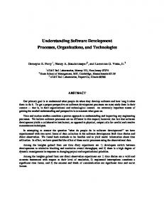
Naming accuracy data for each of the two tasks performed inside the MSI scanner were submitted to an ANOVA with task (letter sound, pseudoword) and grade (Kindergarten, Grade 1) as the within subjects factors, and group (Low Risk, High Risk-Responders) as a between subjects factor. There were significant differences between groups in the degree of developmental change on decoding accuracy, as indicated by a Group by Grade by Task interaction, F(1,28) = 5.09, p < .032. As shown in Figure 1, there was significant improvement between Kindergarten and Grade 1 on letter sound and pseudoword naming accuracy that was slightly greater in degree for the Low Risk group, t(16) = 4.37, p < .0001, than for the High Risk-Responders, t(12) = 3.97, p < .002. Inspection of Figure 1 reveals that all three High Risk-Non-Responders
Figure 1. Accuracy measures of reading performance during the MSI tasks for the two subgroups of non-reading impaired children in Kindergarten (K) and Grade 1. Individual scores for each of the three High Risk Non-Responders are indicated by different symbols. brain regions listed above were submitted to separate ANOVAs with Task, Hemisphere, and Grade as within subjects factors and Group (High Risk- Responders, Low Risk) as a between subjects factor. As will be described in more detail below, group differences were found in three areas, Tmp, IFG, and Oc-T. In contrast, reliable developmental effects on the number of activity sources were not noted. Figure 2 displays the main hemisphere and group effects for each area.
Early development of reading mechanisms 5 For Tmp the presence of group differences was initially indicated by a Group by Hemisphere interaction, F(1,28) = 14.54, p < .001, suggesting that the left hemisphere predominance was stronger for the Low Risk group, t(16) = 3.42, p < .003, than for the group of High Risk-Responders (p = .06) in both grades. As shown in Figure 2, group differences were more systematic in the right, F(1,28) = 7.03, p < .013 (High Risk Responders > Low Risk children), than in the left hemiKindergarten Low Risk
Responders
60 10
5
rs R es po nd e
Lo w
Ri sk
0
# Activity Sources
# Activity Sources
15
LETTER SOUND TASK
* 40
20
0 Left
Inferior Frontal (L+R)
Right
Left
Right
Temporoparietal
Grade 1 Low Risk
30
Responders
# Activity Sources
# Activity Sources
60 20
10
0 Low Risk
Responders
Occipito-temporal (R)
*
40
PSEUDOWORD TASK 20
0 Left
Right
Left
Right
Temporoparietal
Figure 2. Differences in the relative amount of regional activity between the Low Risk and High Risk-Responders at two time points (end of Kindergarten and end of Grade 1). Given that group differences were similar across tasks data were collapsed. Significant differences, indicated by asterisks, were found in three major components of the reading mechanism: temporoparietal (BA 22/40), occipito-temporal (BA 19/37), and inferior frontal cortices (BA 44/45/6). In Grade 1 group differences persisted in temporoparietal areas for both tasks, but were no longer statistically significant in frontal regions bilaterally (L+R). Notice, however, that Low Risk children displayed greater activity in the right (R) occipito-temporal region than High Risk-Responders, a difference that was not present a year earlier. sphere (p = .07). The proportion of children in the Low Risk group who showed predominantly left hemisphere temporoparietal activity (across tasks) was 71% in Kindergarten and slightly higher in Grade 1 (88%), compared to only 23% in
Figure 3. Schematic representation of spatiotemporal activation profiles from the two subgroups of non-impaired readers during performance of the Letter Sound (left-hand columns) and the more demanding pseudoword reading task (right-hand columns). Three sets or regions are outlined from back to front: occipito-temporal, temporo-parietal (including the angular gyrus), and inferior frontal cortices. Regional differences in onset latency are indicated by different colors within each template. Arrows indicate significant group differences in the temporal profile of regional activity that were present in Kindergarten for the more demanding pseudoword reading task. Low Risk children showed a pattern that is typical of older non-impaired readers, featuring early activity in occipitotemporal regions, followed by activity in temporoparietal regions, and then activity in inferior frontal regions. In contrast, there was little temporal differentiation in the onset latency between occipito-temporal and temporo-parietal regions in the group of High Risk Responders.
6 Kindergarten (39% in Grade 1) of the High Risk Responders (Χ2 [1, N = 30] = 6.90, p < .009 and Χ2 [1, N = 30] = 4.34, p < .035, for Kindergarten and Grade 1, respectively). For Oc-T activity group and hemisphere differences varied with grade and task. There was a significant Group by Task by Hemisphere by Grade interaction, F(1,28) = 6.85, p < .014. Follow up two-way ANOVAs (Group by Grade), performed for each task and hemisphere separately, revealed a two-way interaction for the letter sound task in the left hemisphere, F(1,28) = 4.74 p < .038, and a main effect of Group in the right hemisphere, F(1,28) = 4.55, p < .042. As a group, Low Risk children showed greater Oc-T activity in the right hemisphere during performance of the letter sound task than High Risk Responders. A marginally significant trend in the same direction (Low Risk > High Risk Responders) was noted in the right hemisphere for the pseudoword task (p = .024; nonsignificant trends were found in the left hemisphere for both tasks). Finally, a Group by Grade by Hemisphere interaction was found for the number of activity sources in IFG, F(1,28) = 4.22, p < .05, which was mainly due to a significant Grade by Group interaction in the left hemisphere (regardless of task), F(1,28) = 5.18, p = .031. Follow up comparisons revealed that an overall tendency for greater IFG activity for the High Risk Responders, as compared to Low Risk children, approached statistical significance only in Kindergarten (p = .05). Overall analysis: Onset latency of activity. The onset latency data yielded a significant Group by Task by Grade by Hemisphere by Area interaction, F (3,84) = 8.92, p < .0001. To follow this interaction we evaluated: (1) group differences within grade, (2) grade differences for each group, and (3) regional differences for each grade and group (reflecting the temporal sequence of activity). Group main effects and interactions. Follow up three-way ANOVAs performed separately for each grade indicated a significant three-way interaction, F(3,84) = 3.0, p < .035, for Kindergarten. Further analyses focused on group differences at each of the levels of the other independent variables and on main effects and interactions between task, hemisphere, and area, separately for each group. The former effects were not systematic, as the only difference between groups that approached significance was a tendency for the Low Risk group to show earlier onset latencies for activity in the left occipitotemporal region during the Pseudoword reading task (p = .05). The patterns of task-related variations in onset latency were, however, noticeably different across groups. Thus, task differences in the left Tmp region (later onset in response to pseudowords than letters) were only found in the Low Risk group, t(16) = 3.3, p < .005. A similar trend in IFG was apparent only for High Risk-Responders, bilaterally (LH: t[12] = 2.6, p < .023, RH: t[12] = 2.9, p < .014). Grade main effects and interactions. Follow up ANOVAs revealed a significant Grade by Group interaction in the occipito-temporal region, F(1,28) = 15.99, p < .0004. Pairwise comparisons indicated a significant reduction in onset latency in both hemispheres, regardless of task, for the group of High Risk-Responders, t(12) = 4.80, p < .0004. The mean onset latency for the group of High Risk Responders was 320±24 ms in Kindergarten and 210±14 ms in Grade 1. Corresponding
values for the group of Low Risk children were 245±17 and 234±21 ms. A significant interaction involving grade was also found for the onset of activity in IFG (Group by Task by Hemisphere by Grade), F(1,28) = 15.26, p < .0001, which was due to a Group by Grade interaction in the left IFG during performance of the letter sound task, F(1,28) = 7.68, p < .001. Follow up tests indicated that the onset latency in this region actually increased in Grade 1 for the group of High Risk Responders (from 603±50 ms to 786±40 ms), t(12) = 2.98, p < .01. Regional differences. Significant Area by Hemisphere effects were found for both groups, grades, and tasks (p < .0001
Figure 4. Sources of magnetic activity projected on the lateral surface of the brain for three representative participants during performance of the pseudoword reading task in Grade 1.in all cases). Regional differences in onset latency of activity were further assessed using pairwise comparisons (evaluated at α = .001). These tests failed to reveal hemisphere differences between homotopic regions (e.g., earlier onset in the left Tmp compared to the right Tmp). However, several differences were found between areas within the same hemisphere (e.g., earlier onset in the left Oct-T as compared to the left Tmp region), and interhemispherically for different regions (e.g., earlier onset in the left Tmp region as compared to the right IFG). Differences in onset latency between areas that belong to the three major reading circuits outlined in the Introduction are summarized in Figure 3. In general, there was a distinct progression of activity from ventrolateral occipito-temporal to temporoparietal and, finally, to inferior frontal areas across tasks and grades and for both groups, with one exception: dur-
Early development of reading mechanisms 7 ing performance of the pseudoword task in Kindergarten, there was significant temporal overlap in onset latency between occipito-temporal and temporoparietal regions for the group of High Risk Responders. The range of onset latencies (95% confidence intervals) was: 180-328 ms for occipito-temporal activity, 187-591 ms for temporo-parietal activity, and 580-825 for inferior frontal activity. Corresponding values for the Low Risk group were: 150-210, 288-536, and 612-727 ms, showing little or no overlap in the onset latency of activity between occipito-temporal and temporo-parietal regions. To summarize the data presented above, there were conspicuous differences between the two subgroups of non impaired readers. In general, activation profiles were similar for both groups of children when retested a year later, with the exception of the profiles associated with the more demanding pseudoword reading task for the group of High Risk Responders. The typical spatiotemporal profile of a Low Risk reader featured early activation of occipito-temporal cortices in the left hemisphere (including both ventral and lateral extrastriate areas), followed by activity in homotopic areas of the right hemisphere. Next activation was noted in temporo-parietal regions bilaterally. Finally, activity progressed to inferior frontal cortices in both hemispheres. With the exception of Tmp, where there was predominantly left hemisphere activation, activity was bilaterally symmetric in all other areas. The typical activation profile of a High Risk Responder was temporally similar to that of Low Risk readers in Grade 1 but substantially different in Kindergarten. It involved early activity in occipito-temporal areas bilaterally, followed by right Tmp activity. This was, in turn, followed by activity in the left Tmp, right IFG and, finally, the left IFG. In contrast to the Low Risk children, the degree of activity was bilaterally symmetric in all areas (including temporoparietal cortex). On average, High Risk Responders showed greater activation in the right temporo-parietal and inferior frontal regions than children in the Low Risk group. In contrast, children in the Low Risk group showed greater activity in occipito-temporal (predominantly in the right hemisphere) and left temporoparietal regions (although the latter did not reach statistical significance). The relative timing of regional activity was similar for each of the three High Risk Non-Responders as compared to the group of High Risk Responders. However, hemispheric asymmetries in the degree of Tmp activity were notably different, featuring weaker left and even stronger right hemisphere activity. As shown in Figure 3, the spatio-temporal profile of activity in each of the three High Risk-NonResponders at the end of Kindergarten already closely resembled the profile found in older children with significant reading disabilities (Simos et al., 2000a, b, 2002b). Inspection of individual activation profiles did not reveal systematic changes in the degree or onset of activity during the year-long interval between the two scans.
Discussion The findings of the present study pertain to two main issues in the study of brain mechanisms that support reading: (1) the normal individual variability in reading skill during the early
stages of reading acquisition and (2) changes in regional brain activity that accompany the acquisition of reading skills.
Individual differences in brain activity in beginning readers In a previous study of Kindergarten children, we reported that those who were screened as being at risk for developing reading problems had brain activation profiles on a lettersound pronunciation task that were different from those of low risk children (Simos et al., 2002a). However, group differences may have been obscured by the presence of children who represented false negative or false positive screening errors. On the basis of comprehensive achievement testing a year later, we were able to identify children who did not respond to instruction and could be classified as reading disabled using standard criteria. The activation profiles of the three children who met these criteria showed all the characteristic features previously reported by our group and others (Shaywitz et al., 2002; Simos et al., 2000a,b), namely hypoactivation of the left temporoparietal region and increased activity in the homotopic right hemisphere region. They also displayed pronounced activation of inferior frontal cortex bilaterally. While initial differences in the profile of activity between the group of High Risk children, who made significant gains in reading skills during Grade 1 (Responders), and the group of Low Risk children, were still noticeable in the present study, they were far more limited than previously estimated. Although children in the High Risk-Responder group did not show the expected (leftward) hemispheric asymmetries in the degree of temporoparietal activity, this was primarily due to increased activity in the right hemisphere, rather than left temporoparietal hypoactivation. The precise role of the right temporoparietal region in the brain circuit involved in reading, which persisted through Grade 1, can only be speculated upon at present. First, it is possible that right hemisphere activity is triggered by transcallosal input from the contralateral hemisphere, and therefore does little to satisfy task demands. Given that the onset of activity in the right temporoparietal region slightly precedes the onset of activity in the homotopic left hemisphere region, this explanation is unlikely. Alternatively, activity in the right temporoparietal region may be an integral component of the brain mechanism that supports decoding in this population of readers. This activity may reflect engagement of neurophysiological processes specialized for the phonological analysis of print, a function for which the left temporoparietal region is necessary, at least in older proficient readers (Simos et al., 2000c; 2002c). If that were the case, increased activity in the right temporoparietal region should be also apparent in the context of auditory, phonological processing tasks. A third possibility is that increased right hemisphere activity may reflect the engagement of neurophysiological processes serving a fundamentally different component process, such as visual-configural analysis of the printed stimuli. Recent findings utilizing fMRI on a pseudoword reading task with adults, who showed some evidence for compensation of childhood reading deficits, also indicated reliance on compensatory systems in the right hemisphere (Shaywitz et al., 2003). Similarly, Shaywitz et al. (2003) found that compensated poor
8 readers were engaging left temporoparietal areas to a lesser degree than adults who never experienced reading difficulties. As the compensated adults did not read as well as the nonimpaired readers, the results imply that increased right hemisphere activity does not fully support proficient reading. High Risk children also showed increased inferior frontal activation during reading in Kindergarten, yet by the end of Grade 1 these group differences diminished in those who made significant progress in the development of reading skills (High Risk-Responders). It is commonly held that inferior frontal activation in reading tasks reflects the engagement of articulatory recoding processes serving a compensatory role in the case of non-fluent readers (Shaywitz et al., 2002, 2003). The present data also suggest that the two most commonly reported compensatory mechanisms in children who are at risk for developing reading problems have distinct developmental trajectories and can become uncoupled during the course of the early stages of reading acquisition. Although frontal lobe hyperactivity subsided by the end of Grade 1, increased right hemisphere temporoparietal activity persisted through Grade 1 in the group of High Risk- Responders. As these children were younger than those usually studied in functional neuroimaging studies of children, additional follow-up of this and similar cohorts is clearly warranted.
Emergence of the brain mechanism for reading Generally, the brain mechanism that supports phonological decoding appeared to be rather stable during the initial stages of reading acquisition. There were no apparent changes in the degree (or total duration) of activity in regions that constitute major components of the reading circuit for either group of non-impaired readers. The most notable developmental changes noted in the present study were those concerning the temporal features of the brain mechanism for reading. Significant changes were found in (a) the onset latency of activity in occipito-temporal and inferior frontal areas, and (b) the relative timing of regional activity for the group of High Risk Responders. One of these changes, the reduction in the onset latency of activity in occipito-temporal regions, can probably be viewed as reflecting increased “efficiency” of the ventral component of the reading mechanism in the group of High Risk Responders. On average, onset of activity in this region was delayed in this group in Kindergarten, but was similar across groups in Grade 1. This finding complements reports of reduced hemodynamic responsiveness of visual association cortices during reading tasks in children with persistent reading difficulties (Temple et al., 2001). In the same group, onset latency in inferior frontal cortices increased in Grade 1. Assuming that engagement of this region plays a compensatory role within the reading mechanism, this change could reflect the declining role of neurophysiological processes hosted by inferior frontal cortices. This change is consistent with reports of unusually early frontal activity in adults with persisting reading disability (Salmelin, Service, Kiesila, Uutela, & Salonen, 1996). In nonimpaired readers the inferior frontal region presumably hosts neurophysiological processes involved in articulatory recoding of print. If this is the case in poor readers, then enhanced engagement of this region (in both degree and latency) may indi-
cate increased reliance upon recoding strategies in order to assemble phonological representations of unfamiliar stimuli. A third notable developmental change in the temporal profile of regional activity involved the relative timing of activity in occipito-temporal and temporoparietal areas for the High Risk Responders. The presence of significant temporal overlap in the onset latency of occipito-temporal and temporoparietal areas in Kindergarten resolved by Grade 1. At that time the pattern of temporally distinct activations in these regions emerged, which characterizes Low Risk readers in the present study, and older proficient readers in previous studies (Simos et al., 2001). This change among High Risk Responders was probably due to: (a) a trend for reduced onset latency in occipito-temporal regions, and (b) reduced individual variance in the onset latency of both occipito-temporal and temporoparietal regions. There was also a trend for earlier onset of activity in the right temporoparietal region as compared to the homotopic region in the left hemisphere that was all but eliminated in Grade 1. Notably, developmental changes in the relative timing of regional activation for the High Risk Responders were observed only during performance of the pseudoword reading task, which places significant demands for phonological decoding. Therefore, the observed changes appear to be mainly functional, depending on task demands, and not reflecting generalized alterations in the brain mechanism for reading, for instance the recruitment of a brain region that is not normally involved in the brain mechanism for reading. Hemispheric asymmetries for the degree of activity in extrastriate areas were not apparent, similar to findings for 8-12 year-old non-impaired readers (Simos et al., 2001). Given that MSI data from studies with proficient, adult readers reveal clear left hemisphere predominance in the degree of occipitotemporal activity during a variety of reading tasks (Breier, Simos, Zouridakis, & Papanicolaou, 1998, 1999b; Simos et al., 2001), it may be that these asymmetries develop sometime later in childhood. It is possible that increasing hemispheric asymmetries in the degree of activity and progressively earlier onset latencies imply increased specialization of this region for task-specific neurophysiological processes. Assuming that this is the case it follows that specialization of the left occipitotemporal region develops relatively late in the course of reading acquisition. While hemispheric asymmetries in inferior frontal activity were also lacking, predominant left hemisphere activity was noted in temporoparietal areas, a finding limited to children who had shown early mastery of essential prereading skills (Low Risk group). In contrast, children who had not shown evidence of mastery of these skills in Kindergarten (High Risk Responders) displayed evidence of increased activation of the temporoparietal cortex in the right hemisphere. Closer examination of individual brain activation profiles revealed that 88% of Low Risk children showed left hemisphere predominance in the degree of temporoparietal activity during performance of reading tasks in Grade 1. This estimate is in agreement with the rate of left-hemisphere lateralized profiles of activity (87%), obtained in the context of a reading comprehension task from a group of 7 year old children using fMRI (Gaillard, Balsamo, Ibrahim, Sachs, & Xu, 2003). All children in that study had standard scores over 99 on the Woodcock-Johnson
Early development of reading mechanisms 9 Reading Composite (with a mean of 125) and were therefore comparable, in terms of basic reading abilities, to the Low Risk children in the present study (see Table 1). Although the percentage of children demonstrating this profile in the present study was significantly lower among children in the High Risk Responder group, these children still scored within the normal range on standardized reading achievement tests in Grade 1. Finally, a comment regarding the significance of hemispheric asymmetries in temporoparietal activity during performance of reading tasks is in order here. During the short developmental period covered by the present study (end of Kindergarten to end of Grade 1) there were only slight changes in this feature regardless of risk status. Specifically, 1
NI
Laterality Index
0.5 Low Risk
0
High Risk
RD
-0.5
-1 Kindergarden
Grade 1
Grades 2-4
the brain mechanism for reading may be facilitated by enhanced inhibitory signals from the left temporoparietal region onto the right homologous region. This process may in term become facilitated by the continuing development of the corpus callosum (Hellige, Taylor, Lesmes, & Peterson, 1998). Longitudinal data collected over a longer period of time are required to assess individual differences in the developmental trajectory of hemispheric asymmetries in temporoparietal activity. At present we can only suggest that symmetric activation, to the extent that it indicates only weak specialization of the left temporoparietal region for phonological decoding processes, is not a predictor of later reading failure. It is tempting to speculate that reliance on the right temporoparietal and left inferior frontal cortices reflects a normal, yet relatively uncommon variation of development. Provided that language-critical areas in temporoparietal regions of the dominant hemisphere (Wernicke’s area) are fully functional when the process of reading acquisition begins, and can readily assume a key role in phonological decoding functions, then any "interference" induced by the engagement of these right hemisphere/anterior circuit areas does not lead to poor reading outcomes. When Wernicke's area is prevented (for reasons as yet unknown) from becoming an integral component of the brain mechanism that emerges to support reading (as in the case of the three High Risk Non-Responders), then persistent difficulty with the acquisition of reading skills is the most likely outcome.
References Figure 5. Longitudinal (Kindergarten to Grade 1) and crosssectional data (Grades 2-4) regarding changes in the degree of hemisphere asymmetry in temporoparietal cortex activity during the early stages of reading acquisition and later. The vertical axis displays values on a laterality index ([LH-RH] / [LH+RH]) for the number of activity sources in the posterior portion of the superior temporal gyrus and in the adjacent anterior portion of the supramarginal gyrus (BA 22/40). Positive values indicate greater left than right hemisphere activity. Data were first normalized with respect to total brain activity, to enable comparisons across groups. Brain activity measures were obtained in the context of pseudoword reading tasks (three letter tokens were used in the Kindergarten-Grade 1 longitudinal study and 4-6 letter pseudowords with the older children). Data for older non-impaired (NI) and reading disabled children (RD) children were adapted from Simos et al., 2000b and Papanicolaou et al., 2003. children in the Low Risk group continued to show a significant left hemisphere predominance in the duration of temporoparietal activity, while High Risk Responders continued to show a slight (marginally significant) right hemisphere predominance. Cross-sectional data obtained during an equally demanding pseudoword reading task from older readers (Simos et al., 2000b), indicate a further increase in the relative degree of left temporoparietal activation accompanied with a steep reduction of activity in the homotopic right hemisphere region (see Figure 5). In addition to practice- and skilldependent increases in the functional specialization of the left temporoparietal region, the predominance of this region within
Blachman, B.A. (1997). Early intervention and phonological awareness: A cautionary tale. In: B.A. Blachman (Ed.), Foundations of Reading Acquisition and Dyslexia: Implications for Early Intervention. Mahwah, NJ: Lawrence Erlbaum Associates, pp. 409-430. Black, S.E., & Behrmann, M. (1994). Localization in alexia. In: A. Kertesz (Ed.), Localization and neuroimaging in neuropsychology. New York: Academic Press, pp. 331-376. Breier, J.I., Simos, P.G., Zouridakis, G., & Papanicolaou, A.C. (1998). Relative timing of cortical activation during a word recognition task. Journal of Clinical and Experimental Neuropsychology, 20, 782-790. Breier, J.I., Simos, P.G., Papanicolaou, A.C., Zouridakis, G., Wilmore, L.J., Wheless, J.W., Constantinou, J.C., & Maggio, W.W. (1999a). Language dominance determined by magnetic source imaging: a comparison with the Wada procedure. Neurology, 53, 938-945. Breier, J.I., Simos, P.G., Zouridakis, G., & Papanicolaou, A.C. (1999b). Temporal course of regional activation associated with phonological decoding. Journal of Clinical and Experimental Neuropsychology, 21, 465-476. Breier, J.I., Simos, P.G., Wheless, J.W., Constantinou, J.E.C., & Papanicolaou, A.C. (2001). Hemispheric language dominance in children determined by magnetic source imaging. Journal of Child Neurology 16, 124-130. Castillo, E.M., Simos, P.G., Venkataraman, V., Breier, J.I., & Wheless, J.W., & Papanicolaou, A.C. (2001). Mapping of expressive language cortex using Magnetic Source Imaging. Neurocase, 7, 419-422.
10 Clay, M.M. An Observation Survey of Early Literacy Achievement. Heinemann, 2002. Damasio, A.R. & Damasio, H. (1983). The anatomic basis of pure alexia. Neurology, 33, 1573-1583. Eden, G.F. & Zeffiro, T.A. (1998). Neural systems affected in developmental dyslexia revealed by functional neuroimaging. Neuron, 21, 279-282. Eden, G.F., VanMeter, J.W., Rumsey, J.M., & Zeffiro, T.A. (1996). The visual deficit theory of developmental dyslexia. NeuroImage, 4, 108-117. Fiez, J.A. & Petersen, S.E. (1998). Neuroimaging studies of word reading. Proceedings of the National Academy Science (USA), 95, 914-921. Filipek, P. (1996). Structural variations in measures in the developmental disorders. In R. Thatcher, G. Lyon, J. Rumsey, N. Krasnegor (Eds.), Developmental Neuroimaging: Mapping the Development of Brain and Behavior (pp. 169-186). San Diego, CA: Academic Press. Gaillard W.D., Balsamo L.M., Ibrahim Z., Sachs B.C., & Xu, B. (2003). fMRI identifies regional specialization of neural networks for reading in young children. Neurology, 60, 94100. Hellige, J.B., Taylor, K.B., Lesmes, L., & Peterson, S. (1998). Relationships between brain morphology and behavioral measures of hemispheric asymmetry and interhemispheric interaction. Brain & Cognition, 36, 158-92. Henderson, V. (1986). Anatomy of posterior pathways in reading: A reassessment. Brain and Language, 29, 119-133. Hillebrand, A. & Barnes, G.R. (2002). A quantitative assessment of the sensitivity of whole-head MEG to activity in the adult human cortex. Neuroimage, 16, 638-50. Horwitz, B., Rumsey, J.M., & Donohue, B.C. (1998). Functional connectivity of the angular gyrus in normal reading and dyslexia. Proceedings of the National Academy of Science (USA), 95, 8939-44. Jenner, A.R., Rosen, G.D., & Galaburda, A.M. (1999). Neuronal asymmetries in primary visual cortex of dyslexic and nondyslexic brains. Annals of Neurology, 46, 189-96. Klingberg, T., Hedehus, M., Temple, E., Salz, T., Gabrieli, J.D., Moseley, M.E., & Poldrack, R.A. (2000). Microstructure of temporo-parietal white matter as a basis for reading ability: evidence from diffusion tensor magnetic resonance imaging. Neuron, 25, 493-500. Liberman, A.M. (1998). Why is speech so much easier than reading? In C. Hulme & R.M. Malatesha (Eds.), Reading and Spelling: Development and Disorders. Mahwah, NJ: Lawrence Erlbaum Associates. Logan, .G. (1997). Automaticity and reading: perspectives from the instance theory of automatization. Reading and Writing Quarterly: Overcoming Learning Disabilities, 13, 123-146. Lukatela, G. & Turvey, M.T. (1998). Reading in two alphabets. American Psychologist, 53, 1057-1072. Lyon, G.R., Fletcher, J.M., Shaywitz, S.E., Shaywitz, B.A., Torgesen, J.K., Wood, F.B., Schulte, A., & Olson, R. (2001). Rethinking learning disabilities. In C.E. Finn, Jr., RA.J. Rotherham, & C.R. Hokanson, Jr. (Eds.) Rethinking special education for a new century (pp. 259-287). Wash-
ington, DC: Thomas B. Fordham Foundation and Progressive Policy Institute. Maestú, F., Ortiz, T., Fernández, A., Amo, C., Martin, P., & Fernandez, S. (2002). Spanish language mapping using MEG: A validation study. Neuroimage, 17, 1579-1586. Mathes, P.G., Denton, C.A., Fletcher, J.M., Anthony, J.L., Francis, D.J., & Schatschneider, C. (in press). An evaluation of two reading interventions derived from diverse models. Reading Research Quarterly. National Reading Panel (2000). Report of the National Reading Panel: Teaching students to read: An evidence-based assessment of the scientific research literature on reading and its implications for reading instruction. Bethesda, MD: National Institute of Child Health and Human Development. Oldfield, R.C. (1971). The assessment and analysis of handedness: the Edinburgh inventory. Neuropsychologia, 9, 97113. Papanicolaou, A.C., Simos, P.G., Breier, J.I., Zouridakis, G., Wilmore, L.J., Wheless, J.W., Constantinou, J.C., Gormley, W., & Maggio, W.W. (1999). Magnetoencephalographic mapping of the language-specific cortex. Journal of Neurosurgery, 90, 85-93. Papanicolaou, A.C., Simos, P.G., Breier, J.I., Fletcher, J.M., Foorman, B.R., Francis, D.J., Castillo, E.M., & Davis, R. (2003). Brain mechanisms for reading in children with and without dyslexia: A review of studies of normal development and plasticity. Developmental Neuropsychology, 24, 593-612. Papanicolaou, A.C., Simos, P.G., Castillo, E.M., Breier, J.I., Sarkari, S., Pataraia, E., Billingsley, R.L., Wheless, J.W., Maggio, V., & Maggio, W.W. (2004). Magnetoencephalography: A non-invasive alternative to the Wada procedure. Journal of Neurosurgery, 100, 867-76. Pugh, K.R., Mencl, W.E., Jenner, A.R., Katz, L., Frost, S.J., Lee, J.R., Shaywitz, S.E., & Shaywitz, B.A.. (2000). Functional neuroimaging studies of reading and reading disability (developmental dyslexia). Mental Retardation and Developmental Disabilities Research Reviews, 6, 207-213. Rumsey, J.M., Andreason, P., Zametkin, A.J., Aquino, T., King, A.C., Hamburger, S.D., Pikus, A., Rapoport, J.L., & Cohen, R.M. (1992). Failure to activate the left temporoparietal cortex in dyslexia. An oxygen-15 positron emission tomographic study. Archives of Neurology, 49, 527-534. Rumsey, J.M., Nace, K., Donahue, B., Wise, D., Maisog, J.G., & Andreason, P. (1997). A positron emission tomographic study of impaired word recognition and phonological processing in dyslexic men. Archives of Neurology, 54, 562-573. Salmelin, R., Service, E., Kiesila, P., Uutela, K., & Salonen, O. (1996). Impaired visual word processing in dyslexia revealed with magnetoencephalography. Annals of Neurology, 40, 157-62. Sarkari, S., Simos, P.G., Fletcher, J.M., Castillo, E.M., Breier, J.I., & Papanicolaou, A.C. (2002). The emergence and treatment of developmental reading disability: Contributions of functional brain imaging. Seminars in Pediatric Neurology, 9, 227-236. Sarvas, J. (1987). Basic mathematical and electromagnetic concepts of the biomagnetic inverse problem. Physics in Medicine and Biology 32, 11-22.
Early development of reading mechanisms 11 Schatschneider, C., Francis, D.J., Foorman, B.F., Fletcher, J.M., & Mehta, P. (1999). The dimensionality of phonological awareness: An application of item response theory. Journal of Educational Psychology, 91, 467-478. Shaywitz, S.E., Shaywitz, B.A., Pugh, K.R., Fulbright, R.K., Constable, R.T., Mencl, W.E., Shankweiler, D.P., Liberman, A.M., Skudlarski, P., Fletcher, J.M., Katz, L., Marchione, K.E., Lacadie, C., Gatenby, C., & Gore, J.C. (1998). Functional disruption in the organization of the brain for reading in dyslexia. Proceedings of the National Academy of Science, 95, 2636-2641. Shaywitz, B., Shaywitz, S., Pugh, K.R., Mencl, W.E., Fulbright, R.K., Skudlarski, P., Constable, R.T., Marchione, K.E., Fletcher, J.M., Lyon, G.R., & Gore, J.C. (2002). Disruption of posterior brain systems in children with developmental dyslexia. Biological Psychiatry, 52, 101-110. Shaywitz, S.E., Shaywitz, B.A., Fullbright, R.K., Skudlarski, P., Mencl, W.E., Constable, R.T., Constable,, R.T., Pugh, K.R., Holahan, J.M., Marchione, K.E., Fletcher, J.M., Lyon, G.R., & Gore, J.C. (2003). Neural systems for compensation and persistence: Young adult outcome of childhood reading disability. Biological Psychiatry 54, 25-33. Simos, P.G., Papanicolaou, A.C., Breier, J.I., Wheless, J.W., Constantinou, J.E.C., Gormley, W.B., & Maggio, W.W. (1999). Localization of language-specific cortex by using magnetic source imaging and electrical stimulation mapping. Journal of Neurosurgery, 91, 787-796. Simos, P.G., Breier, J.I., Fletcher, J.M., Bergman, E., & Papanicolaou, A.C. (2000a). Cerebral mechanisms involved in word reading in dyslexic children: A Magnetic Source Imaging approach. Cerebral Cortex, 10, 809-816. Simos, P.G., Papanicolaou, A.C., Breier, J.I., Fletcher, J.M., Foorman, B.R., Bergman, E., Fishbeck, K., & Papanicolaou, A.C. (2000b). Brain activation profiles in dyslexic children during nonword reading: A magnetic source imaging study. Neuroscience Letters, 290, 61-65. Simos, P.G., Breier, J.I., Wheless, J.W., Maggio, W.W., Fletcher, J.M., Castillo, E.M., & Papanicolaou, A.C. (2000c). Brain mechanisms for reading: The role of the superior temporal gyrus in word and pseudoword naming. NeuroReport, 11, 2443-2447. Simos, P.G., Breier, J.I., Fletcher, J.M., Foorman, B.R., Mouzaki, A., & Papanicolaou, A.C. (2001). Age-related changes in regional brain activation during phonological decoding and printed word recognition. Developmental Neuropsychology, 19, 191-210. Simos, P.G., Fletcher, J.M., Foorman, B.R., Francis, D.J., Castillo, E.M., Davis, R.N., Fitzgerald, M., & Papanicolaou, A.C. (2002a). Brain activation profiles during the early stages of reading acquisition. Journal of Child Neurology, 17, 159-163. Simos, P.G., Fletcher, J.M., Bergman, E., Breier, J.I., Foorman, B.R., Castillo, E.M., Fitzgerald, M., & Papanicolaou, A.C. (2002b). Dyslexia-specific brain activation profile becomes normal following successful remedial training. Neurology, 58, 1203-13. Simos, P.G., Breier, J.I., Fletcher, J.M., Foorman, B.R., Castillo, E.M., & Papanicolaou, A.C. (2002c). Brain mechanisms
for reading words and pseudowords: An integrated approach. Cerebral Cortex, 12, 297-305. Snow, C.E., Burns, M.S., & Griffin, P. (1998). Preventing reading difficulties in young children. Washington, DC: National Academy Press. Szymanski, M.D., Perry, D.W., Gage, N.M., Rowley, H.A., Walker, J., Berger, M.S., & Roberts, T.P. (2001). Magnetic source imaging of late evoked field responses to vowels: toward an assessment of hemispheric dominance for language. Journal of Neurosurgery, 94, 445-53. Temple, E., Poldrack, R.A., Salidis, J., Deutsch, G.K., Tallal, P., Merzenich, M.M., & Gabrieli, J.D. (2001). Disrupted neural responses to phonological and orthographic processing in dyslexic children: an fMRI study. Neuroreport, 12, 299-307. Torgesen, J.K. (2000). Individual differences in response to early interventions in reading: The lingering problem of treatment resisters. Learning Disabilities Research and Practice, 15, 55-64 Torgesen, J.K., Wagner, R.K., & Rashotte, C.A. (1999). Test of Word Reading Efficiency. Austin TX: Pro- Ed. Vellutino, F.F., Fletcher, J.M., Scanlon, D.M., & Snowling, M.J. (2004). Specific reading disability (dyslexia): What have we learned in the past four decades? Journal of Child Psychiatry and Psychology, 45, 2-40. Wechsler, D. (1999). Wechsler Abbreviated Scale of Intelligence (WASI). San Antonio, TX: The Psychological Corporation. Woodcock, R.W., McGrew, K.S., & Mather, N. (2001). Woodcock-Johnson III Tests of Achievement. Itasca, IL: Riverside Publishing.
Neuropsychology. 2005 Nov;19(6):787-98
Table 1 Sample demographics and psychoeducational profiles. High Risk-Responders (n = 13)
Low Risk (n = 17) Mean (SD) Gender (female/male) Age: Kindergarten st
1 grade)
Range
Mean (SD)
6/11
Range
High Risk-Non-Responders (n = 3) Mean (SD)
6/7
6.3
5.6-6.5
Range
1/2
6.5
6 -7.2
6.4
6.1-6.5
7.2
6.4-7.5
7.6
7.2-8.1
7.1
7-7.3
WASI
102 (17)
78-130
92 (16)
70-130
95
71-119
WJ-Basic Reading Composite
117 (10)
103-142
105 (9)
90-120
80 (9)
73-89
WJ-Passage Comprehension
114 (8)
102-129
108 (9)
98-130
88 (10)
76-95
TOWRE
109 (12)
96-144
98 (7)
89-112
78 (8)
68-89
Note. WASI = Wechsler Abbreviated Scale of Intelligence; WJ = Woodcock Johnson PsychoEducational Test Battery III; TOWRE = Test of Word Reading Efficiency.