4 HeartID: Method and Application Frameworks. 67. 4.1 Problem ..... F. Agrafioti, J
. Gao, D. Hatzinakos, Heart Biometrics: Theory, Methods and Ap- plications, in ...
ECG in Biometric Recognition: Time Dependency and Application Challenges
by
Foteini Agrafioti
A thesis submitted in conformity with the requirements for the degree of Doctor of Philosophy Graduate Department of Electrical and Computer Engineering University of Toronto
c 2011 by Foteini Agrafioti Copyright
Abstract ECG in Biometric Recognition: Time Dependency and Application Challenges Foteini Agrafioti Doctor of Philosophy Graduate Department of Electrical and Computer Engineering University of Toronto 2011 As biometric recognition becomes increasingly popular, the fear of circumvention, obfuscation and replay attacks is a rising concern. Traditional biometric modalities such as the face, the fingerprint or the iris are vulnerable to such attacks, which defeats the purpose of biometric recognition, namely to employ physiological characteristics for secure identity recognition. This thesis advocates the use the electrocardiogram (ECG) signal for human identity recognition. The ECG is a vital signal of the human body, and as such, it naturally provides liveness detection, robustness to attacks, universality and permanence. In addition, ECG inherently satisfies uniqueness requirements, because the morphology of the signal is highly dependent on the particular anatomical and geometrical characteristics of the myocardium in the heart. However, the ECG is a continuous signal, and this presents a great challenge to biometric recognition. With this modality, instantaneous variability is expected even within recordings of the same individual due to a variety of factors, including recording noise, or physical and psychological activity. While the noise and heart rate variations due to physical exercise can be addressed with appropriate feature extraction, the effects of emotional activity on the ECG signal are more obscure. This thesis deals with this problem from an affective computing point of view. First, ii
the psychological conditions that affect the ECG and endanger biometric accuracy are identified. Experimental setups that are targeted to provoke active and passive arousal as well as positive and negative valence are presented. The empirical mode decomposition (EMD) is used as the basis for the detection of emotional patterns, after adapting the algorithm to the particular needs of the ECG signal. Instantaneous frequency and oscillation features are used for state classification in various clustering setups. The result of this analysis is the designation of psychological states which affect the ECG signal to an extent that biometric matching may not be feasible. An updating methodology is proposed to address this problem, wherein the signal is monitored for instantaneous changes that require the design of a new template. Furthermore, this thesis presents the enhanced Autocorrelation- Linear Discriminant Analysis (AC/LDA) algorithm for feature extraction, which incorporates a signal quality assessment module based on the periodicity transform. Three deployment scenarios are considered namely a) small-scale recognition systems, b) large-scale recognition systems and c) recognition in distributed systems. The enhanced AC/LDA algorithm is adapted to each setting, and the advantages and disadvantages of each scenario are discussed. Overall, this thesis attempts to provide the necessary algorithmic and practical framework for the real-life deployment of the ECG signal in biometric recognition.
iii
Acknowledgements First and foremost, I would like to sincerely thank my advisor Prof. Dimitrios Hatzinakos. He gave me guidance and direction that was needed to produce this work. Without his generous help, this research would not have been possible. I would also like to thank the Department of Electrical and Computer Engineering for their financial support. Thank you to my proposal and defence committees for taking the time to provide useful insight. I am also grateful to the Communications Group faculty members for teaching, offering technical advice and inspiration in the beginning of this thesis work. Financial support for this thesis was provided by the Natural Sciences and Engineering Research Council (NSERC) of Canada. Last but not least, I would like to thank my family and friends for their constant encouragement during my studies.
iv
Contents Abstract
ii
List of Acronyms
viii
List of Tables
xi
List of Figures
xii
1 Introduction
1
1.1
Introduction to Biometric Recognition . . . . . . . . . . . . . . . . . . .
1
1.2
ECG Biometrics: Motivation and Challenges . . . . . . . . . . . . . . . .
4
1.3
Research Goals and Contributions . . . . . . . . . . . . . . . . . . . . . .
7
1.4
Publications and Patents . . . . . . . . . . . . . . . . . . . . . . . . . . .
9
1.5
Thesis Outline . . . . . . . . . . . . . . . . . . . . . . . . . . . . . . . . .
12
2 Background and Prior Art
13
2.1
Taxonomy of Errors in Biometric Recognition . . . . . . . . . . . . . . .
13
2.2
Electrocardiogram Fundamentals . . . . . . . . . . . . . . . . . . . . . .
15
2.2.1
Inter-individual variability . . . . . . . . . . . . . . . . . . . . . .
18
2.2.2
Cardiovascular reactivity to emotion . . . . . . . . . . . . . . . .
19
ECG Biometric Recognition: Literature Survey . . . . . . . . . . . . . .
23
2.3.1
24
2.3
Fiducial Based Approaches . . . . . . . . . . . . . . . . . . . . . . v
2.3.2 2.4
2.5
Fiducial Independent Approaches . . . . . . . . . . . . . . . . . .
28
ECG in Affective Computing: Literature Survey . . . . . . . . . . . . . .
34
2.4.1
Discrete Emotional Models . . . . . . . . . . . . . . . . . . . . . .
35
2.4.2
Affective Dimensional Models . . . . . . . . . . . . . . . . . . . .
49
Chapter Summary . . . . . . . . . . . . . . . . . . . . . . . . . . . . . .
57
3 ECG Databases
58
3.1
Experimental Protocols . . . . . . . . . . . . . . . . . . . . . . . . . . . .
58
3.2
Short-Term Recording Experiments . . . . . . . . . . . . . . . . . . . . .
59
3.3
Long-Term Recording Experiments . . . . . . . . . . . . . . . . . . . . .
61
3.4
Passive Arousal Experiment . . . . . . . . . . . . . . . . . . . . . . . . .
61
3.5
Active Arousal Experiment . . . . . . . . . . . . . . . . . . . . . . . . . .
63
4 HeartID: Method and Application Frameworks
67
4.1
Problem Statement . . . . . . . . . . . . . . . . . . . . . . . . . . . . . .
67
4.2
Pattern Recognition for ECG Biometrics . . . . . . . . . . . . . . . . . .
68
4.2.1
The AC/LDA Algorithm . . . . . . . . . . . . . . . . . . . . . . .
69
4.2.2
Quality Assessment with the Periodicity Transform . . . . . . . .
72
Application Frameworks . . . . . . . . . . . . . . . . . . . . . . . . . . .
77
4.3.1
Scenario A. Small-scale Recognition Environments . . . . . . . . .
78
4.3.2
Scenario B. Large-scale Recognition Environments . . . . . . . . .
80
4.3.3
Scenario C. Security in Distributed Systems . . . . . . . . . . . .
82
Performance Evaluation . . . . . . . . . . . . . . . . . . . . . . . . . . .
86
4.4.1
Quality Assessment Results . . . . . . . . . . . . . . . . . . . . .
86
4.4.2
Training on the Generic Pool . . . . . . . . . . . . . . . . . . . .
89
4.4.3
Personalized Recognition . . . . . . . . . . . . . . . . . . . . . . .
90
4.4.4
Template Destabilization . . . . . . . . . . . . . . . . . . . . . . .
92
Chapter Summary . . . . . . . . . . . . . . . . . . . . . . . . . . . . . .
94
4.3
4.4
4.5
vi
5 Affective Patterns of ECG 5.1
Problem Statement . . . . . . . . . . . . . . . . . . . . . . . . . . . . . . 5.1.1
5.2
5.3
5.4
98 98
Signal Processing for Emotion Detection . . . . . . . . . . . . . . 101
ECG-driven Empirical Mode Decomposition . . . . . . . . . . . . . . . . 102 5.2.1
ECG Synthesis . . . . . . . . . . . . . . . . . . . . . . . . . . . . 103
5.2.2
Signal Decomposition . . . . . . . . . . . . . . . . . . . . . . . . . 106
5.2.3
Feature Extraction . . . . . . . . . . . . . . . . . . . . . . . . . . 110
Performance Evaluation . . . . . . . . . . . . . . . . . . . . . . . . . . . 114 5.3.1
Valence classification . . . . . . . . . . . . . . . . . . . . . . . . . 116
5.3.2
Arousal classification . . . . . . . . . . . . . . . . . . . . . . . . . 118
5.3.3
Active vs passive arousal . . . . . . . . . . . . . . . . . . . . . . . 120
Chapter Summary . . . . . . . . . . . . . . . . . . . . . . . . . . . . . . 121
6 Continuous Authentication
123
6.1
Problem Statement . . . . . . . . . . . . . . . . . . . . . . . . . . . . . . 123
6.2
Template Updating . . . . . . . . . . . . . . . . . . . . . . . . . . . . . . 124
6.3
Performance Evaluation . . . . . . . . . . . . . . . . . . . . . . . . . . . 126
6.4
6.3.1
Effect of Template Updating Frequency on System Performance . 127
6.3.2
Biometric Template Updating on Affect Data . . . . . . . . . . . 130
Chapter Summary . . . . . . . . . . . . . . . . . . . . . . . . . . . . . . 136
7 Conclusion
138
7.1
Thesis Summary . . . . . . . . . . . . . . . . . . . . . . . . . . . . . . . 138
7.2
Future Work . . . . . . . . . . . . . . . . . . . . . . . . . . . . . . . . . . 140 7.2.1
Online State Detection and Prediction. . . . . . . . . . . . . . . . 141
7.2.2
Investigation of Acceptable Waiting Periods . . . . . . . . . . . . 141
7.2.3
Addressing Privacy Concerns. . . . . . . . . . . . . . . . . . . . . 142
7.2.4
Fusion of Medical Biometrics vii
. . . . . . . . . . . . . . . . . . . . 142
A The Empirical Mode Decomposition
144
B Human Emotion Models
148
C Affective Computing Features in Prior Art
150
D Heart Rate Variability
153
E Template Update Results
156
Bibliography
160
viii
List of Acronyms AC
Autocorrelation
ADM
Affective Dimensional Model
ANS
Autonomic Nervous System
AV
Arousal-Valence
BVP
Blood Volume Pressure
CRF
Conditional Random Field
DBNN
Decision Based Neural-Network
DCT
Discrete Cosine Transform
DEM
Discrete Emotional Model
DFA
Discriminant Function Analysis
DTW
Dynamic Time Warping
ECG
Electrocardiogram
EEG
Electroencephalogram
EER
Equal Error Rate
EMD
Empirical Mode Decomposition
EMG
Electromyogram
FAR
False Acceptance Rate
FLDA
Fisher Linear Discriminant Analysis
FP
Fisher Projection
ix
FRR
False Rejection Rate
GMM
Gaussian Mixture Model
GSR
Galvanic Skin Response
HMM
Hidden Markov Model
HR
Heart-Rate
HRV
Heart-Rate Variability
IADS
International Affective Digitized Sounds
IAPS
International Affective Picture System
ICA
Independent Component Analysis
LDA
Linear Discriminant Analysis
MAUI
Multimodal Affect User Interface
MBP
Marquardt Backpropagation
MLP
Multilayer Perceptron
PCA
Principal Component Analysis
PPG
Phonocardiogram
PT
Periodicity Transform
SAM
Self-Assessment Manikin
SFFS
Sequential Floating Forward Search
SIMCA Soft Independent Modelling of Class Analogy SVM
Support Vector Machine
x
List of Tables 2.1
Summary of related to ECG based recognition works . . . . . . . . . . .
32
2.2
Summary of related to ECG based recognition works (Continued) . . . .
33
2.3
Biosignals analyzed in [1]. . . . . . . . . . . . . . . . . . . . . . . . . . .
38
2.4
Typical classification features used in [1]. . . . . . . . . . . . . . . . . . .
40
2.5
Affective computing using biosignals: Comparison Milestones . . . . . . .
53
2.6
Affective computing using biosignals: Comparison Milestones (continued)
54
2.7
Affective computing using biosignals: Comparison Milestones(continued2)
55
3.1
Summary of ECG Databases . . . . . . . . . . . . . . . . . . . . . . . . .
66
4.1
Basic steps of the proposed framework. . . . . . . . . . . . . . . . . . . .
77
4.2
EER in Scenario C for individual subjects in the testing set. The average EER is 10% and the standard deviation 7.13
. . . . . . . . . . . . . . .
91
5.1
List of classification experiments performed. . . . . . . . . . . . . . . . . 115
6.1
Template updating with variable-length durations . . . . . . . . . . . . . 126
6.2
State confidence (q) per burst for subjects 1-21. . . . . . . . . . . . . . . 132
6.3
State confidence (q) per burst for subjects 22-43. . . . . . . . . . . . . . . 133
6.4
Equal error rate for each individual in the active arousal database, after template updating. Mean equal error rate is 3.96% . . . . . . . . . . . . 136
C.1 Features for classification used by Scheirer et al. in [2]. . . . . . . . . . . 150 xi
C.2 Typical features for classification used by Picard et al. in [1]. . . . . . . . 151 C.3 Features for Analysis I classification used by Healey et al. in [3]. . . . . . 152 E.1 Equal error rate for cth = 0.76. Mean equal error rate is 3.77% . . . . . . 157 E.2 Equal error rate for cth = 0.78. Mean equal error rate is 3.08% . . . . . . 157 E.3 Equal error rate for cth = 0.8. Mean equal error rate is 2.92% . . . . . . . 158 E.4 Equal error rate for cth = 0.81. Mean equal error rate is 2.8% . . . . . . . 158 E.5 Equal error rate for cth = 0.83. Mean equal error rate is 2.51% . . . . . . 159 E.6 Equal error rate for cth = 0.88. Mean equal error rate is 2% . . . . . . . . 159
xii
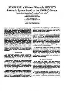
List of Figures 1.1
Heart Beats of the same individual recorded a few years apart. Heart beats have been drawn from the PTB diagnostic database [4] . . . . . . . . . .
2.1
4
Main components of an ECG heart beat. Each wave describes a distinct phase of the cardiac cycle. . . . . . . . . . . . . . . . . . . . . . . . . . .
16
2.2
Configuration of Leads I II and III. . . . . . . . . . . . . . . . . . . . . .
18
2.3
Variability surrounding the QRS complex among heart beats of the same individual. . . . . . . . . . . . . . . . . . . . . . . . . . . . . . . . . . . .
3.1
ECG samples from the short-term recordings database. Every subject was recorded during two sessions. . . . . . . . . . . . . . . . . . . . . . . . . .
3.2
23
60
Data labeling for the passive arousal experiment. Every picture of the IAPS photo-set is assigned to a unique number which indicates the beginning of the respective emotion on the data. . . . . . . . . . . . . . . . . .
63
3.3
Game and face video playback, used for self-assessment of arousal. . . . .
63
3.4
Data labeling for the active arousal experiment. The FEELTRACE is a continuous arousal indication. . . . . . . . . . . . . . . . . . . . . . . . .
4.1
64
Flow diagram of the proposed method. Every input is assigned with a quality measure that contributes to matching. The AC is divided into a number of sections each favored with a predefined weight. The weights WA−D cab be chosen to decrease linearly. . . . . . . . . . . . . . . . . . . xiii
76
4.2
The two stages of an ECG based recognition system in closed environments i.e., cases where the pool of enrollees is known prior to LDA training. . .
79
4.3
The three distinct stages of general access control. . . . . . . . . . . . . .
82
4.4
The three distinct stages of ECG-based recognition in distributed systems. 83
4.5
The enrollment pipeline for the distributed verification framework. . . . .
4.6
Performance of the PT quality factor. AC segments screened with a quality threshold (0.8). . . . . . . . . . . . . . . . . . . . . . . . . . . . . . . . .
4.7
4.9
87
Three principal components of AC for 5 different subjects A) before and B) after quality screening. . . . . . . . . . . . . . . . . . . . . . . . . . .
4.8
84
87
Tradeoff between false acceptance and rejection rates for various decision thresholds. . . . . . . . . . . . . . . . . . . . . . . . . . . . . . . . . . . .
88
ROC plot depicting the performance of Scenario B. The EER is 45%. . .
89
4.10 ROC plot when imposing universal recognition thresholds in Scenario C.
90
4.11 Correlation coefficient values for two different subjects, with five different reference (starting) points. Corresponding coherence durations (illustrated with arrows) are determined with a threshold or tolerance range of 2.3%, with respect to the starting point. . . . . . . . . . . . . . . . . . . . . . .
96
4.12 Maximum and Minimum correlation found within 2-hour recordings for every subject. Correlation is computed between the first 5 second segment and all the following. . . . . . . . . . . . . . . . . . . . . . . . . . . . . .
97
4.13 Verification performance under template destabilization. . . . . . . . . .
97
5.1
2-D trajectory movement and P Q R S, T typical locations. . . . . . . . . 104
5.2
A real and a synthetic ECG. The two signals are synchronized but xS (t) has no anatomical uniqueness or psychological variability. . . . . . . . . . 105
5.3
EMD analysis for a standardized synthetic ECG signal. IMFs of order higher than three do not exhibit oscillatory activity. The first IMF has three oscillatory components, the second has two and the third has one. xiv
107
5.4
BEMD example on a complex ECG signal, formed using a real ECG segment and a synthetic one. Low order IMFs show fast rotating components. 109
5.5
Simultaneous decomposition of real and a synthetic ECG signal using the BEMD. . . . . . . . . . . . . . . . . . . . . . . . . . . . . . . . . . . . . 110
5.6
Comparison of Univariate and driven Bivariate EMD decomposition on the same ECG signal. For the BEMD case the IMFs exhibit less mode mixing as well as the oscillation structure follows the properties of ECG decomposition in the absence of noise i.e., IMF 1 is tricomponent, IMF 2 is bicomponent and IMF 3 is monocomponent.
5.7
. . . . . . . . . . . . . . 111
Local oscillation for a synthetic ECG. A,C,E) First three IMFs of the synthetic signal B,D,F) ρi (t) for the previous IMFs G) Dominant oscillations for the three IMFs. With increasing order of IMF, the strength of the oscillation (time-scale) decreases. . . . . . . . . . . . . . . . . . . . . . . 113
5.8
Per subject classification performance for each of the 32 individuals in the database. While classification among five classes is promising for certain individuals, valence separation is feasible with respect or irrespective of arousal (erotica vs gore or erotica vs disgust) . . . . . . . . . . . . . . . . 116
5.9
Classification performance for all subjects in experiments A-H. . . . . . . 117
5.10 Active arousal detection performance for 42 subjects. . . . . . . . . . . . 119 5.11 Subject specific arousal detection for the two experiments. The average rate for all subjects under passive arousal is 52.41%. Similarly for active arousal the average is 78.43%. . . . . . . . . . . . . . . . . . . . . . . . . 120 6.1
Verification performance for highly correlated training and testing ECG windows. . . . . . . . . . . . . . . . . . . . . . . . . . . . . . . . . . . . . 128
6.2
Verification performance for moderate correlation between the training and testing set.
. . . . . . . . . . . . . . . . . . . . . . . . . . . . . . . . . . 128 xv
6.3
Verification performance for minimum correlation between the training and testing set. . . . . . . . . . . . . . . . . . . . . . . . . . . . . . . . . 129
6.4
Verification performance for the active arousal database, without template updating (EER = 15.1%). . . . . . . . . . . . . . . . . . . . . . . . . . . 134
6.5
Verification performance with template updating for 9 individuals of the active arousal database. . . . . . . . . . . . . . . . . . . . . . . . . . . . 135
A.1 EMD steps on an ECG signal. . . . . . . . . . . . . . . . . . . . . . . . . 145 A.2 EMD analysis for a real ECG segment (5 out of 16 IMFs). . . . . . . . . 147 B.1 The Arousal-Valence (AV) plane. Reproduced from [5]. . . . . . . . . . . 149 D.1 R peak detection for the HRV estimation. . . . . . . . . . . . . . . . . . 154 D.2 R-R time series used for HRV estimation. . . . . . . . . . . . . . . . . . . 154 E.1 Equal error rates of the template updating algorithm for various coherence thresholds cth . As the threshold increases, better coherence is imposed on the bursts which leads to more frequent template updating and subsequently smaller EER. . . . . . . . . . . . . . . . . . . . . . . . . . . . . . 156
xvi
Chapter 1 Introduction 1.1
Introduction to Biometric Recognition
Automatic and accurate identity validation is becoming increasingly critical in several aspects of our every day lives, such as in financial transactions, access control, traveling, healthcare and many others. Traditional strategies for automatic identity recognition include items such as PIN numbers, tokens, passwords and ID cards. Despite the wide deployment of such tactics, the means for authentication is either entity-based or knowledge-based which raises serious concerns with regard to the risk of identity theft. According to the latest US Federal Trade Commission report [6], in 2009 identity theft was the number one complaint category (a total of 721,418 cases of consumer complaints). Identity theft can take different forms - credit card fraud (17%), falsification of government documents (16%), utilities fraud (15%), employment fraud (13%) and others. Among these cases, true-identity theft constitutes only a small portion of the complaints, while ID falsification appears to be the greatest threat. Unfortunately, the technology for forgery advances without analogous improvements on the security side. Biometric recognition was introduced as a more secure means of identity establishment. Biometric modalities are characteristics of the human body that are unique for 1
Chapter 1. Introduction
2
every individual and that can be used to establish his/her identity in a population. These characteristics can be either physiological or behavioral. For instance, the face, the iris and the fingerprints are physiological biometric modalities. Keystroke dynamics, the gait and the voice are examples of behavioral biometric modalities. The fact that biometric modalities are directly linked with the users presents an extraordinary opportunity to bridge the security gaps caused by traditional recognition strategies. Biometric modalities are difficult to steal or counterfeit when compared to PIN numbers or passwords. In addition, the convenience of not having to carry a piece of ID or remember a password makes biometric systems more accessible and easy to use. Each biometric modality has unique characteristics. For instance, face pictures may be acquired from distance which makes them suitable for surveillance. On the other hand, fingerprints need direct contact with the sensing device. Every biometric feature has its own strengths and weaknesses and deployment choices are based on the characteristics of the envisioned application environment. There is a major drawback with biometric recognition - as opposed to static PIN numbers or passwords, biometric recognition may present false rejection since usually no two readings of the same biometric modality are identical. Anatomical, psychological or even environmental factors affect the appearance of the biometric modality at any particular instance. For instance, faces may be presented to the recognizers under various expressions, different lighting settings or with occlusion (glasses, hats etc). This may introduce significant variability (commonly referred to as intra-subject or intra-class variability), and the challenge is to design algorithms that are robust to it. Provided intra-subject variability can be addressed with appropriate feature extraction, another important consideration is the robustness to circumvention and replay attacks. Circumvention is a form of biometric forgery - for example, falsified fingerprints that are reproduced from an original fingerprint. A replay attack is the presentation to the system of the original biometric feature from an illegitimate subject, for example
Chapter 1. Introduction
3
pre-recorded voice playbacks in speaker recognition systems. Biometric obfuscation is another prominent risk with this technology. There are cases where biometric features are intentionally removed to avoid establishment of the true identity (for example asylumseekers in Europe [7] removed their fingerprints to avoid identification). With the wide deployment of biometrics, these attacks are becoming frequent and concerns are being raised on the security levels that this technology can offer. Concentrated efforts have been made for the development the next generation of biometric characteristics that are inherently robust to the above mentioned attacks. Characteristics that are internal to the human body have been investigated such as vein patterns, the odour and cognitive biometrics. The medical biometrics constitutes another category of new biometric modalities that encompasses signals which are typically used in clinical diagnostics. Some examples of medical biometric signals are the electrocardiogram (ECG), phonocardiogram (PPG), electroencephalogram (EEG), blood volume pressure (BVP) and electromyogram (EMG). Medical biometrics have been actively investigated only within the last decade. Although the biometric property of these signals had been observed before, the complicated signal acquisition process and the waiting times were restrictive for application in access control. However, with the development of dry recoding sensors that are easy to attach even by non-trained personnel, the medical biometrics field flourished. The rapid advancement over the last decade was supported by the fact that signal-processing tools had already been developed for diagnostic purposes. The main advantage of medical biometrics is the robustness to circumvention, replay and obfuscation attacks. If established as biometrics, then the respective systems are empowered with an inherent shield to such threats. Another advantage of medical biometrics is the possibility of utilizing them for continuous authentication, since they can provide a fresh biometric reading every couple of seconds. In addition, medical biometrics are one dimensional physiological signals, which ensures low computational effort.
4
Chapter 1. Introduction
Subject 1
Subject 2
Subject 5
Subject 6
Subject 3
Subject 7
Subject 4
Subject 8
Figure 1.1: Heart Beats of the same individual recorded a few years apart. Heart beats have been drawn from the PTB diagnostic database [4] In this work, we study the ECG signal. The concepts presented herein, however, may be extended to all medical biometric modalities.
1.2
ECG Biometrics: Motivation and Challenges
The ECG signal describes the variation of electrical activity of the heart over time. It is recorded non-invasively with electrodes attached at the surface of the body. Traditionally, physicians use the ECG to gain insight on heart conditions, while usually complementary tests are required to finalize a diagnosis. However, from a biometrics perspective, it has been demonstrated that the ECG has sufficient detail for identification. Among the desirable properties of the ECG biometric modality are universality, permanence, uniqueness, robustness to attacks, liveness detection, continuous authentication and data minimization. More precisely, 1. Universality refers to the ability of collecting the biometric sample from the general population. Since the ECG is a vital signal, this property is satisfied naturally. 2. Permanence refers to the ability of performing biometric matches against templates
Chapter 1. Introduction
5
that have been designed earlier in time. This essentially requires that the signal is stable over time. Figure 1.1 shows an example of signal stability from the PTB database [4], which offers signals for the same individual collected a few years apart. As will be discussed later, the ECG is affected by both physical and psychological activity. Even though the specific local characteristics of the pulses may change, the overall diacritical waves and morphologies are still observable. 3. Uniqueness is guaranteed in the ECG signal because of its physiological origin. While ECG signals of different individuals conform to approximately the same pattern, there is large inter-individual variability due to the various electrophysiological parameters that control the generation of this waveform. 4. Robustness to attacks. The particular appearance of the ECG waveform is the outcome of several sympathetic and parasympathetic factors of the human body. Controlling the waveform or attempting to mimic somebody else’s ECG signal is extremely difficult, if not impossible. To the best of our knowledge, there is currently no means of falsifying an ECG waveform and presenting it to a biometric recognition system. Obfuscation is also addressed naturally. 5. Liveness detection. ECG offers natural liveness detection, being only present in a living subject. With this modality the recognizer can trivially ensure sensor liveness. Other biometric modalities, such the iris or the fingerprint require additional processing to establish the liveness of the reading. 6. Continuous authentication. As opposed to static iris or fingerprint images, the ECG is a dynamic biometric modality that evolves with time. When deployed for security in welfare monitoring environments, a fresh reading can be obtained every couple of seconds to re-authenticate an identity. This property is unique to medical biometrics.
Chapter 1. Introduction
6
7. Data minimization. Privacy intrusion is becoming increasingly critical in environments of airtight security. One way to address this problem is to utilize as few identifying credentials as possible. Data minimization is possible with ECG biometrics because there are environments where the collection of the signal is performed irrespective of the identification task. Examples of such environments are tele-medicine, patient monitoring in hospitals, field agent monitoring (fire-fighters, policemen, soldiers etc). Despite the advantages, notable challenges arise with this technology when large-scale deployment is envisioned: 1. Time dependency. With time-varying biosignals there is high risk of instantaneous changes which may endanger biometric security. Recordings of the cardiac potential at the surface of the body are very prone to noise due to body movements. However, even in the absence of noise, the ECG signal may destabilize with respect to a biometric template that was constructed some time earlier. The reason for this is the direct effect that the body’s physiology and psychology have on the cardiac function. Therefore, a central aspect of the ECG biometrics research is the investigation of the sources of intra-subject variability. 2. Collection periods. As opposed to biometrics such as the face, the iris or the fingerprint, where the biometric information is available for capturing at any time instance, this is not the case with the ECG signal. Every heart beat is formed within approximately a second, which essentially means that longer waiting times are expected with this technology, especially when long ECG segments are required for feature extraction. The challenge is to minimize the number of pulses that an algorithm uses for recognition, as well as the processing time. 3. Privacy implications. When collecting ECG signals a large amount of sensitive information is inevitably collected. The ECG signal may reveal current and past
Chapter 1. Introduction
7
medical conditions, as well as hints about the emotional state of the monitored individual. Traditionally, the ECG is available to physicians only. Thus, the possibility of linking ECG samples to identities raises serious privacy issues. 4. Cardiac Conditions. Although cardiac disorders are not as frequent a damaging factor as injuries for more conventional biometrics (fingerprint, face), they can limit ECG biometric methods. Disorders can range from an isolated irregularity (atria and ventricle premature contractions) to severe conditions which require immediate medical assistance. The biometric challenge is therefore to design algorithms that are invariant to everyday ECG irregularities [8].
1.3
Research Goals and Contributions
There are a number of technical challenges that have motivated the work presented in this thesis. Overall, the necessary framework for real life deployment of ECG biometric recognition is provided from an algorithmic and implementation point of view. While all application possibilities are discussed, the main interest of this work is in securing welfare monitoring environments, where the user’s identity is authenticated continuously. It is anticipated that security within such settings will be one the most prominent application of this technology. The contributions of the present work can be summarized as follows:
• Design of an efficient algorithm for ECG-based recognition. Our prior work in this field [9] was the basis for the development of signal quality assessment methodologies, which preprocess the ECG signal in-hand before biometric matching. In addition, the original AC/LDA algorithm has been improved to further address physiological variations of the signal namely the Heart Rate Variability (HRV). The enhanced algorithm is evaluated using databases of ECG signals that were collected at the Biometrics Security Laboratory, at the University of Toronto.
Chapter 1. Introduction
8
• The application frameworks that this technology can fit in are defined and the respective technical challenges are addressed. Three distinct application environments are identified namely, A) small scale access control, B) large scale recognition and C) recognition in distributed systems. The recognition algorithm has been adjusted to every setting. • The third contribution of this thesis is the identification of psychological factors that may compromise an ECG biometric template. In welfare monitoring, the timedependent property of the ECG biometric is two-fold. While a new reading can be collected and used for continuous authentication, emotional factors may destabilize the signal. A first step of this analysis was to demonstrate the perils of ignoring time-dependency. Experiments that simulate real life monitoring environments were performed, and it was observed that in the absence of noise and physical activity, the waveform of the ECG signal may still vary due to the effects of the autonomic nervous system (ANS). • The above issue is extensively investigated in this thesis by examining the feasibility of detecting human emotion from the ECG signal (affective computing). A new approach to emotional pattern recognition is proposed based on the Empirical Mode Decomposition (EMD). The decomposition is first refined for the ECG case, while the analysis is performed on signals that were collected by inducing both active and passive arousal, as well as valence (see Appendix B for definitions). This study indicates that ECG emotional variation is subject-specific as well as most prominent under active arousal. These findings are of great importance for real life deployment of ECG biometrics. • A method to automatically detect the destabilization of the ECG template is also proposed in this work, by incorporating the above findings into the recognition framework. It is demonstrated that the detected emotional states correspond to
Chapter 1. Introduction
9
portions the signal characterized by sufficient biometric stability.
1.4
Publications and Patents
The work presented in this thesis has been published in the following journal and conference papers. ECG recognition algorithms and frameworks (enhanced AC/LDA) • F. Agrafioti, D. Hatzinakos, Signal Validation for Cardiac Biometrics, IEEE 35th International Conference on Acoustics, Speech, and Signal Processing (ICASSP 2010), March 14-19, 2010, Dallas, Texas, USA • F. Agrafioti, J. Gao, D. Hatzinakos, Heart Biometrics: Theory, Methods and Applications, in Biometrics: Book 3, J. Yang, Eds., Intech (In publication) • J. Gao, F. Agrafioti, H. Mohammadzade, D. Hatzinakos, ECG for Blind verification in Distributed Systems, IEEE 36th International Conference on Acoustics, Speech, and Signal Processing (ICASSP 2011), May 22-27, Prague • F. Bui, F. Agrafioti, and D. Hatzinakos, Electrocardiogram (ECG) biometric for robust identification and secure communication , in Biometrics: Theory, Methods and Applications, N. Boulgouris, E. Micheli-Tzanakou, and K. Plataniotis, Eds. Wiley • F. Agrafioti, F. M. Bui, D. Hatzinakos, Medical Information Management with ECG Biometrics: A Secure and Effective Framework, in Handbook on Ambient Assisted Living for Healthcare, Well-being and Rehabilitation, Paul McCullagh, IOS Press (In publication)
Chapter 1. Introduction
10
Affective Computing using the ECG • F. Agrafioti, D. Hatzinakos, A. K. Anderson, ECG Pattern Analysis for Emotion Detection, IEEE Transactions on Affective Computing, 14 pages, submitted January 2011 (Under second review with minor revisions) • F. Agrafioti, D. Hatzinakos, An Enhanced EMD Algorithm for ECG Signal Processing, IEEE 17th International Conference on Digital Signal Processing (DSP 2011), July 6-8, Corfu, Greece ECG biometrics in monitoring • F. Agrafioti, F. M. Bui, D. Hatzinakos, Medical Biometrics in Mobile Health Monitoring, Wiley’s Security and Communication Networks Journal, Special Issue on Biometric Security for Mobile Computing, vol. 4, no. 5, pp. 525-539, July 2010 • F. Agrafioti, F. M. Bui, D. Hatzinakos, Medical Biometrics: The Perils of Ignoring Time Dependency, IEEE Third International Conference on Biometrics: Theory, Applications and Systems (BTAS 2009), Sept. 28-30, 2009, Washington DC, USA ECG biometrics in anonymous frameworks • F. Agrafioti, F. M. Bui, D. Hatzinakos, On Supporting Anonymity in a BAN Biometric Framework, IEEE 16th International Conference on Digital Signal Processing (DSP 2009), July 5-7, 2009, Santorini, Greece Patent Filing of the following patent is anticipated: • F. Agrafioti, F. M. Bui, and D. Hatzinakos, ”ECG-Based Recognition Frameworks for Small and Large Scale Applications”, US Provisional Patent # 61484470 Note that the proposed frameworks rely heavily on this author’s prior work on ECG biometrics as well as on related publications on the Empirical Mode Decomposition, published in:
Chapter 1. Introduction
11
• F. Agrafioti, J. Gao, H. Mohammadzade, D. Hatzinakos, A 2D Bivariate EMD Algorithm for Image Fusion, IEEE 17th International Conference on Digital Signal Processing (DSP 2011), July 6-8, Corfu, Greece • H. Mohammadzade, F. Agrafioti, J. Gao, D. Hatzinakos, BEMD for Expression Transformation in Face Recognition, IEEE 36th International Conference on Acoustics, Speech, and Signal Processing (ICASSP 2011), May 22-27, Prague • Z. S. Fatemian, F. Agrafioti, D. Hatzinakos, HeartID: Cardiac Biometric Recognition , IEEE Fourth International Conference on Biometrics: Theory, Applications and Systems (BTAS 2010), Sept. 27-29, 2010, Washington DC, USA • F. Agrafioti, D. Hatzinakos, ECG Biometric Analysis in Cardiac Irregularity Conditions , Signal, Image and Video Processing, Springer, vol. 3, no. 4 pp 329-343, 2009 • F. Agrafioti, D. Hatzinakos, Fusion of ECG sources for human identification, IEEE 3rd International Symposium on Communications, Control and Signal Processing (ISCCSP 2008), March 12-14, 2008, Malta • F. Agrafioti, D. Hatzinakos, ECG based recognition using second order statistics, IEEE 6th Annual Conference on Communication Networks and Services Research (CNSR 2008), May 5-8, 2008, Halifax, Canada • Y. Wang, F. Agrafioti, D. Hatzinakos and K. N. Plataniotis, Analysis of Human Electrocardiogram for Biometric Recognition, EURASIP, Journal on Advances in Signal Processing, Special Issue on Advanced Signal Processing and Pattern recognition Methods for biometrics, Article ID 148658, May 2007.
Chapter 1. Introduction
1.5
12
Thesis Outline
The remainder of this thesis is organized as follows. Chapter 2 covers the background information and prior art in the areas of ECG biometric recognition as well as affective computing. Appendix B contains a detailed discussion on emotion modeling. Chapter 3 presents the experimental protocols that have been designed for ECG data collection. Chapter 4 describes the proposed algorithm for ECG quality assessment and feature extraction (HeartID). This chapter also presents the application frameworks for this technology along with the customization of the recognition algorithm for every setting. Simulation results on biometric recognition are reported at the end of this chapter. Chapter 5 presents the analysis for emotion detection from ECG signals. This analysis relies heavily on the Empirical Mode Decomposition, a description of which can be found in Appendix A. In addition, chapter 5 presents the performance of the emotion classification system evaluated over ECG signals acquired with customized experimental setups. Chapter 6 brings the reader back to the biometric recognition topic by incorporating the ECG affective computing findings into the HeartID system. The thesis concludes with Chapter 7.
Chapter 2 Background and Prior Art 2.1
Taxonomy of Errors in Biometric Recognition
Matching two biometric feature vectors does not have a single positive or negative answer. In biometrics, even though features originate from the same subject, significant intra-class variability is usually observed that renders classification very difficult. The error of such systems is directly linked to the mode under which they operate. For this reason, the biometric modes of operation are herein presented first. 1. Enrollment. During this mode of operation the biometric system collects the recognizing modality (ex. ECG, face, iris), performs some quality check, processes to extract discriminative features, and stores the result in the gallery set. 2. Identification. During this mode of operation, the system uses an input biometric reading to perform one-to-many matches with the gallery set. The purpose of this operation is to answer the question: What is the identity of this user? 3. Verification (or Authentication). During this mode of operation, the user submits to the system a biometric sample along with an identity claim. The system compares the input sample with the corresponding record from the gallery set, and 13
Chapter 2. Background and Prior Art
14
a Good/Poor match decision is returned. The purpose of this operation is to answer the question: Is the user who he/she claims to be? Instead of the credential information, the output of a biometric system is often a match score, revealing the degree of resemblance for a given pair of biometric samples. In essence, the match score expresses the degree of certainty (or uncertainty) about a user’s identity. Match scores can take the form of a probability, similarity, or distance, and authentication is then carried out by setting a threshold empirically. In order to distinguish the types of errors that a biometric system can make, it is important to outline the following states as the system’s possible conditions: 1. Identify an individual correctly, which is measured in identification rates. 2. Misidentify an enrolled individual, which is measured in mis-identification rates. 3. For more complex systems, authentication of legitimate subjects is referred to as sensitivity and measured in authentication (or verification) rates. 4. Deny identity authentication to a legitimate subject, measured in false rejection rates (FRR). 5. Deny identity authentication to intruders, referred to as specificity of the system. 6. Authenticate intruders, which is measured in false acceptance rates (FAR). Specifically, the false acceptance and rejection statistics are computed as:
FAR =
Number of falsely authenticated subjects Total number of intruders
(2.1)
Number of rejected legitimate subjects Total number of ID attempts
(2.2)
FRR =
Chapter 2. Background and Prior Art
15
The equal error rate (EER) is also defined as the point in the FAR and FRR curves, where false acceptance is equal to false rejection i.e., EER = F AR = F RR. The lower the equal error rate, the better the authentication performance of the system. Depending on the employed similarity measure, the appearance of the FAR and FRR distribution can vary. When distance is used to associate two records, suggesting that the higher the score the less the resemblance, FAR is expected to increase as the distance threshold increases. This way, for a higher selection of the threshold, intruder authentication is rendered more likely. Similarly, the false rejection percentage is expected to fall as the distance threshold increases, because more legitimate subjects will be rejected. Obviously, there is a trade-off between false acceptance and rejection cases, and even though ideally a biometric system would demand both of them to be low, it is usually left up to the designer to decide on the specifics of the application.
2.2
Electrocardiogram Fundamentals
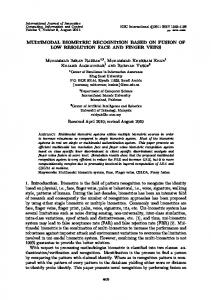
The electrocardiogram (ECG) is one of the most widely used signals in healthcare. Recorded at the surface of the body, with electrodes attached in various configurations, the ECG signal is studied for diagnostics even at the very early stage of a disease. In essence, this signal describes the electrical activity of the heart over time, and pictures the sequential depolarization and repolarization of the different muscles that form the myocardium. The first recording device was developed by the physiologist Williem Einthoven in the early 20th century, and for this discovery he was rewarded with the Nobel Prize in Medicine [10]. Since then ECG became an indispensable tool in clinical cardiology. However, the deployment of this signal in biometric recognition and affective computing is relatively young. Figure 2.1 shows the salient components of an ECG signal i.e., the P wave, the QRS
16
Chapter 2. Background and Prior Art
complex and the T wave. The P wave describes the depolarization of the right and left atria. The amplitude of this wave is relatively small, because the atrial muscle mass is limited. The absence of a P wave typically indicates ventricular ectopic focus. This wave usually has a positive polarity, with a duration of approximately 120 ms, while its spectral content is limited to 8-10 Hz, i.e., low frequencies.
R
Inter-beat interval
R
0.6-1.2ms (at 60-100 bpm)
T P
Q Duration Freq.
80 ms 8-10Hz
S
120 ms 80-120ms
160 ms
10-40 Hz
5-8Hz
-
Figure 2.1: Main components of an ECG heart beat. Each wave describes a distinct phase of the cardiac cycle. The QRS complex corresponds to the largest wave, since it represents the depolarization of the right and left ventricles, which are the chambers with the most substantial mass in the heart. The duration of this complex is approximately 70-110 ms in a normal heartbeat. The anatomic characteristics of the QRS complex depend on the origin of the pulse. Due to its steep slopes, the spectrum of a QRS wave is higher compared to that of other ECG waves, and is mostly concentrated between 10 and 40 Hz. Finally, the T wave depicts the ventricular repolarization. It has a relatively small amplitude and is usually observed about 300 ms after the QRS complex. However, its precise position depends on the heart rate, e.g., appearing closer to the QRS wave at rapid heart rates.
Chapter 2. Background and Prior Art
17
There is more than one approach to ECG recording, such as the orthogonal leads and synthesized leads [10]. However, the most widely applied system is the standard 12-lead ECG where there are three main sets of lead orientations. The bipolar limb leads are usually denoted as I, II and III and they track the electrical potential of the heart when three electrodes are attached at the right and left wrist and left ankle [10]. By convention, lead I measures the potential difference between the two arms. In lead II, one electrode is attached on the left leg and the other one on the right hand as depicted in Figure 2.2. Finally, in lead III configuration, the measured potential is between the left leg and right hand. Following the electrode position as pictured in Figure 2.2, the limb leads are measured in the following combinations:
I = VLH − VRH
(2.3)
II = VLL − VRH
(2.4)
III = VLL − VLH
(2.5)
The preceding equations suggest that, having recorded any two of the bipolar limb lead signals, the third one can be directly derived. The augmented unipolar limb leads fill the 60o gaps in the directions of the bipolar limb leads. Using the same electrodes, the augmented unipolar leads are measured as: VLH + VLL 2 VRH + VLL aV L = VLH − 2 VLH + VRH aV F = VLL − 2 aV R = VRH −
(2.6) (2.7) (2.8)
The third category of lead orientation involved in the conventional 12-lead system comprises the precordial leads (V1, V2, V3, V4, V5, V6). These signals are recorded with 6 electrodes attached successively on the left side of the chest, thus capturing more detailed information in the electrocardiogram [10].
18
Chapter 2. Background and Prior Art
I
VLH
VRH
II
III VLL
Figure 2.2: Configuration of Leads I II and III. Apart from the well known physiological process that generates the ECG, the signal is also affected by various sympathetic and parasympathetic processes. From a signal processing point of view, this is the reason why the ECG signal is not perfectly periodic. In the subsequent discussion the purpose is to differentiate between the anatomical properties of the heart, which encourage its deployment in biometrics, and the sources of variability due to the autonomic nervous system that support its analysis in affective computing.
2.2.1
Inter-individual variability
This section will briefly discuss the physiological rationale for the use of ECG in biometric recognition. Overall, healthy ECG signals from different people conform to roughly the same repetitive pulse pattern. However, further investigation of a person’s ECG signal can reveal notably unique trends which are not present in recordings from other individuals. The inter-individual variability of ECG has been extensively reported in the literature [11, 12, 13, 14, 15, 16, 17]. More specific, the ECG signal depicts the various electrophysiological properties of the cardiac muscle. Model studies have shown that physiological factors such as the heart mass orientation, the conductivity of various areas, and the activation order of the heart, are sources of significant variability among individuals [16, 17].
Chapter 2. Background and Prior Art
19
Furthermore, geometrical attributes such as the exact position and orientation of the myocardium, and torso shape designate ECG signals with particularly distinct and personalized characteristics. Other factors affecting the ECG signal are the timing of depolarization and repolarization and lead placement. In addition, except for the anatomic idiosyncrasy of the heart, unique patterns have been associated to physical characteristics such as the body habitus and gender [11, 15, 16, 17, 18]. The electrical map of the area surrounding the heart may also be affected by variations of other organs in the thorax [17]. In fact, various methodologies have been proposed to eliminate the differences among ECG recordings. The idea of clearing off the inter-individual variability is typical when seeking to establish healthy diagnostic standards [12]. Automatic diagnosis of pathologies using the ECG is infeasible if the level of variability among healthy people is high [16]. In such algorithms, personalized parameters of every subject are treated as random variables and a number of criteria have been defined to quantify the degree of subjects’ similarities on a specific feature basis.
2.2.2
Cardiovascular reactivity to emotion
The central and peripheral nervous systems (CNS, PNS) of the human body are responsible for physical and psychological behavior. The CNS is located in the cranial cavity and the spinal cord, and is the information processing unit of the body. The PNS acts as the communication channel between the CNS and the organs, and consists of information transferring nerves. Depending on whether the individual has control over these nerves or not, the PNS is divided into the somatic and the autonomic nervous systems (SNS, ANS). Various essential involuntary activities of the human body such as heart pulses, respiration, salivation etc. come under the ambit of the ANS. The ANS is further classified into the sympathetic and parasympathetic subsystems, which operate in an antagonistic manner. The resulting manifestation depends on which subsystem is dominant
Chapter 2. Background and Prior Art
20
at a particular instance. The opposing nature of the two results in a mannered balance within the body, called homeostasis. The ANS nerve-endings within the cardiac muscle play a major role in the cardiac output because they affect the rhythm at which the muscle pumps blood. The fibers of the sympathetic system run along the atria and the ventricles, and when activated stimulate the cardiac muscle to increase the heart rate. On the other hand, the parasympathetic system reduces the cardiac workload. Specifically, in the presence of a mental stressor, the sympathetic system dominates the parasympathetic, resulting in the following reactivity effects [19]: 1. Automaticity. The intrinsic impulse firing (automaticity) of the pacemaker cells increases, which translates directly to an increased heart rate. 2. Contractility. During every contraction the fibers of the heart shorten more, compared to the case during homeostasis, thereby increasing the force of contraction. 3. Conduction rate. The natural pacemaker, the SA node, is forced to conduct faster. 4. Excitability. During sympathetic stimulation, the person has increased perceptiveness to internal and external stimuli, which increases the irritability of the cardiac muscle and possibly lead to ectopic beats. 5. Dilation of coronary blood vessels. The diameter of the coronary blood vessels increases, followed by increased blood flow to the cardiac muscle. Depending on the intensity of a particular emotion, the sympathetic system is stimulated to prepare the body for vigorous activity. Apart from the well established cardiac reaction to ANS, there, there are questions that are yet to be answered. For instance, it is not clear whether the sympathetic and parasympathetic branches of ANS affect the heart in the same way i.e., whether there exist independent ANS drives on the cardiac muscle or not.
Chapter 2. Background and Prior Art
21
Shouldice et al. [20] provided evidence that there is separate ANS innervation to the two pacemakers (SA and VA nodes). By analyzing PP and PR intervals in transitions from supine to stand postures and vice versa, the authors showed a decoupling of the SA and VA modulation. This suggests that the cardiac reactivity may contain more detail about the stimulus than just the intensity (arousal). Another interesting finding in [20] is that there is large inter-subject autonomic innervation variability, meaning that one cannot generalize how the excitability of the SA and VA nodes is affected over a population. Their work leads one to suspect the specificity of emotion with regard to particular people. Furthermore, signals such as the heart rate variability (HRV) and blood volume pressure (BVP) have been associated with the development of coronary artery disease. The cardiac reactivity to stress has been studied in relation to feelings such as anxiety, hostility or challenge [21, 22]. Despite the dependence of the HRV time series on posture (standing/supine), increased vagal modulation of the cardiovascular system was statistically correlated with the aforementioned emotions. Similarly, the difficulty levels of memory tasks were shown to impact cardiac reactivity [23]. Furthermore, Blascovich et al. [24] were able to differentiate challenge from threat using cardiac traits such as the HR, ventricular contractility and cardiac output. As already discussed, the most widely studied trait of cardiovascular activity is the HR, or HRV [25, 26, 27]. Based on the effects of sympathetic stimulation on the cardiac muscle, the HR is the most natural choice for arousal detection using comparison of sympathetic and parasympathetic frequency bands of the time series [27]. However, it is highly dependent on the position of the body during monitoring [28, 21]. Emotion specific reactivity has been observed on the ECG signal itself, without collapsing the embedded information to arbitrary interval measurements (HRV). In fact, Andrassy et al. [29] observed QT prolongation (typical for stress) without significant R-R interval changes.
Chapter 2. Background and Prior Art
22
Folino et al. [30] recorded measurements of features such as the duration of the QRS complex, low amplitude waves at the terminal portions of the complex, and the root mean square voltage of the QRS while the subject was performing various mental arithmetic tasks. They observed that with increasing levels of difficulty, the energy of the QRS complex increased significantly, while its duration was reduced (not a result of increasing HR). The T wave amplitude was cross examined with mental stress in [31, 32]. An interesting observation brought to light by these works was that manifestations of mental stress on ECG depends on whether the stressor is active or passive. Contrary to the popular belief that mental stress response is generic, no significant effects were observed during passive tasks (i.e. stressors without the active involvement of the subject). Apart from mental stress, cardiovascular responses have been examined for other particular emotions. Sinha et al.[33] experimented with five emotional states namely joy, sadness, fear, anger and neutral. The HR, BVP, stroke volume, pre-ejection period and cardiac output were measured, employing imagery tasks for emotion elicitation. Pairwise comparison performance (for every two emotions) was reported for every measurement, with the HR being among the signals that exhibit the least discriminative power. In summary, there is significant evidence in the literature that cardiovascular reactivity can differentiate not only the intensity of the stimulus but also the valence (see Appendix B), depending on how the stimulus is presented. Although in the affect research ECG is primarily used for HR estimation, we argue that it is not used to its full extend, as this signal is yet one of the most illustrative and detailed records of cardiac activity. Furthermore, ECG exhibits idiosyncratic patterns due to the unique anatomy of people’s cardiac muscles. Along these lines, in this work we investigate the dynamics of the ECG signal and their association with emotion specific ANS activity, from a subject specific point of view.
Chapter 2. Background and Prior Art
23
Figure 2.3: Variability surrounding the QRS complex among heart beats of the same individual.
2.3
ECG Biometric Recognition: Literature Survey
Prior works in the ECG biometric recognition field can be categorized as either fiducial points dependent or independent. Fiducials are specific points of interest on an ECG heart beat such as the ones shown in Figure 2.1. Fiducial based approaches rely on local features of the heart beats for biometric template design, such as the temporal or amplitude difference between consecutive fiducial points. On the other hand, fiducial points independent approaches treat the ECG signal or isolated heart beats holistically and extract features statistically based on the overall morphology of the waveform. This distinction has a direct analogy to face biometric systems, where one can operate locally and extract biometric features such the distance between the eyes or the size of the mouth. Alternatively, a holistic approach would be to analyze the facial image globally. Both approaches have advantages and disadvantages. While fiducial oriented features risk to miss identifying information hidden behind the overall morphology of the biometric modality, holistic approaches deal with a large amount of redundant information that
Chapter 2. Background and Prior Art
24
needs to be eliminated. The challenge in the later case, is to remove this information in a way that the intra-subject variability is minimized, and the inter-subject is maximized. For the ECG case, detecting fiducial points is a very obscure process due to the high variability of the signal. Figure 2.3 shows an example of aligned ECG heart beats which belong to the same individual. Even though the QRS complex is perfectly aligned, there is significant variability surrounding the P and the T waves which renders the localization of these waves’ onsets and offsets very difficult. In fact, there is no universally acknowledged rule that can guide this detection [17]. This section provides an overview of fiducial dependent and independent approaches that are currently found in the literature. A comparison is also provided in Tables 2.1 and 2.2.
2.3.1
Fiducial Based Approaches
Among the earliest works in the area is Biel et al.’s [34] proposal, in 2001, for a fiducial feature extraction algorithm, which demonstrated the feasibility of using ECG signals for human identification. The standard 12 lead system was used to record signals from 20 subjects of various ages. Special experimentation was carried out to test variations due to lead placement in terms of the exact location and the operators who carry out the procedure. Out of 30 clinical diagnostic features that were estimated for each of the 12 leads, only 12 features were retained for matching, by inspection of the correlation matrix. These features pictured local characteristics of the pulses, such as the QRS complex and T wave amplitudes, P wave duration and other. This feature set was subsequently fed to SIMCA for training and classification. Results of combining different features were compared to demonstrate that, in the best case, classification rate was 100% with just 10 features. Kyoso et al., [35], also proposed fiducial based features for ECG biometric recognition.
Chapter 2. Background and Prior Art
25
Overall, four feature parameters were selected i.e., the P wave duration, PQ interval, QRS complex and QT durations. These features were identified on the pulses by applying a threshold to the second order derivative. The subject with the smallest Mahalanobis distance between each two of the four feature parameters was selected as the output. The highest reported performance was 94.2% for using just the QRS and QT intervals. In 2002, Shen et al. [36] reported an ECG based recognition method with seven fiducial based features that relate to the QRS complex. The underlying idea was that this wave is less affected by varying heart rates, and thus is appropriate for biometric recognition. The proposed methodology encompassed two steps [36]: During a first step, template matching was used to compute the correlation coefficient among the QRS complexes in the gallery set in order to find possible candidates and prune the search space. A decision based neural network (DBNN) was then formed to strengthen the validation of the resulting identity. While the first step managed to correctly identify only 85% of the cases, the neural network resulted in 100% recognition. More complete biometric recognition tests were reported in 2004, by Israel et al. [37]. This work presented the three clear stages of ECG biometric recognition i.e., preprocessing, feature extraction and classification. In addition, a variety of experimental settings are described in [37] such as, examination of variations due to electrode placement and physical stress. The proposed system employed only temporal features. A filter was first applied to retain signal information in the band 1.1- 40 Hz and discard the rest of the spectral components which were attributed to noise. By targeting to keep discriminative information while applying a stable filter over the gallery set, different filtering techniques were examined to conclude to a local averaging, spectral differencing and Fourier band-pass filter. The highest identification rate achieved was close to 100% which generally established the ECG signal as a biometric modality.
Chapter 2. Background and Prior Art
26
A similar approach was reported in the same year by Palaniappan et al., [38]. In addition to commonly used features within the QRS complex, a form factor, which is a measure of signal complexity, was proposed and tested as input to a neural network classifier. An identification rate of 97.6% was achieved over recordings of 10 individuals, by training a MLP-BP with 30 hidden units. Kim et al. [39], proposed a method to normalize time domain features by up-sampling the heart beats. In addition, the P wave was excluded when calculating the features, since it disappears when heart rate increases. With this strategy, the performance improved significantly when the testing subjects were performing physical activities. Another work that addressed the heart rate variations was by Saechia et al., [40] in 2005. The heart beats were normalized to healthy durations and then divided into three sub-sequences: P wave, QRS complex and T wave. The Fourier transform was applied on a heart beat itself and all three sub-sequences. The spectrums were then passed to a neural network for classification. It was shown that false rate was significantly lower (17.14% to 2.85%) by using the three sub-sequences instead of the original heart beat. Zhang et al. [41], suggested 14 commonly used features from ECG heart beats on which a PCA was applied to reduce dimensionality. A classification method based on the Bayes Theorem was proposed to maximize the posterior probability given prior probabilities and class-conditional densities. The proposed method outperformed Mahalanobis’ distance by 3.5% to 13% depending on the particular lead that was used. Singh et al. [42], proposed a way to delineate the P and T waveforms for accurate feature extraction. By examining the ECG signal within a preset window before the Q wave onset and apply a threshold to its first derivative, the precise position of the P was revealed. In addition to the onset, the peak and offset of the P wave were detected by tracing the signal and examining the zero crossings of its first derivative. The accuracy of this system was reported as 99%, tested over 25 subjects. In 2009, Boumbarov et al. [43], investigated different models such as HMM-GMM
Chapter 2. Background and Prior Art
27
(Hidden Markov model with Gaussian mixture model), HMM-SGM (Hidden Markov model with single Gaussian model) and CRF (Conditional Random Field), to determine different fiducial points in an ECG segment, followed by PCA and LDA for dimensionality reduction. A neural network with a radial basis function was realized as the classifier and the recognition rate was between 62% to 94% for different subjects. Ting et al. [44], described in 2010 a nonlinear dynamical model to represent the ECG in a state space form with the posterior states inferred by an Extended Kalman Filter. The Log-likelihood score was used to compare the estimated model of a testing ECG to that of the enrolled templates. The reported identification rate was 87.5% on the healthy beats of 13 subjects from the MIT Arrhythmia database. It was also reported that the method was robust to noise for SNR above 20 dB. The Dynamic time warping or FLDA were used in [45], together with the nearest neighbor classifier. The proposed system was comprised of two steps as follows: First the FLDA and nearest neighbor operated on the features and then a DTW classifier was applied to additionally boost the performance (100% over a 12-subject database). For verification, only features related to QRS complex were selected due to their robustness to heart rate variability. The same two-stage setting was applied together with a threshold and the reported performance was 96% for 12 legitimate subjects and 3 intruders. Another fiducial based method was proposed by Tawfik et al. [46]. In this work, the ECG segment between the QRS complex and the T wave was first extracted and normalized in the time domain by using Framingham correction formula or by assuming constant QT interval. The DCT was then applied and the coefficients were fed into a neural network for classification. The identification rate was 97.72% for that of the Framingham correction formula and 98.18% for the case of constant QT interval. Interestingly, using only the QRS complex without any time domain manipulation yielded a performance of 99.09%. In summary, although a number of fiducial based approaches have been reported for
Chapter 2. Background and Prior Art
28
ECG based biometrics, accurate localization of fiducial points remains a big challenge. This ambiguity risks the accuracy of the respective recognizers which require the precise location of such points. In the likely event of failing to adequately determine the locations of these points, fiducial approaches would rather reject the heart beat and require an extra reading, rather than risking the accuracy of their decision. This however, results in increased rejection rates that subsequently increase thw waiting time of the system. It is important to note, that in order to to acquire one clear pulse the sensors need to be attached/ held by the user for some time, until no muscular movements take place. Therefore, additional recording sessions due to rejection increase the inconvenience of use of such systems.
2.3.2
Fiducial Independent Approaches
On the non-fiducial methodologies side, the majority of the works were reported after 2006. Among the earliest is Plataniotis et al.’s [47] proposal for an autocorrelation (AC) based feature extractor. With the objective of capturing the repetitive pattern of ECG, the authors suggested the AC of an ECG segment as a way to avoid fiducial points detection. It was demonstrated that the autocorrelation of windowed ECG signals embeds highly discriminative information in a population. However, depending on the original sampling frequency of the signal, the dimensionality of a segment from the autocorrelation was considerably high for cost efficient applications. To reduce the dimensionality the discrete cosine transform (DCT) was applied. The method was tested on 14 subjects, for which multiple ECG recordings were available, acquired a few years apart. The identification performance was 100%. However, the DCT reduces the dimensionality from a signal processing point of view, which is sub-optimal for classification problems. Wubbeler et al. [48], have also reported an ECG based human recognizer by extracting biometric features from a combination of Leads I, II and III i.e., a two dimensional heart vector also known as the characteristic of the ECG. To locate and extract pulses a thresh-
Chapter 2. Background and Prior Art
29
olding procedure was applied. For classification, the distance between two heart vectors as well as their first and second temporal derivatives were calculated. A verification functionality was also designed by setting a threshold on the distances. Authenticated pairs were considered those which were adequately related, while in any other case, input signals were rejected. The reported false acceptance and rejection rates were 0.2% and 2.5% corresponding to a 2.8% equal error rate (EER). The overall recognition rate of the system was 99% for 74 subjects. A methodology for ECG synthesis was proposed in 2007 by Molina et al. [49]. A heart beat was normalized and compared with its estimate, which was previously constructed from itself and the templates from a claimed identity. The estimated version was produced by a morphological synthesis algorithm involving a modified dynamic time warping procedure. The Euclidean distance was used as the similarity measure and a threshold was applied to decide the authenticity. The highest reported performance was 98% with a 2% EER. In 2008, Chan et al. [50], reported ECG signal collection from the fingers by asking the participants to hold two electrode pads with their thumb and index finger. The Wavelet distance was used as the similarity measure with a classification accuracy of 89.1%, which outperformed other methods such as the percent residual distance and the correlation coefficient. Furthermore, an additional recording session was conducted for several misclassified subjects which overall improved the system’s performance to 95%. In the same year, Chiu et al. [51], proposed the use of DWT on heuristically isolated pulses. More precisely, every heart beat was determined on the ECG signal, as 43 samples backward and 84 samples forward from the R peaks. The DWT was used for feature extraction and the Euclidean distance as the similarity measure. When the proposed method was applied to a database of 35 healthy subjects, a 100% verification rate was reported. The author also pointed out that false rate would increase if 10 subjects with cardiac arrhythmia were included in the database.
Chapter 2. Background and Prior Art
30
Fatemian et al. [52], also suggested the Wavelet transform to denoise and delineate the ECG signals, followed by a process wherein every heart beat was resampled, normalized, aligned and averaged to create one strong template per subject. A correlation analysis was directly applied to test heart beats and the template since the gallery size was greatly reduced. The reported recognition rate was 99.6% for a setting where every subject has 2 templates in the gallery. The Spectogram was employed in [53] to transform the ECG into a set of timefrequency bins which were modeled by independent normal distributions. Dimensionality reduction was based on Kullback-Leibler divergence where a feature is selected only if the relative entropy between itself and the nominal model (which is the spectrogram of all subjects in database) is larger than a certain threshold. The log-likelihood ratio was used as a similarity measure for classification and different scenarios were examined. For enrollment and testings over the same day, a 0.37% ERR was achieved for verification and a 99% identification rate. For different days, the respective performance was 5.58% ERR and 76.9%. Ye et al. [54], applied the discrete wavelet transform (DWT) and independent component analysis (ICA) on ECG heart beat segments to obtain 118 and 18 features respectively. The feature vectors were concatenated. The dimensionality of the feature space was subsequently reduced from 136 to 26 using PCA which retained 99% of the data’s variance. An SVM with Guassian radial basis function was used for classification with a decision level fusion of the results from the two leads. A rank-1 classification rate of 99.6% was achieved for healthy heart beats. Another observation was that even though dynamic features such as the R-R interval proved to be beneficial for arrhythmia classification, they were not as good descriptors for biometric recognition.
Chapter 2. Background and Prior Art
31
Coutinho et al. [55], isolated the heart beats and performed an 8-bit uniform quantization to map the ECG samples to strings from a 256-symbol alphabet. Classification was based on finding the template in the gallery set that results in the shortest description length of the test input (given the strings in the particular template) which was calculated by the Ziv-Merhav cross parsing algorithm. The reported classification accuracy was 100% on a 19-subject database, in presence of emotional state variation. Autoregressive modeling was used in [56]. The ECG signal was segmented with 50% overlap and an AR model of order 4 was estimated so that its coefficients are used for classification. Furthermore, the mean PSD of each segment was concatenated as add-on features which increased the overall performance to 100% using a k-Nearest Neighbor classifier. Li et al. [57], proposed a method to model the ECG signal in both the temporal and cepstral domain. The hermite polynomial expansion was employed to transform heart beats into Hermite polynomial coefficients which were then modeled by an SVM with a linear kernel. Cepstral features were extracted by simple linear filtering and modeled by GMM/GSV (GMM super-vector). The highest reported performance was 98.26% with a 5% ERR corresponding to a score level fusion of both temporal and cepstral classifiers. It is clear from the above, that a large variety of fiducial independent techniques have been proposed for ECG biometric analysis. While some approaches are more computational intensive than other, or they operate on heart beats rather than finite ECG segments, there are practically a number of open issues in the literature with regard to ECG biometrics. Among the most prominent ones is the question of signal stability, or permanence, with time. The majority of prior works did not examine the evolution of the ECG signal with time. To some extent, the sources of intra-subject variability of the ECG signal have been ignored. We advocate that the study of the factors that affect the ECG waveform and may render the biometric template less accurate is very important for real
Principle
Performance
Number of subjects
Kyoso et. al [35]
Analyzed four fiducial based features from heart beats, to
94.2%
9
100%
20
100%
20
100%
29
97.%
10
N/A
10
97.15%
20
97.4%
502
100%
14
99%
74
determine those with greater impact on the identification performance Biel et. al [34]
Use a SIEMENS ECG apparatus to record and select appropriate medical diagnostic features for classification
Shen et. al [36]
Use template matching and neural networks to classify QRS complex related characteristics
Israel et. al [37]
Analyze fiducial based temporal features under various stress conditions
Palaniappan et. al [38]
Use two different neural network architectures for classification of six QRS wave related features
Kim et. al [39]
By normalizing ECG heartbeat using Fourier synthesis the performance under physical activities was improved
Saechia et. al [40]
Examined the effectiveness of segmenting
Chapter 2. Background and Prior Art
Method
ECG heartbeat into three subsequences Zhang et. al [41]
Bayes’ classifier based on conditional probability was used for identification and was found supurior to Mahalanobis’ distance.
Plataniotis et. al [47]
Analyze the autocorrelation of ECGs for feature extraction and apply DCT for dimensionality reduction
Wubbeler et. al [48]
Utilize the characteristic vector of the electrocardiogram for fiducial based feature extraction out of the QRS complex
Table 2.1: Summary of related to ECG based recognition works
32
Molina et. al [49]
Morphological synthesis technique was
10
95%
50
proposed to produced a synthesized ECG heartbeat between the test sample and template Chan et. al [50]
Wavelet distance measure was introduced to test the similarity between ECGs
Singh et. al [42]
A new method to delineate P and T waves
99%
25
Fatemian et. al [52]
Less templates per suject in gallery set to speed up
99.6%
13
86.1%
9
87.5%
13
76.9%
269
100%
15
99.09%
22
99.6%
36 normal
computation and reduce memory requirement Boumbarov et. al [43]
Neural network with radial basis function was employed as the classifier
Ting et. al [44]
Use extended Kalman filter as inference engine to estimate ECG in state space
Odinaka et. al [53]
Time frequency analysis and relative entropy to classify ECGs
Venkatesh et. al [45]
Apply dynamic time warping and Fisher’s discriminant analysis on ECG features
Tawfik et. al [46]
Examined the system performance when
Chapter 2. Background and Prior Art
98%
using normalized QT and QRS and using raw QRS Ye et. al [54]
Applied Wavelet transform and Independent component analysis, together with support vector machi
and 112 arrhythmic
as classifier to fuse information from two leads Coutinho et. al [55]
Treat heartbeats as a strings and using
100%
19
100%
12
98.3%
18
Ziv-Merhav parsing to measure the cross complexity Ghofrani et. al [56]
Autoregressive coefficient and mean of power spectral density were proposed to model the system for classification model the system for classification
Li et. al [57]
Fusion of temporal and cepstral features
33
Table 2.2: Summary of related to ECG based recognition works (Continued)
Chapter 2. Background and Prior Art
34
life deployment of this technology. Furthermore, the one-dimensional representation of the ECG signal presents significant advantages to fast identity computation, and complex processing algorithms (neural networks, warping etc.) may compromise this asset. Overall, the prior art in ECG biometrics recognition, as summarized in Tables 2.1 and 2.2, did not discuss the way that the respective solutions will fit in real-life settings.
2.4
ECG in Affective Computing: Literature Survey
Emotion recognition in the field of human computer interaction has drawn enormous attention as early as the 1980s. To date, there are numerous works in literature focusing on facial expression or speech analysis for the purpose of emotion detection. Irrespectively of the employed modality, each reported work contributes to the overall understanding of emotions and to the way they are expressed through the human body. Nevertheless, this section places the emphasis on physiological signal processing for detection and classification of emotions. Although the approaches found in literature vary algorithmically and with respect to the experimental procedures employed, current methodologies can be classified in to two categories based on the way the emotional models are conceived and deployed. The first category includes discrete emotional models (DEM), where the main objective of the proposed frameworks is to recognize and label standard emotional states (for instance joy, sadness or fear) depending on the application. These methods rely heavily on the assumption that any affective state is physiologically well defined and completely distinguishable from the rest. On the other hand, the affective dimensional models (ADM) relax the conditions for discrete emotions and treat any affective state as a combination of two parameters, namely arousal and valence. Arousal is a measure of emotional stimulation which varying
Chapter 2. Background and Prior Art
35
from low to high. Valence is a measure of pleasantness for the experienced emotion, ranging from very pleasant to unpleasant. These two measures define a two dimensional space (the AV plane), and classification is carried out among predefined areas of the plane, which do not correspond necessarily to a discrete emotion. More information pertaining to emotion definitions can be found in Appendix B. Both methodologies have advantages and disadvantages. For instance, the discrete emotional models might be easily conceptualized by people and fitted to applications, however their performance is risked as there is no well defined physiological borders among emotions. The rest of this section provides a literature survey for both discrete and dimensional emotional models.
2.4.1
Discrete Emotional Models
To the best of our knowledge, the first documented effort for emotion recognition using physiological signals is by Ekman et al. [58] in 1983. This work was considered a breakthrough as it opposed to the dogma dictates that no activity of the autonomic nervous system (ANS) is emotion specific. In other words, Ekman et al.’s work provided evidence that emotions can be distinguished among the various reactions of the human body. Using both physiological signals and videos of facial expressions, six emotions were studied: surprise, disgust, sadness, anger, fear and happiness. The bio signals considered were the heart rate, left and right hand temperature, skin resistance and forearm muscle tension. The respective feelings were induced in a different manner for two recording modalities. For facial gestures, the subjects were asked to mimic emotional expressions that they had practiced before using a mirror, and following the instructions of an operator concerning the facial muscles they were required to contract. On the other hand, for the stimulation of physiological signals an experience reliving approach was chosen. For this imagery task, every subject was asked to relax and bring to memory a personal experience that related to each of the studied emotions. Upon
Chapter 2. Background and Prior Art
36
completion, the subjects were asked to rank the intensity of their feelings on a predefined scale. For this experiment, 16 subjects were examined, 12 of which were professional actors. It is important to note that using actors to induce and analyze feelings, was a standard practice in the beginning of the affective computing research, as they were expected to manifest emotions naturally. The signal analysis performed in [58], was based on the change scores principle. Data during emotional instances were averaged and subtracted from data of non-emotional phases. These differences were used as features which were then examined to evaluate organization in clusters based on tree decisions (for example, if the heart rate change was high and temperature was low, this would be assigned to the fear cluster). This technique although simplistic, demonstrated for the first time that physiological information can be organized in emotional clusters. Ekman et al.’s findings have drawn the attention of the medical field (both physiological and psychological medicine), which has contributed significantly in the field. In 1992, Sinha et al. [33], reported a statistical analysis concerning the cardiovascular differentiation of emotions. Six heart related indexes were recorded from 27 male subjects. The signals were the heart rate, blood pressure, stroke volume, cardiac output, peripheral vascular resistance and an index of myocardial contractility. The objective was to examine and identify patterns of the cardiovascular activity under the expression of 4 main emotions: fear, anger, joy and sadness. The experimental procedure in [33] involved a careful screening of the subjects participating in the study. This selection is generally prominent as the volunteers should be persons who not only experience various emotional states, but are also able to describe them verbally with accuracy. The physiological reaction of humans has been shown to be relative to their imagery ability [59]. For this reason, the subjects were selected with a questionnaire. The main experimental part involved two phases, one for the volunteers to familiarize with the imagery procedure (training), and one for the data collection.
Chapter 2. Background and Prior Art
37
The methodology for emotion analysis described in [33], resembles Ekman et al.’s proposal. Change scores were calculated for every cardiovascular measure by subtracting the baseline mean scores from the mean score during an emotional phase. With this kind of analysis it was demonstrated that the heart rate is very sensitive to anger compared the less valent feelings of joy or sadness. Another significant finding was that even though the emotions of fear and anger can increase the heart rate, systolic blood pressure and mascular blood flow, they can be differentiated by carefully examining the diastolic blood pressure, which decreases for fear only. In other words, peripheral vascular resistance decreases under fear. A similar study [60] in 1996 has expanded this analysis to non cardiovascular features, and analyzed skin conductance under mental stress to show very high correlation between this measure and a chosen psychological state. In 2001 Picard (the pioneer of this field) et al. [1], proposed an emotionally intelligent system that utilizes information of 4 physiological signals. This work was the first to define and analyze the challenges of affective computing. The authors emphasize the fact that data collection for the purpose of emotion detection is a sensitive process compared to any standard experimental setup, and requires special attention. This is because in order to gather valid affective data, one has to make sure that first, the desired emotion was induced and second, that the subject labeled it accurately [61]. This is a non trivial procedure, first because as explained earlier some subjects are suppressive, thus any attempt to cause an emotional disturbance might fail, and second because people might have experienced feelings but not being aware of them or not feeling comfortable to declare them. Accordingly, Picard et al. [1] enumerated five parameters to be considered when designing such experiments:
1. An emotion can be self elicited (imagery), or controlled by external factors like audio and visual stimuli. 2. The experimental environment plays a major role and the level of familiarity with
38
Chapter 2. Background and Prior Art
Signal
Annotation
Sensor Placement
Electromyogram (EMG)
E
Masseter muscle (Face)
Blood Volume Pressure (BVP)
B
Tip of ring finger (left hand)
Skin Conductance
S
Index and middle fingers (left hand palm)
Respiration Rate
R
Diaphragm
Heart Rate
H
Measured from BVP peaks
Table 2.3: Biosignals analyzed in [1]. the subject should be defined. 3. There are several different modalities to explore (face, voice, gesture, biosignals). 4. The subject can be aware or unaware that some signal is being monitored. 5. The subject can be aware or unaware of the purpose of the experiment. In addition, one of the major contributions in [1] is that data collection was carried out over multiple days for the same individual. This was novel not only from a data perspective, but also because it moved the interest of the community to examining also person-dependent recognition methodologies instead of modeling emotion by averaging population data. Furthermore, the stability of the emotional expression was examined over a period of time. There were 8 emotions under consideration: anger, hate, grief platonic love, romantic love, joy and reverence. Because this list has several non traditional emotions, the authors argued that the exact selection of emotions was not important, as the only requirement is for the subject to understand and identify them consistently. The physiological signals measured and analyzed in [1] are listed in Table 2.3 along with the respective sensor placement. From the sensor orientation, one can observe that the aim was to stabilize the left hand while leaving some freedom of movement to the right hand. The experiment lasted for 30 days and every day the subject was recorded
Chapter 2. Background and Prior Art
39
for 25 minutes. Emotion elicitation was based on imagery exercises. The volunteer was asked to pick scenes or situations from her own experience that could induce the desired emotion with adequate valence. Picard et al. [1] suggested for classification features picturing the statistical properties of the signals rather than just their changing rate. These statistical features are listed in Table 2.4. In addition, and in order to account for the effects of non emotional factors, an extra set of physiology related features was calculated. These features along with a description can be found in Appendix C. Having acquired a feature set, the next step of the training procedure is to select the appropriate ones and transform them. The authors described a combination of the Sequential Floating Forward Search algorithm (SFFS) in conjunction with Fisher Projection (FP) [62]. SFFS is a statistical searching methodology, which performs an nonexhaustive search by continuously adding and removing features from a set by checking their classification power. FP is a method for dimensionality reduction, which can lower the dimensions of the feature space down to C − 1, where C is the number of classes, i.e., emotions the system can identify. These two methods were combined hierarchically in one framework, by using the output of SFFS as input to FP. For classification, a k -Nearest Neighbor scheme was employed with k varying between 1 and 20. The highest classification performance reported in [1] was 83.3 % for the identification of three emotions. Scheirer et al. [2], reported a novel experimental procedure to induce and measure frustration. This new approach had considerable advantages to the synchronization problem. More precisely, instead of deploying imagery or audio visual induction techniques, they have created a computer game which aimed to frustrate the user. At random interval of times the game failed (mouse clicking did not work) this way spoiling the players’ pleasure, who got eventually frustrated [63]. Utilizing a computer game to provoke emotion has significant advantages for signal
40
Chapter 2. Background and Prior Art
Mean Standard Deviation Mean of first differences or raw samples Mean of first differences or normalized samples Mean of second differences or raw samples
σx = δx =
µx = s
1 N −1
1 N −1
1 N
N P
Xn
n=1
N P
(Xn − µx )2
n=1 NP −1
|Xn+1 − Xn |
n=1
δex = σδxx NP −2 γx = N 1−2 |Xn+2 − Xn | n=1
Mean of second differences or normalized samples
γx = e
γx σx
Table 2.4: Typical classification features used in [1].
labeling. In the affective computing research, one of the major challenges is generating accurate datasets, and this is first because it is extremely difficult to elicit emotions and second because the ground truth is not always well defined. To this end, the ground truth represents the true labeling of the signals with respect to the onset of an emotional phase. What Scheirer et al. suggested in [2], was that using a computer game to frustrate the user, the time instances of frustration could be easily labeled. However, that alone is not very accurate and latency periods should be taken into account as there is usually a short time interval before a human actually experiences a feeling internally (three seconds for galvanic skin response [2]). Scheirer et al.’s work [2] although focusing on human frustration could have been deployed for any other emotion by accordingly changing the computer game. In [2], frustration is defined as a manifestation of negative arousal when something impedes the subject’s progress toward a goal. To quantify this arousal and distinguish between frustration and non-frustration states, two physiological signals were considered, the galvanic skin response and blood volume pressure. Once more, this selection of signals was not compulsory and the authors explain that this choice might not even be optimal. However, for the purpose of their experimental setup, GSR and BVP were suitable because
Chapter 2. Background and Prior Art
41
they can be measured unobtrusively by occupying only one of the subject’s hands. For the analysis in [2], data from 24 subjects were utilized. The data were modeled with a Hidden Markov Model (HMM) because of its success in describing dynamic systems like speech recognition. A set of features was proposed for the discrimination of frustration based on the variability of GSR and BVP. More details pertaining to these features can be found in Appendix C. During training, the highest classification performance achieved was 81.87%, which dropped to 67.4% for the testing set. Given that only two classes were considered this result is slightly better than random guessing. The major novelty of [2] is in setting the standards for the experimental procedures in the affective computing research. The authors explained that the poor performance reported, could be addressed with more sophisticated pattern recognition and machine learning techniques. In 2003, Nasoz et al. [64], introduced the idea of adapting a software interface according to a user’s current emotional state. A multimodal framework was proposed, based on the combination of facial, verbal, gesture and physiological recordings. A synthesizer was designed to combine all this information and provide the user with feedback. The emotional states explored were six: sadness, anger, surprise, fear, frustration and amusement. They were elicited using visual stimuli, movies particularly chosen to relate to the above mentioned psychological states. The biosignals measured were the galvanic skin response, heart rate and body temperature. Data from 31 subjects were divided into training and testing and two algorithms were used for classification, namely the k -NN and and the discriminant function analysis. (DFA). The two algorithms were tested independently to show that DFA outperforms k -NN with a maximum recognition performance of 90% for fear. A later and more detailed report by the same authors [65] placed the emphasis to presence technologies. The MAUI (Multimodal Affect User Interface) prototype system was presented to enhance social presence using physiological signals. Facial expression recognition was also central to MAUI, as one of its main features was an avatar who could
Chapter 2. Background and Prior Art
42
mimic the user’s expressions. This is because when characters (personas) are employed in virtual environments, their ability to communicate emotions increases significantly the social presence and co-presence. The list of emotions considered was the same as in [64]. Their study involved 14 subjects who were asked to watch movie clips from a predefined list of films that had been shown to elicit specific emotions successfully [66]. Physiological signals (galvanic skin response, heart rate and temperature) were measured non invasively with a wireless armband. An important step in feature processing was normalization of the measurements with respect to the respective relaxation value. This was done in order to avoid the individual differences of participants. The main analysis in [64] included the classification methods explored previously; k -NN, DFA plus a newone, namely the Marquardt Backpropagation (MBP). The MBP method was reported to outperform the rest in recognizing all emotions (for instance 92% for sadness). In 2004, Kim et al. [67] reported large scale experiments for physiological emotion recognition with more than 100 subjects. The methodology relied on features out of three physiological measurements i.e., skin temperature, heart rate and galvanic skin response. A strong justification of the relationship between these signals and the autonomic nervous system’s reaction to stimuli can be found in [67]. The analysis was based on two experiments; the first involved 125 subjects (5-8 years old) under a variety of audio and visual stimuli. The second experiment was carried out a year later to record 50 more subjects (7-8 years old). The heart rate variability (HRV) information was obtained through ECG with peak detection (using the Teager energy operator) and calculation of the R-R intervals. The authors suggested the spectrum of HRV as a feature source. Given that the spectrum estimation should be accurate even for a short term signal (so that real-time approximations can be obtained), time-series models for frequency estimation were used [68]. More specific, two sub-bands were considered, a low frequency one at 0.03-0.15 Hz and
Chapter 2. Background and Prior Art
43
a high frequency at 0.15-0.4 Hz. For the GSR, feature extraction comprised detection of response occurrences which are waves of various amplitudes and lengths depending on the stimuli. To do that, the GSR signals were differentiated and smoothened in order to apply a threshold for the detection of the waves’ onsets and offsets . Four features were used for classification out of GSR i.e., the mean DC level, mean values of GSR detected wave amplitudes and duration, and the number of event (interesting wave) occurrences in a 50 second segment. Finally, the skin temperature was used as a feature without special processing. The signals were divided into training and testing sets for classification among three or four discrete emotional states i.e., sadness, anger, stress (and optionally surprise). The recognition performance for three clusters of data from the first experiment was 55.2%, and 78.4% for the second experiment. According to [67] the later dataset included signals of higher quality. Among four emotional states, the correct classification rate of the second dataset was reported to be 61.8%. In 2005, Healey et al. [3] suggested the use of physiological signals for stress detection under real world driving tasks. The purpose of the proposed system was to analyze a driver’s psychological status in order to provide feedback or take appropriate action. An adaptive framework was envisioned, where the computer would cope with the driver’s stress by diverting cell phone calls to voice mail, suggest appropriate music and reduce workload by manipulating the navigation systems. Four signals were considered in [3], ECG, EMG, GSR and respiration (R). Among the challenges of that implementation was for the system to react to stress detection in real time. However, none of the monitored biosignals responds in time for appropriate adjustment of vehicle control (1-3 min lag) [3]. To that end, two experimental setups were considered: 1. Analysis I. The first analysis attempted to recognize general stress levels (defined as low, medium and high), by utilizing data of 5 minutes long. In addition, this
Chapter 2. Background and Prior Art
44
module was designed to detect stress based on a systematic fusion of all four signals using standard pattern recognition techniques. Overall, 21 features were estimated and used for classification, listed in Table C.3 in Appendix C. 2. Analysis II. The second analysis aimed at a more detailed examination of the physiological signals. For this reason, the signals were processed independently, and at a shorter time span i.e., every second. A special continuous stress metric was designed to judge the environmental stressors at every second of the experiment, in order to investigate the correspondence between this measure and the biosignals. The stress metric was computed in a way that it revealed the instantaneous workload of the driver, according to videos taken throughout the drives.
Classification on features from the first analysis indicated high accuracy, with low level stress being correctly recognized for all testing data (100%), medium and high stress recognition rates were 94.7% and 97.4% respectively. For Analysis II, continuous time features were designed out of each of the biosignals, in the same range as the stress metric, for the correlation coefficient (in the range [−1, 1]) to be used for matching. The highest performance was achieved for a GSR metric picturing the instantaneous variation of the mean, with the correlation coefficient reaching 0.99. Driving applications of affective computing were revisited later by H¨onig et al. [69]. By recording 24 subjects in a driving simulation environment, the authors attempted to recognize the various stress levels provoked by driving related tasks such as changing lanes, along with memory task such as question answering and arithmetic. Six physiological signals were monitored during the simulation: ECG, EMG, GSR, BVP, temperature and respiration along with some of their derivatives (ECG or BVP heart rate, reaspiration rate and so on). During the simulations, the subjects’ facial expressions were recorded with a camera to be used for evaluation of stress elicitation. Extra subjects were used as labelers i.e.,
Chapter 2. Background and Prior Art
45
to watch the video recordings and assess the stress levels based on the facial reaction of the volunteers. This labeling, along with the standard stress onset timing designation provided by the system (for instance, the instance when the driver is required to change lane), were used in order to establish the data ground truth. To cover a range of short to long period reactions, feature extraction operated on signals of length varying between 1 and 60 seconds. A large number of features was computed in [69] based on the mean, standard deviation and slope of the various windows. The Fisher linear discriminant analysis was used for dimensionality reduction and classification was carried out based on Gaussian mixture model probabilities. Overall, and by fusing information from all physiological signals, a correct stress recognition rate of 88.8% was achieved. Lee et al. [70], suggested the use of a neural network that would be trained on emotional cues and would then be tested on data not previously seen. Interestingly, only 6 subjects participated in the experimental set up. According to the authors that was because the number of subjects involved is independent from the overall performance as emotions are experienced in a different physiological manner by every individual. Two physiological signals were considered: the galvanic skin response and the heart rate, which was acquired with QRS peak detection from the electrocardiogram. The heart rate was processed in the frequency domain, by estimating the ratio between low and high frequency coefficients of the spectrum. A multilayer perceptron (MLP) neural network was trained for the recognition of four emotional states: sadness, calm pleasure, interesting pleasure and fear. All emotions were induced by video clips, that the volunteers had chosen themselves. After watching each video, the subjects were asked to describe their emotional experience using the self assessment manikin (SAM). The overall performance for all emotional clusters was 80.2%, with fear being the best physiologically distinguishable feeling. Anttonen et al. [71], focused in designing technologies which are not only unobtrusive
Chapter 2. Background and Prior Art
46
to the user but also offer no distractions. Traditional equipment for physiological signal measurements are wired sensors which need to be attached to the human body by trained personnel. This limits significantly the range of applications as mobility is restricted to a small area. In addition, such sensors fail a lot because they get easily detached, and distract the subject who is expected to focus in experiencing an affective state. For this reason, the authors in [71] suggested the use of a chair which is equipped with electromechanical film (EMFi chair) to be able to record physiological measurements while someone is sitting on it. An initial step in Anttonen et al.’s [71] experimental setup was to ensure that the chair can collect sufficiently accurate data. Therefore, they used a simple photoplethysmography (PPG) sensor attached to the earlobe of the subject while he/she was sitting on the chair. Both the chair and the PPG sensor offered heart rate measurements, which were compared with each other to demonstrate consistency in the chair recordings (the correlation between the two was 0.99). This allowed the authors to rely on the chair recordings for the rest of their experiments. The objective of the main experimental procedure in [71] was to classify among positive, neutral and negative emotional states. 26 subjects were recorded sitting on the EMFi chair, while their psychological state was stimulated via audio and visual stimuli (International Affective Picture System (IAPS) [72] and International Affective Digitized Sounds (IADS)). Both positive and negative stimuli resulted in heart rate deceleration compared to the neutral reaction. What is more, the deceleration under negative effects was greater than the positive in magnitude and in length. Heart rate changes in positive stimuli were observed only during the first two seconds and right before returning to a normal rhythm. The latency period for the manifestation of heart rate changes under negative reactions was approximately six seconds from the onset of the stimuli. Nakasone et al. [73], presented a framework for machine emotion recognition which concentrated on the implementation challenges of this technology. More precisely, the
Chapter 2. Background and Prior Art
47
authors looked at gaming scenarios between humans and humanoid agents, who were envisioned to ”read” the player’s psychological state and respond appropriately. The system was designed upon five main layers namely, the synchronization, the device, signal categorization, bayesian network and interface layers. The synchronization layer was set responsible for the initialization of emotion assessment and communication among the rest of the layers. For instance, data acquisition, storage and analysis where synchronized in this module. The device layer controlled the physiological recording devices, which measured EMG and GSR. At the categorization layer, the signals were subjected to feature extraction, which involved a simple calculation of their sample mean and subtraction from a baseline value (where the baseline was considered to be the physiological response at rest). The bayesian network layer would utilize the classification results in order to finalize the emotion decision and finally, the interface layer adapted the game to the particular player status. Although no specific experiments were carried out, Nakasone et al. [73], addressed problems related to the actual realization of affective computing. Overall, three challenges were outlined: 1) The baseline problem, which relates to the standard resting conditions against which instantaneous physiological measurements should be compared, 2) The timing problem, i.e., detecting the exact onset of an emotional phase and 3) The intensity of a particular experienced emotion. To make sure baseline signals were obtained for comparison, the authors suggested an initial relaxation period, where the subject would listen to calm music. Furthermore, to ensure that emotions were captured in the signals, the authors suggested sampling every 50 msec. In 2008, Benovoy et al. [74], proposed another novel application for affective recognition systems. The main idea was for emotion assessment to be used in a performance setting, for instance a musical act, were the performer leads an instrument to compose an artistic output based on the instantaneous physiological reaction of a subject. This application had two main design phases:
Chapter 2. Background and Prior Art
48
1. Biosignal recording and mapping with emotional states. The blood volume pressure (BVP), galvanic skin response (GSR), electromyography (EMG) and respiration were considered for classification among four emotions (joy, anger, sadness and pleasure). 2. The second stage was meant to provide a rich external manifestation of a person’s internal emotional status [74]. The purpose was to build a musical device which would be mapped to the user’s feelings but also controlled by him/her.
Given the specifics of the application, a direct interaction with the experimental subject was preferred over standard emotion induction methodologies used previously (audio visual). A professional actor guided by an experienced theatre director was monitored while experiencing a variety of characters and situations, particularly designed to elicit the desired emotions. By the end of each experiment the actor was asked to fill in a questionnaire concerning the intensity of the previously experienced emotion. A large variety of features was estimated from the recorded biosignals. Overall 225 features were used, similar to [1]. A list of these features can be found in Appendix C. The next step was to select the optimal for classification features using the sequential forward selection (SFS) algorithm. The overall dimensionality was reduced with Fisher discriminant analysis, and finally classification was carried out with either the linear discriminant analysis (LDA), or k -NN or a multilayer perceptron (MLP) neural network. It was demonstrated that the LDA outperforms the rest of the classification schemes, with a classification rate of 90% for four emotions. This notable result was attributed to the fact that a professional actor was recorded, thus emotion elicitation was more successful compared to prior trials. In summary, prior DEM approaches have established biosignals as a carriers of affective information. The simplicity in the conception of emotion within such models (i.e., the discretization to distinct emotional conditions) makes them easily employable in real
49
Chapter 2. Background and Prior Art
life systems. However, prior works require the collection of more than one physiological measurements in order to assess emotion, which limits real life applications due to increased obtrusiveness. In addition, the ECG signal has been primarily used for HR estimation while the evolution of the waveform under emotional activity has not been studied.
2.4.2
Affective Dimensional Models
In 2004, Mandryk et al. [75], established the applicability of physiological emotion recognition in the evaluation of entertainment technologies. The breakthrough of this work lies in proving that there is a different physiological response when users play against other players, as opposed to playing against the computer. As a matter of fact, there is a different reaction when playing against friends compared to strangers [76]. In addition, it was shown that people tend to immerse and enjoy a game more, when they play against humans [75]. These observations and the respective technology apply directly to the entertainment industry for the design of virtual environments and computer games. Mandryk outlined the three challenges of this research in [77]: • What is the definition for a successful game?
A game can not be characterized
successful based on the user performance, but on the user’s affective experience [77]. A prosperous game environment enhances interaction between the players (co-presence), and only this can offer a truly immersive user experience. Thus, success is measured in terms of the psychological engagement of the players. • How can the entertainment variables be measured? A means to quantify user engagement, is by measuring his/her emotional involvement. The interest is not in defining which emotion is experienced, but the level of subject’s arousal in general. The biosignals considered for this purpose are the galvanic skin response, various cardiovascular measurements (electrocardiogram, heart rate, blood pressure),
Chapter 2. Background and Prior Art
50
respiration rate and amplitude, and electromyography. • How can a measurement be associated to the player’s experience? The game events were monitored to label the signal recordings. This approach is similar to the Scheirer et al.’s [2] idea for controlled emotion elicitation which allows for better signal segmentation. The features used for discrimination of arousal were simply the time average of the signals. This kind of features diminished the variability of each psychological state, but this was intentional since the authors were interested only in detecting the presence of emotions and not in defining which emotion was experienced. The experimental set up involved 10 male subjects and it was shown that the arousal was greater for all physiological measurements when playing against humans. In 2007, the same researchers [78] managed to design a consistent mapping between physiological measurements and emotional states i.e., the arousal-valence (AV) plane. This time the authors were interested in identifying the exact emotional state (fun, excitement, frustration, challenge and boredom). A fuzzy approach was adopted to systematically combine information from four physiological measurements (galvanic skin response, heart rate, smiling and frowning electromyograph) and then project it to meaningful valence and arousal dimensions. Accordingly, a second fuzzy scheme established the correspondence between the AV axes and the desired emotional states. The four biosignals served as inputs to the first fuzzy framework. A preprocessing step was required to ensure that the measurements were comparable with each other (this was required by the fuzzy model). Preprocessing involved smoothening with a moving average window and then normalization into a percentage between 0 and 100. The experimental setup in [78] included 12 subjects. The physiological readings from the first 6 were used for training i.e., to analyze emotional response and design the fuzzy logic, and the other 6 subject recordings were used for testing. To evaluate the performance of the algorithm, the subjective reports (questionnaires) were compared
Chapter 2. Background and Prior Art
51
against the objective recognition results (from the fuzzy scheme) to reveal similar trends for most emotional classes. Even though the performance of the framework was not presented analytically, Mandryk et al. were the first to attempt a direct match between physiological signals and the AV plane. The reader should note that prior to this work, researchers have concentrated in modeling specific emotional states. Haag et al. [79] proposed an affective dimensional model that can classify information on an arousal and valence basis. A collection of physiological signals was used for this purpose, each processed independently and according to its special characteristics. The biosignals were the electromyogram (EMG), galvanic skin response (GSR), skin temperature, blood volume pulse (BVP), electrocardiogram (ECG) and respiration. The authors in [79] comment strongly on the importance of the emotion induction methodologies and to the effects on the performance. To elicit emotions appropriately, strong realism is required which is usually not in accordance with the experimental ethics (provoking severe negative emotions is ethically questionable). For this reason they used a collection of pictures from the International Affective Picture System (IAPS) [72]. The pictures were classified into six sets, each consisting of 5 photos. Every set was labeled as low, medium or high valence, or low, medium or high arousal. The combination of the two extensions (arousal and valence) defined a two dimensional space for the classification of the physiological features. The statistical features employed for classification were: • The standard deviation of the heart rate, estimated with peak detection out of the ECG signal. • The standard deviation of the heart rate, estimated with peak detection out of the BVP signal. • Standard deviation of BVP amplitudes. • The EMG amplitude.
Chapter 2. Background and Prior Art
52
• Standard deviation of skin conductivity (GSR). • Standard deviation of respiration amplitude. • Standard deviation of respiration rate. It should be mentioned that the above mentioned list of features was the optimal selection, and more features were tested and rejected in [79] because of poor performance. One subject was tested on different days and also various times of the day. The recognition rate was reported for correct classification considered within 10% and 20% distance from the true class on the AV plane. The second option increases the bandwidth thus greater performance was expected. More specific, for the large bandwidth a recognition rate of 96.58% and 89.93% was achieved for arousal and valence respectively. Jones et al. [80], proposed a system for recognition of affective states based on dimensional modeling. The main idea is that an emotional state can be sufficiently characterized by its arousal and valence magnitudes, without mapping them to distinct emotions (sadness, happiness etc). The two dimensional AV model was used as a basis for projection of information collected from physiological signals. The plane was divided into 25 areas, each representing an emotional region. Three physiological signals are considered for recognition, the blood volume pressure (BVP), skin conductance (GSR) and the respiration rate. Emotions were induced with 21 images from IAPS [72] since these images have already been rated in valence and arousal by psychologists. Each picture was displayed to the subject for 15 seconds after which there was an interval of rest for 25 seconds. As with any pattern recognition problem, collection was followed by feature extraction and classification. The features used in [80] were simple statistical measures of the signals like the mean and variance. For classification, two multi layer perceptron neural networks were used, one for each descriptive dimension (AV). Overall, 13 subjects were recorded. The data of 10 subjects were used for training and the rest for testing. The authors
53
Chapter 2. Background and Prior Art
Emotional Model
Ekman et al.
Sinha et. al
Picard et al.
Scheirer et al.
Nasoz et al.
Kim et al
Type
DEM
DEM
DEM
DEM
DEM
DEM
Emotions Induced
Surprise, Disgust, Sadness, Anger, Fear, Happiness
Fear, Anger, Joy, Sadness
Frustration
Sadness, Anger, Surprise,Fear, Frustration, Amusement
Sadness, Anger, Stress, Surprise
Elicitation Method
Imagery Task
Imagery Task HR, BVP, and other cardiovascul ar measures
Anger, hate, Grief, Platonic love, Romantic love, Joy, Reverence Imagery Task
Computer Game GSR, BVP
Movie clips GSR, HR, T
Audio, Visual stimuli T, HR, GSR
Signals
Biosignal Processing
Feature Extraction
HR, T, GSR, Muscle tension
EMG, BVP, GSR, R, HR
Change Scores
Change Scores
Mean and std analysis with SFFS and Fisher projection.
Variance and pinch measures.
Signal Average for each emotion.
HR spectrum, GSR wave features
Decision Tree
MANOVA
k-NN
HMM
k-NN, MBP, discriminant analysis.
SVM
Classification
Performance
Number of subjects
Classification Rate
Not reported Professional actors were recorded.
Comments
Not reported Fear and anger can be differentiate d through the diastolic blood pressure.
14
24
1
27
16
83.3% for 3 emotions Evaluation of subject dependent method. Experiments with the same subject over several days.
50
67.4%
92% for sadness
61.8%
The experimental process was controlled with the use of a specially designed computer game.
Emphasized the use of affective computing in presence technologies.
Special properties of the biosignals were considered for appropriate feature extraction.
Table Notation DEM Discrete Emotional Model
ADM Affective Dimensional Model
HR Heart Rate
T Temperature
GSR Galvanic Skin Response
BVP Blood Volume Pressure
R Respiration
ECG Electrocardiogram
EMG Electromyogram
Table 2.5: Affective computing using biosignals: Comparison Milestones
54
Chapter 2. Background and Prior Art
Emotional Model
Lee et al
Anttonen et al.
Benovoy et al.
Mandryk et al.
Haag et al.
Jones et al.
Type
DEM
DEM
DEM
ADM
ADM
ADM
Emotions Induced
Sadness, Calm Pleasure, Interesting pleasure, fear Video clips
Positive, neutral and negative status
Joy, Anger, Sadness, Pleasure
AV plane
AV plane
AV plane
Audio, Visual stimuli
Imagery Task
Visual stimuli
Visual stimuli
HR, GSR
HR
BVP, GSR, EMG, R
Computer games against a friend or the computer GSR, HR, smile EMG, frown EMG
EMG, GSR, R, T, BVP, ECG
BVP, GSR, R
Signals used for classification directly
ANOVA
Signal histogram analysis
Standard deviation features
Mean and variance features
MLP Neural Network
-
Mean and variance features selected with SFS and Fisher discriminant k-NN, MLP, LDA
Fuzzy logic Method
Neural Network
6
26
1
12
1
Two MLP neural networks for Arousal and Valence. 13
80.2%
62.5%
90%
96.58%
Experime nts with unobstrusi ve sensors.
Emphasized the use of affective computing in artistic performance settings.
*Arousal 3% Valence 6% Defined the standards for the application of affective computing in the entertainment industry
Elicitation Method
Signals
Biosignal Processing
Feature Extraction
Classification
Performance
Number of subjects
Classification Rate Comments
62% Valence 67% Arousal
* Performance measured in terms of distance between the AV values obtained with the fuzzy scheme and the subject reports.
Table 2.6: Affective computing using biosignals: Comparison Milestones (continued)
55
Chapter 2. Background and Prior Art
Emotional Model
Healey et al.
Nakasone et al.
Honig et al.
Type
DEM
DEM
DEM
Emotions Induced Elicitation Method
Low, Medium, High Stress Driving Task
Relaxation, Joy, Frustration Computer card game
Relaxation, Stress
ECG, EMG, R, GSR
EMG, GSR
Statistical and Spectral features
Signal mean
Mean, Standard deviation, Slope and LDA
Discriminant Analysis
Bayesian network
GMM
Driving simulations combined with oral stressors ECG, EMG, GSR, R, T, BVP
Signals
Biosignal Processing
Feature Extraction
Classification
Performance
Number of subjects
Classification Rate
24
24
100 % Low stress 94.7% Medium stress
Comments
No experiments reported
97.4% High stress Performed experiments for real time stress detection in real life scenarios.
No experiments reported
Proposed a layer design for real affective computing applications in gaming scenarios.
88.8 %
The biosignals were labeled with special attention leading to impressive stress detection rates.
Table 2.7: Affective computing using biosignals: Comparison Milestones(continued2)
Chapter 2. Background and Prior Art
56
described two performance measures, based on the location of the system’s output on the AV plane. The first measure counted as correct classification cases only outputs which conformed exactly with the true class on the AV plane. With this arrangement, the performance was 30% and 35% for the valence and arousal dimensions respectively. The second performance measure was less strict and considered successful a classification within +/- 1 unit of the true class. With this arrangement, the system achieved 62% and 67% correct classification for the valence and arousal dimensions respectively. In conclusion, prior works in this field have managed to establish the idea of affective computing along with its connection to psychophysiology. Various biosignals have been examined and used for recognition based either on discrete or dimensional modeling of emotions. Affective computing is a multi-disciplinary field inviting works from the computer science, statistics, psychology, engineering, social and medical fields. Even though the emotional intelligence problem with biosignals has been studied for years, no affective modeling methodologies has been proposed to perform pattern recognition using the signals’ inherent properties. Tables 2.5 2.6 and 2.7 outline some comparison milestones among previous works in the field. It can be inferred from these Tables, that even though the experimental procedures and emotion modeling have been advancing throughout the years, there are limited attempts to design affective pattern recognition techniques, and feature extraction usually operated on standard statistical measurements (mean, standard deviation etc.).
Chapter 2. Background and Prior Art
2.5
57
Chapter Summary
This chapter presented a discussion of the ECG signal from two different perspectives i.e., from a physiological (anatomical) and psychological point of view. These interact on the ECG signal in a non-linear fashion. The essence of biometric recognition is the detection of physiological patterns that are robust to psychological variations. Similarly, the observation of psychological cues, irrespective of subject-specificity, is the foundation of affective computing. Therefore, biometric recognition and affective computing have contradicting interests on the ECG signal. To ensure strong biometric templates, one needs to guarantee robustness to psychological variations that may affect the signal. Ideally, we need to isolate the physiological aspect of the ECG. However this may not be mathematically feasible given the complex nature of the signal. Moreover, psychology itself is a very complex science wherein one needs to define the exact conditions that may threaten the biometric template. The first step in this thesis is to present the problem of biometric recognition, with psychological factors not taken into account. The objective of this analysis is to demonstrate the perils of ignoring time dependency from an identity verification point of view. The next step is to examine the signal from a psychological point of view. While prior works in the affective computing literature have marginally used the ECG waveform, this thesis argues that under particular conditions this signal can exhibit emotion specific patterns which are associated with instances of biometric template destabilization. Finally, a solution to this problem is presented to accommodate biometric recognition in monitoring settings wherein template destabilization is most prominent.
Chapter 3 ECG Databases 3.1
Experimental Protocols
This chapter presents the experimental protocols that have been designed for the collection of ECG signals. The data from the subsequent experiments have been organized, anonymized and securely stored. In addition, all databases have been made publicly available, by the Biometric Security Laboratory, and can be provided upon request. When evaluating performance on real ECG signals, it is very important to establish experimental procedures which ensure that the necessary information is captured and in the data. Although a number of ECG databases are publicly available, they are not suitable for biometric testing. Since the signal is typically used in medical diagnostics, existing databases offer ECG signals which depict a number of cardiac conditions. For biometric testing the database needs are different than for clinical testing. First of all, the subjects are required to be healthy or at least to not exhibit prominent cardiac irregularities. Although it is of interest for the biometric system to tolerate small irregularities, the ECG signal needs to be first established as a biometric modality for healthy individuals. Furthermore, in addition to the uniqueness evaluation of the ECG, one needs to also examine the permanence of the signal with time. A typical way to do 58
Chapter 3. ECG Databases
59
this in the biometrics field is to collect the ECG signal from the same individual during different recording sessions. For the above reasons, two different experimental setups have been designed for biometric testing. In the first experiment (Short-term recording experiments), the objective was to collect ECG signals from as many subjects as possible (to evaluate uniqueness), during two different recording sessions (in order to evaluate permanence). This experiment is referred to as short-term because the collection periods in every session were very short. In the second experiment (Long-term recording experiment), longer ECG readings were acquired for every individual. For the purpose of affective computing, this chapter also presents experimental procedures for ECG signal collection under emotional activity. These experimental procedures are more complex than typical biometric setups, because one needs to guarantee successful emotion elicitation. This chapter presents two such experiments i.e., a passive and an active arousal induction experiments. The difference between the two is that in the first the subjects were passively exposed to an emotional stimulus, while for the second case the subjects participated actively in the experimental procedure.
3.2
Short-Term Recording Experiments
The short-term recording experiments took place at the Biometrics Security Laboratory of the University of Toronto (Ethics protocol # 23018). 52 volunteers participated in this experiment, the majority of which were graduate students. Overall, the age range was between 22 and 50. For this experimental setup, two recording sessions took place several weeks apart, in order to investigate the permanence of the signal in terms of identification performance. All participants were asked to repeat the experiment, however only 16 were recorded at both sessions.
60
Chapter 3. ECG Databases
Session A, Subject 1
Session B, Subject 1
0.5 0 -0.5
0
1
0.5
1.5
2
Session A, Subject 2
0
1
0.5
1.5
2
0.5
1
1.5
2
Voltage (mV)
0
0
0.5
1
1.5
2
2.5
Time (sec)
0.5
1
1.5
2
2.5
Time (sec)
0
0
0.5
1
1.5
2
2.5
Time (sec)
Session B, Subject 4
Session A, Subject 4
-0.5
0
1
-1
2.5
2.5
Time (sec)
2
Time (sec)
0.5
2
Session B, Subject 3 Voltage (mV)
0
1.5
0 -0.5
2.5
0
1
1
Time (sec)
1
0.5
0.5
2
-1
0
Session B, Subject 2 Voltage (mV)
Voltage (mV)
0
Voltage (mV)
0 -0.5
2.5
1
Session A, Subject 3
Voltage (mV)
0.5
Time (sec)
0.5
-0.5
1
Voltage (mV)
Voltage (mV)
1
0.5
0
-0.5
0
0.5
1
1.5
2
2.5
Time (sec)
Figure 3.1: ECG samples from the short-term recordings database. Every subject was recorded during two sessions.
For the short-term recording experiments, a Vernier1 ECG sensor (EKG-BTA) with the Go!Link connector were used. Lead I ECG was collected, by placing the electrodes at the inside of the right and left elbows, and the ground on the right wrist. The sampling frequency was set to the device’s maximum (200Hz) and the recording duration to 3 minutes. The experimental procedure was disclosed to the volunteers in the beginning of the experiment, who also signed consent forms. During the collection, the subjects were given no special instructions, in order to allow for mental state variability to be captured in the data. Figure 3.1 shows some examples of ECG signals from this database, from four different subjects. In order to evaluate the performance of a biometric algorithm, the earliest recording are typically used for enrollment of the user in the system and the latter for testing. 1
http://www.vernier.com
Chapter 3. ECG Databases
61
For the 36 individuals that only one recording is available, different treatment can be performed. For instance, when a generic pool is necessary, the ECG signals from these individuals are used to form it.
3.3
Long-Term Recording Experiments
The purpose of the long-term recording experiment was to explore the dynamic nature of the ECG signal and to investigate in greater detail its instantaneous changes. For this experiment, all subjects were debriefed at the Biometrics Security Laboratory, however the collection was performed in their personal working stations. 10 volunteers participated in this experiment. The recording sessions were approximately 2 hours for every individual. The Hidalgo Equivital EQ-01 monitor2 was used for the collection. This device allows for long term wireless ECG monitoring. It was chosen for this experiment because unobtrusive data collection was necessary. The signals were recorded from the chest area, and wirelessly transmitted to a computer. In order to account for the particular emotional changes, no special instructions were given to the participants. After connection establishment and testing of the equipment, subjects signed consent forms and returned to their personal working stations, to continue their daily activities for 2 hours. ECGs were digitized at 256 Hz.
3.4
Passive Arousal Experiment
The passive arousal experiment was conducted at the Affect and Cognition Laboratory, of the University of Toronto (Ethics protocol # 22059). The purpose of this experiment was to evaluate the response of the ECG signal to passive emotional stimuli. 2
http://www.equivital.co.uk
Chapter 3. ECG Databases
62
The International Affective Picture System (IAPS) [72] was used in this experiment as a passive stimulus for emotion elicitation. This picture set provides ratings of affect for a large set of emotional conditions using a wide range of semantic categories. The pictures have been rated by more than 100 subjects at the University of Florida based on three criteria: arousal, valence and dominance. Efforts have been made to include pictures that cover the range of these norms [72]. The IAPS photo-set is widely deployed in affect research as a passive emotional stimulus. In this experiment, the volunteers were asked to sit conveniently and inspect images of the IAPS photoset, presented to them as a slide-show. The images attempted to induce the following emotional conditions: gore, fear, disgust, excitement, erotica and neutral. These conditions cover the valence spectrum from negative (gore, fear, disgust) to positive (erotica, excitement). Also, with respect to the arousal, the gore and erotica stimuli are considered to have induced the most highly arousing states (see Appendix B). Five pictures of the same emotional target (emotion batch) were displayed in random order. The continuous display of pictures with similar valence and arousal attempted to create an ”emotional state” rather than an instantaneous emotional reaction, in order to account for potential cardiac latencies. Every batch was repeated twice with a different set of pictures. Between batches the subject was asked to perform a simple arithmetic task that would bring the psychological activity to baseline. Lead I ECG was collected during the slide-show, at 1KHz, with the BIOPAC MP 150 system. Upon completion of the experiment, the volunteers were instructed to look at the pictures again, and report subjectively valence and arousal. In total, 44 volunteers, of ages between 21-40 participated in this experiment. Recordings from 12 individuals were discarded due to noise and artifacts. The BIOPAC system allowed for automatic synchronization of the ECG signal with the pictures. Every emotional state is assigned with a unique number. A vector of the same length as the signal, such as the one shown in Figure 3.2, was provided by the system, where these unique
63
Chapter 3. ECG Databases 150
Gore
Fear
Disgust Erotica Excitement Neutral
Labels
100
50
Math
0
0
0.5
Math
1
Math
Math
1.5
2
Math
2.5
3
Time (samples)
Math
3.5
4 5
x 10
Figure 3.2: Data labeling for the passive arousal experiment. Every picture of the IAPS photo-set is assigned to a unique number which indicates the beginning of the respective emotion on the data.
Figure 3.3: Game and face video playback, used for self-assessment of arousal. numbers indicated the onset of the respective emotional state
3.5
Active Arousal Experiment
The active arousal experiment was also conducted at the Affect and Cognition Laboratory, of the University of Toronto (Ethics protocol # 25419). In this experiment, a commercial video game was used to elicit active mental arousal.
64
Chapter 3. ECG Databases
Aro ousal Intensitty
10
Excited
5
0
Neutral
!5
Bored
!10
0
100
200
300
400
500
600
Time (seconds)
Figure 3.4: Data labeling for the active arousal experiment. The FEELTRACE is a continuous arousal indication. The game was designed to present increasing difficulty. The goal was to have the player gradually immersed, by increasingly concentrating in order to meet the game requirements. A popular video game was used, namely the Cube 2: Sauerbraten3 , developed in 2009. This game allows the user to dynamically pre-edit the environment and control the difficulty levels. A pilot game was built on this ground, to assist the needs of the experiment. The subjects got motivated with deception, by letting them know that the purpose of the experiment is to measure game completion time. All participants were seated in front of a computer screen, and presented with a short introduction to the video game. A five minute pilot game will was played, for the participant to learn and be adjusted to the game. During that time, no physiological response was monitored. When the subject felt comfortable with the process, the ECG sensor was placed and the main game was initiated. In total 43 volunteers participated in this study. Data from one person were discarded due to noise. Depending on the familiarity of the subject with game playing, the duration of the experiment varied between 20-45 minutes. During the game, ECG was monitored using Hidalgo’s Equivital sensor, which is portable and wireless. Unobtrusiveness was very important for the subjects to naturally immerse to the game. ECG was recorded from the chest, and digitized at 256Hz. Because of the chaotic nature of the game, and the 3
http://sauerbraten.org
Chapter 3. ECG Databases
65
unforeseeable order of events that can take place, arousal annotation was self-determined. For this reason, a video of the player’s facial expressions was captured during the game (synchronous to ECG). Upon game completion, the subjects were asked to watch a playback video of the game and their facial expressions while continuously reporting arousal using FEELTRACE [81]. Figure 3.3 shows an example of such a self-assessment video. In order to compare the effects of active and passive arousal, we asked the same volunteers to participate in both experiments, to acquire subject specific arousal data for both scenarios. In total, signals from 31 volunteers were common and eligible for processing.
Long-Term
ECG Database
ECG Database
Database
Database
No of Subjects
52
10
44
43
Rec. Duration
3 minutes
2 hours
15 min
20-45 min
Lead I
Chest
Lead I
Chest
Vernier EKG-BTA
Equivital EQ-01
BIOPAC MP 150
Equivital EQ-01
200 Hz
256Hz
1KHz
256Hz
Rest
Office activities
Picture Inspection
Video Game
Two recording sessions
Long ECG recordings
Induction of passive
Induction of active
of the same individuals
during regular every day
arousal using the
arousal using a shooting
(one month apart),
activities, in order to
International Affective
video game of increasing
in order to examine
examine instantaneous
Pictures System [72].
level of difficulty.
the permanence of the
variations of the signal.
Sensor Placement Rec. Device Sampling Freq. Exp. Conditions Principle
Passive Arousal
Active Arousal
Chapter 3. ECG Databases
Sort-Term
biometric features. Table 3.1: Summary of ECG Databases
66
Chapter 4 HeartID: Method and Application Frameworks 4.1
Problem Statement
This chapter presents the HeartID recognition system which is based on the ECG biometric. The core of this system is the Autocorrelation / Linear Discriminant Analysis (AC/LDA) algorithm that has been originally proposed in [9] and later extended to cardiac arrhythmia settings in [8]. Information on the prototype HeartID system can be found in [82]. In HeartID, the AC is used for feature extraction, as it naturally explores the quasiperiodic property of the ECG signal. The LDA is a machine learning algorithm that performs feature selection in a class-optimal way. The two algorithms work in synergy so that the recognizer ”learns” the idiosyncratic properties of every subject’s ECG. However, during collection, noise and recording artifacts may be presented in the signal that the filters are not able to remove. In biometric recognition, it is important that the systems are empowered with a quality assessment tool that allows them to reject a poor reading rather than risking a low confidence decision. Given the properties of the AC/LDA 67
Chapter 4. HeartID: Method and Application Frameworks
68
feature extractor, this chapter presents a solution to automatic quality assessment that boosts the overall accuracy of the system. Furthermore, this chapter discusses the next step to the deployment of the AC/LDA approach in real-life systems. Since machine learning is incorporated to the recognition framework, the overall architecture needs to be designed in a way that ensures viability in real-life settings. Three frameworks are explored, each leading to a distinct application environment: 1) small-scale recognition 2) large-scale recognition and 3) recognition in distributed systems. The AC/LDA algorithm is adapted to each scenario based on the requirements of the application environment. To adapt the algorithm, we first need to answer the following questions: 1. Is the population of the enrollees known at the time of system training? 2. What is the relative size of the gallery set? 3. Do the environment and privacy policy allow the maintainance of a central database of biometric templates? 4. Does the system allow communication with the server where the recognizer is trained? The answers to the above questions have direct impact on the AC/LDA recognition scheme. For instance, if general access control is envisioned (for example for subway access), it is not practically feasible to train the LDA algorithm on the recordings of the particular enrollees. This chapter will address these concerns by introducing the concept of generic training for the ECG signal, as well as by providing solutions that can personalize the recognizer to the intrinsic variability of every individual.
4.2
Pattern Recognition for ECG Biometrics
In this section, the original AC/LDA [9] algorithm is briefly presented in order to extend it to a quality enhanced version. The fact that the autocorrelation is used for feature
Chapter 4. HeartID: Method and Application Frameworks
69
extraction is central to the proposed quality assessment approach. More specific, since the discriminative power of the ECG lies within its second order statistics, a factor that guarantees sufficient repeatability is presented to ensure signal quality.
4.2.1
The AC/LDA Algorithm
ECG biometrics is essentially a pattern recognition problem, comprised of three distinct steps i.e., pre-processing, feature extraction and classification.
Pre-processing. The ECG data in raw format contain both high (powerline interference) and low frequency noise (baseline wander) that needs to be eliminated. Baseline wander is caused by low frequency components that force the signal to extend away from the isoelectric line. The source of this kind of artifacts is respiration, body movement or inadequate electrode attachment. Powerline interference is generated by poor grounding or conflicts with nearby devices. To reduce the effects of noise, a butterworth bandpass filter of order 4 is used. Based on the spectral properties of each wave in the heart beat, the cutoff frequencies of the filter are 1Hz and 40Hz. The order of the filter and the pass-band frequencies are selected based on empirical results [9, 83].
Feature Extraction. As mentioned before, the core of the proposed feature extraction method is the autocorrelation (AC) of ECG signals. The rationale for AC is that it captures the repetitive property of the ECG signal in a way that only significant, iterative components contribute to the waveform i.e., the P wave, the QRS complex and T wave. By analyzing the AC, incidental patterns of low discriminative power are attenuated, while persistent components of discriminative power are brought to light. The syllogism behind AC with respect to fiducial points detection, is that it blends into a sequence of sums of products, ECG samples that would otherwise need to be sub-
Chapter 4. HeartID: Method and Application Frameworks
70
jected to fiducial detection. Furthermore, the AC allows a shift invariant representation of similarity features over multiple cycles. The AC can be computed as: N −|m|−1
bxx [m] = R
X
x[i]x[i + m]
(4.1)
i=0
where x[i] is the windowed ECG for i = 0, 1...(N − |m| − 1), and x[i + m] is the time shifted version of the windowed ECG with a time lag of m = 0, 1, ...(M − 1); M