ECG RECORDER SAMPLING AT THE VARIABLE RATE Piotr Augustyniak♣♠, Andrzej Wrześniowski♣ ♣
♠
ZEM Aspel S.A. 32-080 Zabierzów, Sienkiewicza 33, POLAND University of Mining and Metallurgy, 30-059 Kraków, Mickiewicza 30, POLAND
[email protected]
Abstract: This paper discusses practical aspects of implementation of the signal-dependent adjustable subband coding in a real-world portable long-term digital recorder. Various dependency rules for the local sampling frequency are considered. The most sophisticated one consists in the on-line detection of QRS complexes and adjusting the effective sampling rate to the expected signal bandwidth. The recorded signal is digitized at a constant maximum rate, but immediately afterwards it is split into the mandatory coarse approximation and the facultative, ruledependant upper-band details. Regardless the algorithm uses orthogonal time-frequency decomposition, it is feasible to implement it in the recorder's hardware. The only difference for the hardware using the fixed-point arithmetic is the mandatory use of wavelets that maps integers to integers. The methodological consequences of the use of the fixed-point data representation are focused on in the closing chapter of this paper. Introduction Many discussions were devoted to the topic of the adequate sampling frequency for the ECG data. In the stand-alone 12-lead standard recorders the rule is very simple: using the higher the sampling frequency increases the precision of the signal representation that in consequence enhances the diagnosis performance and reliability [1]. Certainly, some scientifically used longterm recorders (Holter) do not have the memory limitation as well, but in general, the amount of data stored by the device is defined directly by the memory size and indirectly by the energetic aspects or the assumed final production outlay. Making the cardiac long-term recordings accessible for anyone always involves a compromise between the amount of data to be stored and the precision of signal representation. The alternative solution, presented in this paper is the ECG recorder sampling at the variable rate. Usually, digital recorders comply with the general assumption made on signal analysis saying that the occurrence of any probable signal component is possible at any time, hence full bandwidth of the transmission channel is to be provided continuously [2]. This approach is widely used for its generality and careless use of technical resources, however is far from
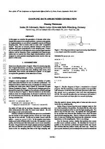
being efficient. It guarantees that the parameters of the channel throughput are time-invariant and thus the transmission features, such as distortions, are related to the amplitude and frequency characteristics of input signal components and not to their occurrence in time. It is worth a remark here, that when the bandwidth of the digitized signal changes in time, the sampling frequency may be locally adapted for satisfy the Shannon theorem. This remark is the foundation of sampling signals at the variable rate and is developed further in this chapter. Fortunately, the ECG signal is predictable in some aspects [3] and has several properties that may be important when considering the local optimization of sampling frequency: - Some extend of regularity may be anticipated, even in case of serious heart diseases and some cooccurrences of signal components are impossible for the physiological reasons. - The full bandwidth is used for short time intervals only (i. e. the QRS complex) and for a large amount of time the local bandwidth is much lower [4], [5]. - The density of medical information is distributed not equally in the signal that makes some parts more important than the remaining sections – this medical point of view converges with the technical notion of information throughput expressed by the local bandwidth (figure 1) [6]. - The ECG signal may be segmented automatically with high reliability as it is done for the diagnostic purpose by commercially available software [7] – the local signal properties highly correlate with the waves start- and endpoints [8] [9]. These interesting properties of the ECG signal motivated us to design an ECG-dedicated recorder that would use the memory resources in more efficient way, but preserve the signal diagnostability typical for devices sampling continuously at high rate. Except for the reasonable use of memory, another important feature provided by such a recorder is its insensitivity to the interference in the frequency band between the current signal bandwidth and the maximum signal bandwidth. For the lack of this interference, the signal sampled at the variable rate differs from the identical signal sampled continuously at the maximum rate, what is sometimes confused with distortions. Actually, there is no reliable measure of distortions for the ECG signal.
Figure 1. Averaged normalized time-frequency planes of main components of heartbeat and the multilead signal in time domain (db5 wavelet, CSE-Multilead database, CSE mo_00001 representative beat)
The Percent Root-mean-square Difference (PRD), used in the evaluation of the numerical experiment, is provided for comparative purpose only. 1
2 n 2 ∑ [x1 (i ) − x 2 (i ) ] PRD = i =1 n ⋅ 100 % 2 [x1 (i ) ] ∑ i =1
(1)
Certainly, for the ECG recorder sampling at the variable rate, the most important issue is the definition of the local sampling rate based on the signal features. Initially, we started with a simple dependency rule successively moving towards the more sophisticated algorithms. Four scenarios were considered as the driving rules for the sampling frequency: - The fixed compromise between the sampling rate and the recording time settled by the doctor at the beginning of the test. This scenario is interesting from the statistical point of view answering the question of the doctors preferences for particular medical cases, but it does not actually involve the change “in fly” of the sampling frequency and thus will not be developed hereafter. - The fixed time interval recorded at exceptionally higher sampling rate triggered by the event button. Unlike the precedent, this scenario may be clinically useful for detailed pursuit of malignant episodes sensed by the patient. Despite the manual settlement, this scenario meets the fundamental criteria on the adaptation of the sampling frequency to the current physiological state. - The intervals of variable duration are recorded at a higher rate accordingly to the results of instantaneous measure of signal bandwidth thresholded by the value of acceptable distortion coefficient (i. e. 5%). This rule involves two stages
typical to automation: the measure of the signal local bandwidth and the control over the sampling frequency. Having a reasonable solution for the correct measure of the instantaneous bandwidth yields a smart algorithm for the management of the distortion. Since the algorithm does not use the knowledge about the temporal distribution of ECG data, it processes any part of the signal equivalently and may be considered as a general-purpose data reduction technique. - The intervals of variable duration corresponding to automatically detected waves are recorded at a higher rate if the signal nature justifies the expectation of wider bandwidth. This rule also involves the measurement and the control. Main novelty is here that the medical information is extracted from the signal by the specialized algorithm as it were used for the diagnostic purpose and applied to influence the sampling parameters. This scenario is believed to be best adapted to the local changes of the signal bandwidth and its clinical importance. It reflects the non-uniform distribution of medical data in the ECG signal and uses the detection of P, QRS and T waves, widely recognized for clinical use, as a foundation of the control over the sampling frequency. Transforming the last scenario into the working device involves many other issues that have to be discussed and optimally solved at the designing stage: - The on-line preprocessing of the ECG aiming at extracting the medical information appropriate for the decision on the local sampling frequency. - The optimal technique of controlling the sampling frequency. - The format of storage and transmission of nonuniformly sampled ECG signals. All these subjects are presented in details in the following section.
Materials and methods Attempting to build the real-world digital recorder sampling the ECG at the variable rate needs to define the fundamental procedures and the data structures suitable for the signal storage and transmission. Measurement of local signal properties: Deriving the medical information representative for the local bandwidth of the signal is the foundation of the correct adaptive adjustment of the sampling frequency. Our previous study on the statistical signal properties [4] [5] and the temporal distribution of medical information in the ECG signal [6] led to the conclusion that the local bandwidth of the signal is best correlated with the occurrence of P, QRS and T waves (figure 1). The physiological background for this approach is also currently studied. The compression algorithm, based on the variable signal bandwidth for P, QRS and T waves, was recently successfully implemented [8] [9]. Another advantage of this approach is that the waves start- and endpoints may be reliably delimited by the time-domain signal segmentation subroutine designed for the purpose of standard ECG diagnostics. The ECG signal segmentation may be performed by any subroutine, but for the reasons of reliability it is important to use the software complying with the diagnostic standards [10]. In our experiment we used a custom-developed subroutine marketed as a firmware of a family of stand-alone automatic 12-lead recorders. Since three years it received wide recognition in the medical world. It was also tested for accuracy against the CSE 12-leads Database [11] and achieved the following positions in the ranking of 20 ECG and VCG reference results: 7-th for T-end delimitation accuracy; 6-th for P-onset and P-end delimitation accuracy; 5-th for QRS-onset and QRS-end delimitation accuracy. The input data was a single lead continuous ECG signal, and the output data was the set of five segmentation points for each recognized heartbeat as described above. For the synchronized occurrence of waves in all simultaneously recorded channels, the segmentation performed for one channel is valid for the others. If multiple recording channels are available, for the reasons of wave detection reliability (particularly Pwave) two channels having best Signal-to-Noise Ratio (SNR) are processed. Controlling the sampling frequency: Controlling the sampling frequency may be performed by the hardware or by the software. In the first case, the sampling frequency changes require a new control word or new trigger rate is to be sent by the master microcontroller to the analog-todigital converter. The sampling frequency may be settled at any frequency with respect to the quantization provided by the frequency divider. Another issue is tuning the anti-aliasing filter each time the sampling frequency changes, but fortunately, thanks to the
integrated switching capacity filters (e. g. MAX 295 [12]), all the operation is sending another control word to the filter chip. However, the oscillatory response of the filter caused by changing the cut-off frequency can hardly, if even, be reduced [13]. For this reason, controlling the sampling frequency by the hardware is not appropriate for the systems, where the sampling frequency is updated relatively frequently. The software control of the sampling frequency consists in the sampling at the maximum constant rate followed by the resampling. The anti-aliasing filters have to be performed by the software aiming at attenuating the frequencies exceeding the half of the target effective sampling rate. In general case, the new frequency is limited only by the condition that the ratio of the old to the new frequencies has to be expressed as the ratio of two integers [14]. However, resampling the signals at any rate, particularly for short intervals, yields the transition effect because the sampling intervals do not fit exactly to the total wave’s length. Considering the above, we decided to perform the sampling frequency control by the software and to limit the possible sampling frequency range to the values being sub-multiples of the maximum rate. This let the medical measurements to be done on the digital signal sampled at the maximum rate immediately before the resampling. Additionally, the computationally expensive resampling may be substituted by subsampling, or saying straight, discarding every odd sample of the original signal. As an adequate and efficient tool for signal decimation we used the orthogonal wavelet decomposition [15]. Four decimation stages are sufficient for cover the range of sampling frequency variability from the maximum 1024 Hz to the minimum 64 Hz. Main advantages of using wavelet filters are the following: - The anti-aliasing filters are no longer required because the filter pair splitting the bandwidth assures each subband to be sampled correctly at a half of initial rate. - The wavelet decomposition does two required tasks (filtering and subsampling) in one step. - The orthogonal decomposition guarantees no data loss and no redundancy thus two resulting subbands complete each other perfectly. Wavelets often make think of heavy mathematics, hence they are not willingly implemented in the realworld devices. To preserve the perfect reconstruction property, the orthogonal decomposition has to be performed without the round-off errors and, if one of the 'classical' wavelets is used, requires a floating-point Digital Signal Processor (DSP). In addition, the use of a floating-point transform causes the real-valued timefrequency representation and the resulting signal sampled at the variable rate is also real-valued or the additional quantization issues the round-off errors. An interesting alternative is the use of wavelets that map integers to integers [16]. For the novelty of this issue the principles of this kind of decomposition are worth to be reminded hereby.
Figure 2. The computing scheme of one stage of wavelet decomposition using M lifting steps The single stage of lifted wavelet signal decomposition (figure 2) starts with splitting the signal into two half-length components, what is called the trivial wavelet transform or the Lazy Wavelet. Next, the half-band properties of these strings are improved using the lifting and the dual lifting alternately. The lifting operation means here increasing the number of vanishing moments of a wavelet without any changes of its properties. The Lazy Wavelet splits the signal into two strings:
s 1( ,0l ) = s 1 , 2 l d 1(,0l ) = s 1 , 2 l + 1
(2)
first, containing only even-indexed samples second, containing only odd-indexed samples
A dual lifting step, despite the name used first, consists of applying a low-pass integer filter p to the even samples and subtracting the results from the corresponding odd samples:
d 1(,il) = d 1(,il−1) −
∑
p k( i ) ⋅ s1(,il−−1k)
(3)
k
A primal lifting step, used immediately thereafter, consists of applying a high-pass integer filter u to the odd samples and subtracting the results from the corresponding even samples:
s 1(,il) = s 1(,il− 1 ) −
∑u
(i) k
⋅ d 1(,il )− k
(4)
k
After M lifting steps, the even samples become lowpass coefficients and the odd samples become high-pass coefficients, with applying the scaling coefficient K:
1 ⋅ s 1( ,Ml K = K ⋅ d 1(,Ml
s1,l =
)
d 1,l
)
(5)
In our application, we used the simplest Haar filters for p and u. The first difference acts as high-pass filter, and the average acts as low-pass filter:
d 1,l = s 0 , 2 l +1 − s 0 , 2 l 1 s 1 ,l = ( s 0 , 2 l + s 0 , 2 l +1 ) 2
(6)
It is worth a remark, that the lifting algorithm generates two subsampled strings: the decimated lowpass coarse signal and the detail high-pass signal, exactly like one decomposition stage of a traditional wavelet transform. The lifting scheme is a reversible process; thus the resulting stings contain complete original information. Thanks to perfect reconstruction property, it corresponds to reversible wavelet decomposition. The whole processing involves the integer-format values only. For the average, the result is rounded towards -∞ or +∞, depending on the difference’s least significant bit, since the sum and difference of two integers may only be even or odd both. The output data format: The statistically largest part, nearly a half of the signal length is sampled at the minimum rate, while less than a quarter only is sampled at the maximum rate. For this reason the decimation is performed continuously yielding an uninterrupted signal being the simplest approximation of the real ECG. Within the P, QRS and T waves, that start- and endpoints are valid also in the time-frequency domain, the signal is completed by the components belonging to high frequency bands until the local cut-off frequency is reached. The high frequency components are thus not continuous because their appearance depends on the local bandwidth of the ECG. The existence of gaps involves an additional synchronization byte referring to the continuous signal. Composing the resulting signal by including or not the adjacent frequency bands is very simple, and may be performed in a lossless way thanks to the orthogonality of decomposition. Adding the high frequency information to the approximation sampled at the low rate increases locally the effective sampling rate expanding the bandwidth accordingly to the Shannon theorem. The output data consists of two components (fig. 3): - Coarse approximation (CA) being a continuous low-frequency time-domain signal. - Separate details (SD) being the complementary information added locally when the extended signal bandwidth is required. This approach has several advantages: - The CA component is a continuous time-domain signal containing all fundamental features of the ECG with the effective sampling rate of 64 Hz. If the SD component is lost due to processing errors, computing the heart rate or distinguishing the morphology of Supraventricular and Ventricular beats is still possible relying on the CA component alone. - The CA component is ready for displaying and when only a general insight is required (i. e. monitoring in an ICU) [17], no further processing is necessary. - The SD component is occasional (i. e. not continuous) that gives the opportunity for individual adjustment of the amplitude discretization scale adaptively to the local signal properties.
a) 256...512 Hz 128..256 Hz
'separate details' (SD) component high frequencies 32 ... 512 Hz occasional
64...128 Hz 32...64 Hz 0...32 Hz
'coarse approximation' (CA) component low frequencies 0 ... 32 Hz continuous
b)
Figure 3. Splitting the ECG signals into two components: coarse approximation (CA) and separate details (SD). a) – the principle of splitting, b) – the components’ behavior for an example real signal -
The SD component is computed only for short sections of the signal that are automatically detected during segmentation: P, QRS and T waves. The complete processing diagram flow: The complete processing diagram flow is displayed in the figure 4. It consists of the previously described subroutines: segmentation of the ECG, time domain to time-frequency domain signal transform and splitting the time-frequency representation into the continuous CA and occasional SD components. For the reason of the signal segmentation performed at the beginning and for easier data management the analog signal is sampled at the constant maximum rate. In the output signal, however, the data stream subsampled to the basic rate is complemented by the additional samples from the SD component. These fall at the fraction of the basic sampling interval and thus the effective sampling frequency increases. This justifies the name of the newly proposed ECG recorder yielding a signal sampled at the variable rate.
sampling (fs = 1024 Hz)
P, QRS and T waves detection
integer time-frequency transform
components split
CA signal
SD strings
Figure 4. The complete processing diagram flow
Results The algorithm was coded in C/Matlab for the testing purpose and than implemented in the AD-21066 DSP chip. During the tests, we used the CSE-Multilead Database [11] with all signals resampled up to 1024 Hz. The database features the statistic reference start- and endpoint for P, QRS and T waves in each signal, but for the use of the segmentation algorithm tested beforehand against the CSE Database, we did not take them in consideration.
analysis is concluded from its wide recognition in the medical world. The bandwidth control procedure relies on the assumed local properties of the ECG. The function defined in the time-frequency plane describes the expected bandwidth at any time point with reference to the waves start- and endpoints computed beforehand. The management of the output signal was implemented in a most straightforward way: a signal sampled at the constant basic rate is accompanied by the high frequency components where needed. Thanks to
Table 1. Results for CSE Database files (PRD differences in %)
CSE-ID 1 2 3 4
data reduction ratio 3.6132 2.8996 3.2676 3.9375
global difference 3.5871 1.6688 6.2218 4.2162
P-wave difference 0.3273 0.2270 1.2060 0.3577
QRS-wave difference 0.3351 0.3615 1.0147 0.3144
T-wave difference 0.4443 0.4321 1.1487 0.3810
extra-wave difference 2.3226 1.1833 4.1033 2.7979
123 124 125 mean value max value
4.3224 4.5840 4.0652 4.2666 8.8867
3.3980 2.7566 5.5942 4.7463 20.485
0.2499 0.2602 0.3888 0.3816 1.4572
0.3225 0.4024 0.2975 0.4028 1.0839
0.2650 0.2613 0.2645 0.4990 2.3704
1.8455 1.0076 3.0423 3.6303 15.938
The numerical experiment we carried out was expected to demonstrate the capability of the algorithm to locally adjust the data stream in correlation to the ECG contents. Additionally the measure of dissimilarity between the signal sampled continuously at full rate and the signal sampled at variable rate was performed using the PRD method. Thanks to simultaneous multilead recording, we can process all traces of the ECG signal as if they were independent, although having common segmentation points. All data reported in Table 1 as a result for a particular CSE file, is in fact a mean value of these 15 traces. Figure 5 displays the statistical results of the memory management. CSE files are sorted accordingly to the data reduction ratio and the PRD difference to the original signal. Apart from global values, additional criteria for sorting were local PRD difference values computed for the waves separately. Discussion The idea of sampling an electrocardiogram at the variable rate has been brought alive. The intelligent recorder's output is a data stream representing the signal digitized at adaptively adjustable time interval. The adaptivity is based on the medical findings, typically used for the diagnostic purpose, derived automatically from the signal itself. The high reliability of this
the perfect reconstruction property of the wavelet transform used, the signal in its conventional form sampled at the constant maximum rate may be synthesized in a lossless way. Although the memory management has been improved significantly with the newly proposed algorithm of sampling at the variable rate, the authors consider this successful implementation as a beginning of new challenging research. There are still several limitations in the system that should be overcome yielding the further improvements in the ECG-dedicated recorders. These limitations are: - The sampling frequency is changed only by the factor of two, provided the decimation performed by the wavelet decomposition is dyadic; this step is suspected to be too coarse for closely follow the bandwidth variability function. - The sampling frequency is adjusted once for the whole wave that simplifies the data management; the time resolution for this adjustment should be increased to the maximum extent satisfying the uncertainty (Heisenberg) theorem. - Apart from the P, QRS and T waves, other complex signal irregularities should be involved to the sampling rate adjustment. Special care must be taken for the P wave aberration such as atrial flutter or nodal beats, because they do not appear like a regular wave.
Figure 5. Compression ratios (upper part) and distortion coefficients (lower part) on CSE files set.
Figure 6. Example of temporal distortions distribution for the CSE-001 signal along with the lead aVL reconstructed (the top-first) and original (the top-second) traces. Below two detailed views focused on the P-onset neighborhood and T-end neighborhood. Note that the vertical axes’ values apply to distortion plots only.
-
For all other cardiac events that cannot be detected in an unambiguous way or that typical bandwidth is undefined, the system must assign the bandwidth that would be sufficient for the lossless storage; the current implementation recognizes only regular waves. The ultimate use of fixed-point data representation is very important advantage of the implemented algorithm. It guarantees the compatibility with any fixed-point DSP and, despite the use of advanced mathematics, opens the possibility of wide-range use. Acknowledgement This work was supported by ZEM Aspel S.C. References [1] MOSS A. J., STERN S. "Noninvasive Electrocardiology – Clinical Aspects of Holter Monitoring", W. B. Saunders Co. Ltd. Cambridge University Press, 1996. [2] PROAKIS J. “Digital communications” 2-nd ed. New York McGraw-Hill 1989 [3] JALALEDDINE S. M. S., HUTCHENS C. G., STRATTAN R. D., COBERLY W. A. “ECG data compression techniques – a unified approach” IEEE Trans. Biomed. Eng. vol. 37 pp 329-343, 1990 [4] AUGUSTYNIAK P, TADEUSIEWICZ R. "The Bandwidth Variability of a Typical Electrocardiogram" in proceedings of European Medical and Biological Engineering Conference EMBEC ’99, Wien Austria, 04-07.11.1999 [5] AUGUSTYNIAK P. "Measuring the local bandwidth of the ECG with noise estimation for lower frequencies" (in Polish) in proceedings of the 2-nd Workshop on Measurements and Modelling in Medicine, Krynica Górska 8-12.05.2000 pp. 191 – 196 [6] AUGUSTYNIAK P. "The assesment of the local information density in an electrocardiogram by cancelling coefficients in time-frequency domain" (in Polish) in proceedings of the 2-nd Workshop on Measurements and Modelling in Medicine, Krynica Górska 8-12.05.2000 pp. 221 - 230
[7] MORLET D. "Contribution a l'analyse automatique des electrocardiogrammes – algorithmes de localisation, classification et delimitation precise des ondes dans le systeme de Lyon" (in French) – PhD these INSA-Lyon 1986 [8] AUGUSTYNIAK P. "Distinct compression of the ECG upper octaves" in proceedings of the 10-th Conference on Biocybernetics and Biomedical Engineering, Warszawa 02-04.12.1999 [9] AUGUSTYNIAK P., TADEUSIEWICZ R. "The ECG-dedicated compression method using high frequency patterns" in proceedings of First International Conference on Advances in Medical Signal and Information Processing, Bristol, United Kingdom, 4-6.09.2000 pp. 257 - 264 [10] WILLEMS J. L., ARNAUD P., VAN BEMMEL J. H. "Assessment of the performance of electrocardiographic computer programs with the use of a reference data base" Circulation, 1985, Vol. 71 No 3, p 523-534. [11] WILLEMS J. L. "Common Standards for Quantitative Electrocardiography" 10-th CSE Progress Report, 1990. Leuven: ACCO publ., 1990, 384p. [12] "Datasheet of the Switching Capacity filters MAX 291, MAX 292, MAX 295 and MAX 296" www.maxim.com [13] RANDALL R. B., Tech B. A. "Frequency analysis", Bruel & Kjaer, 1987 [14] KESTER W. "Mixed-Signal and DSP Design Techniques" Analog Devices, USA, 2000 [15] AKAY M. “Wavelets in Biomedical Engineering” Annals of Biomed. Eng. Vol. 23, 531-542, 1995 [16] CALDERBANK A. R., DAUBECHIES I., SWELDENS W. and YEO B. "Wavelet transforms that map integers to integers", technical report, Princetown Univ., 1996 [17] NORMANN R. A. "Principles of Bioinstrumentation" University of Utah, John Wiley & Sons Inc., New York 1988