ECG SIGNAL CLASSIFICATION BASED ON SVM Radovan Smíšek Doctoral Degree Programme (1), FEEC BUT E-mail:
[email protected]
Supervised by: Jana Kolářová E-mail:
[email protected]
Abstract: Cardiovascular diseases nowadays represent the most common cause of death in Western countries. Long-term ECG recording is modern method, because it allows to detect sporadically occurring pathology. We designed an automatic classifier to detect five pathologies (AAMI standard) by SVM method. The classifier was tested on the entire MIT-BIH Arrhythmia Database with an accuracy of 99.17 %. We also compared the quality of parameters entering the classifier. Keywords: ECG classification, support vector machines, SVM, MIT-BIH database 1. INTRODUCTION Classification of ECG is still the current task in the field of signal processing. Reliable classifier is needed especially for modern long-term ECG (Holter, telemetry). Cardiologist cannot see the entire signal that is often sensed several days. Classifier helps to find pathologies in the signal. The main aim of this study is to propose a reliable classification algorithm and test it on a standard publicly available database. Several classification methods were developed to classify the ECG in other studies. Frequently used methods are support vector machines (SVM) [1, 2], artificial neural network (ANN) [3, 4], nearest neighbor (NN) [5, 6] and decision tree (DT) [7, 8]. In this study, we used SVM method. Cycles of ECG are not usually entering the classifier, but parameters (called features) derived from the signals are entering. Features derived from time domain are usually used. This may be the width and height of the waves, skewness, kurtosis, area under the curves and so on. [1, 3, 5] RR intervals are also frequently used features. Other features may also be used in more advanced algorithms. It can be features derived from the frequency or time-frequency domain, features calculated using wavelet transform and higher order statistics features (HOS). [1, 2, 3, 4, 5] Selection of entering features is very important. The classifier is unable to distinguish categories, if there is only small number of features or features are chosen inappropriately. A large number of features leads to large computational complexity and the resulting success rate may be lower. Universal recommendation of number of features is not easy to create. One of the aims of this study is to compare the various types of features and determine their suitability. MIT-BIH Arrhythmia Database [9, 10] was selected for testing the quality of the classifier. The database contains 48 records, which are about 30 minutes long. Each record has a two leads. Sampling frequency is 360 Hz, the resolution is 11-bit within a 10 mV range. Each QRS complex was classified by cardiologists into one of the fifteen heartbeat types. These groups can be united into 5 groups according to the AAMI standard [11]: supraventricular ectopic beat (S), ventricular ectopic beat (V), fusion beat (F), unknown beat (Q) and any heartbeat not in the S, V, F or Q classes (N). The entire database contains 90,510 N beats, 2774 S beats, 7703 V beats, 802 F beats and 8031 Q beats. In this work, we classified into these five groups. [9, 10]
365
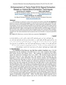
The following section describes the test data, pre-processing, features extraction and description of the selected classifier. In chapter 3 are presented and discussed the results. Finally, the conclusion of the paper is in chapter 4. 2. METHODS Basic steps of the proposed algorithm are shown in the block diagram in Figure 1. Each block will be described in detail in this section.
Figure 1: Block diagram of the proposed algorithm 2.1.
PRE-PROCESSING
During the pre-processing was removed baseline wandering. It was removed by zeroing the spectral components before calculation of features. The advantage of this method compared to conventional filters is that it does not introduce delays. We removed components corresponding to the frequency band 0-0.5 Hz. Removing this band has no significant influence on the useful signal. 2.2.
FEATURES EXTRACTION
Entry into the classifier is a set of features which characterize the QRS complex. Values of each feature should be the most different between groups that we want to separate and on the other hand, the values of feature should be approximately the same for complexes of the same type. Features used in this study were derived from the time domain, frequency domain, time-frequency domain and time-scale domain (calculated using the continuous wavelet transform). Meaningfulness of using these features is described in the following paragraph. Features derived from the time domain to describe the morphology of the QRS complex (such as R wave amplitude, skewness and kurtosis of QRS complex, area under the QRS complex and QRS width) vary depending on place of origin of action potential and method of action potential conduction. QRS complex has sharp edges, and it is narrow at the physiological state. When the activation pulse is formed in the chamber or it is blocked at some point in its transmission, QRS complex becomes wider and has less sharp edges. The features in the frequency domain are thus also changing. If there are less sharp edges of the QRS complex, the energy of high frequency components decreases. Decomposition using wavelet transform (WT) allows detailed analysis the useful low frequency components, while high frequency components are not decomposed. A special group of features are RR intervals before, after and in the wider area of the analyzed QRS complex. Using the RR intervals is necessary to distinguish supraventricular ectopic beats, because these beats do not differ in shape from physiological QRS complex. The difference is only in the previous and subsequent RR interval. The importance of different groups of parameters is discussed in the section 3. All parameters were calculated from both available leads. Features were normalized before entering the classifier. This means that the software centers and scales each feature by the mean and standard deviation [12]. 2.3.
CLASSIFICATION
Support vector machine (SVM) method was used for classification in this work. SVM is a supervised machine learning method. Groups which should be separated are not usually linearly separable in the original n-dimensional space of features. SVM transforms original n-dimensional space into m-dimensional space (m > n), in which groups can be separated. Kernel transformation is used
366
for this transfer. We used polynomial kernel transformation of second order in this work. The aim is to find group separator, when groups distance from the separator is largest possible. SVM use to search for separator support vectors, which are the points near the separator. The basic version of the SVM is applicable for the separation of only two groups. There are several modified methods for applying SVM to separate multiple groups. Method one vs. one is used in this work. The classifier is gradually learned to distinguish each of two groups separately. We need therefore in total (K×(K-1)/2) binary learning, where K is the number of separated groups (in this case 5 and therefore total 10). [13] 3. RESULTS AND DISCUSSION This chapter describes the achieved accuracy of five-category classification (supraventricular ectopic beat (S), ventricular ectopic beat (V), fusion beat (F), unknown beat (Q) and any heartbeat not in the S, V, F or Q classes (N)). In this chapter, it is also compared the importance of using different types of features. Training and testing of the classifier is realized by ten-fold cross-validation. All QRS complexes of all signals are randomly divided into ten equally sized subgroups. Nine subgroups are used for training and one for testing the classifier. Gradually, each of the subgroups is selected as the test (there will be ten testing in total). Classification accuracy is described by variables: accuracy (Acc), sensitivity (Se), specificity (Sp), positive predictive values (PV+) and negative predictive values (PV−). These variables are calculated as follows: Acc = (TPN + TPS + TPV + TPF + TPQ) / Y,
(1)
Sei = TPi / (TPi + FNi),
(2)
Spi = TNi / (TNi + FPi),
(3)
PV+i = TPi / (TPi + FPi),
(4)
PV−i = TNi / (TNi + FNi),
(5)
where TP (true positive) is the number of correctly classified complexes into the analyzed group, TN (true negative) is the number of correctly classified complexes into other groups than the analyzed group, FP (false positive) is the number of incorrectly classified complexes into the analyzed group, FN is the number of incorrectly classified complexes into other groups than the analyzed group, Y is the number of all classified QRS. Subscript indicates the analyzed group (N, S, V, F, Q or i), where i stands for any group. Se, Sp, PV+ and PV− are calculated for each group separately. Achieved accuracy is 0.9917 for the proposed classifier using all the features. Sensitivity, specificity, positive and negative predictive value of the proposed classifier for each group are shown in the Table 1. Hardest classificable groups are S and F, where the sensitivity is low. This means that large number of these complexes was classified into the wrong group. This may be due to small number of the complexes of S and F type compared with other types, so classifier was not learned sufficiently for these types. Confusion matrix is in Table 2. Above conclusions are confirmed. Confusion matrix shows that it is significantly more N complexes compared to other types of complexes. On the other hand, F complexes are very little. Therefore, classification of N complexes has a greater influence on the resulting classification accuracy than other complexes. On the other hand, classification of F complexes has little effect on the calculated accuracy of the classifier.
367
N S V F Q Se 0.9981 0.8641 0.9784 0.7968 0.9951 Sp 0.9695 0.9990 0.9985 0.9996 0.9998 PV+ 0.9935 0.9592 0.9801 0.9342 0.9969 PV− 0.9911 0.9965 0.9984 0.9985 0.9996 Table 1: Sensitivity (Se), specificity (Sp), positive predictive values (PV+) and negative predictive values (PV−) of classifier for each type of QRS complex
Correct groups
N S V F Q
Output groups S V F 83 60 9 2397 24 1 16 7537 35 3 59 639 0 10 0 Table 2: Confusion matrix
N 90342 352 106 101 29
Q 16 0 9 0 7992
Using just certain groups, classification accuracy is shown in Table 3. Group All means all the features; KW is a group in which are used features selected by Kruskal-Wallis test from all features; TM are morphological features derived from the time domain; RR are features including RR intervals around QRS; Fr are features derived from the frequency domain; TF are features derived from the time-frequency domain and TS are features derived from the time-scale domain. Using ALL or KW is the most successful. Morphological features derived from the time domain are the most successful from separate groups. Conversely, classification is inappropriate by features derived only from the frequency domain or by RR intervals. Acc Se Sp PV+ PV−
All KW TM RR Fr TF TS 0.9917 0.9881 0.9770 0.7411 0.7243 0.9599 0.9179 0.9265 0.8989 0.8496 0.3338 0.4239 0.7538 0.6618 0.9933 0.9894 0.9803 0.8330 0.8400 0.9613 0.9377 0.9728 0.9719 0.9266 0.3184 0.4461 0.9206 0.7344 0.9968 0.9960 0.9899 0.8324 0.8335 0.9861 0.9516 Table 3: Success of classifier using only selected group of features
The results of this study can be compared with previous studies. This comparison is shown in Table 4. Accuracy of the classifier described in this paper is approximately in the middle between studies mentioned here. The benefit of this study is comparing the above-mentioned types of features. Number Number Accuracy Features of target Authors of records [%] groups SVM RR, TM 5 33 98.8 Zidelmal et al. [1] SVM Samples 4 48 99.64 Thanapatay et al. [2] ANN Samples 6 32 94.26 Das et al. [3] NN Samples 4 44 99.61 Chang et al. [6] NN TM, Fr, HOS, TS 5 48 93.59 Kutlu et al. [5] SVM TM, RR, Fr, TF, TS 5 48 99.17 This article Table 4: Comparison of the results of previous studies with the results obtained in this work
Classification method
368
4. CONCLUSION This article describes the classifier of QRS complexes in the ECG signal. We described all steps of the classification process. We used SVM method for classification and we used features derived from the time domain, frequency domain, time-frequency domain and time-scaling domain. Classification is realized into five types according to the AAMI standard. We tested the classifier on the entire MIT-BIH Arrhythmia database. The benefit of this study is comparing the types of features. Types of features were compared according to their suitability (it means classification accuracy using these features). Groups TM (Acc = 97.70 %) and TS (Acc = 95.99 %) were successful. Overall, the most successful was a group of all features (Acc = 99.17 %). REFERENCES [1]
ZIDELMAL, Z, AMIROU, A, OULD-ABDESLAM, D and MERCKLE, J. ECG beat classification using a cost sensitive classifier. Computer Methods and Programs in Biomedicine. 2013. Vol. 111, no. 3, p. 570–577.
[2]
THANAPATAY, Dusit, SUWANSAROJ, Chaiwat and THANAWATTANO, Chusak. ECG beat classification method for ECG printout with Principle Components Analysis and Support Vector Machines. ICEIE 2010 - 2010 International Conference on Electronics and Information Engineering, Proceedings. 2010. Vol. 1, no. Iceie, p. 72–75.
[3]
DAS, Manab Kumar, MEMBER, Student and ARI, Samit. Electrocardiogram ( ECG ) Signal Classification using S-transform , Genetic Algorithm and Neural Network. 1st International Conference on Condition Assessment Techniques in Electrical Systems. 2013.
[4]
JIANG, Wei and KONG, Seong G. Block-based neural networks for personalized ECG signal classification. IEEE Transactions on Neural Networks. 2007. Vol. 18, no. 6, p. 1750– 1761.
[5]
KUTLU, Yakup and KUNTALP, Damla. A multi-stage automatic arrhythmia recognition and classification system. Computers in Biology and Medicine. 2011. Vol. 41, no. 1, p. 37– 45.
[6]
CHANG, Kuang-Chiung, WEN, Cheng, YEH, Ming-Feng and LEE, Ren-Guey. Comparison of Similarity Measures for Clustering of Qrs Complexes. Biomedical Engineering: Applications, Basis and Communications. 2005. Vol. 17, no. 06, p. 324–331.
[7]
PARK, Juyoung and KANG, Kyungtae. PcHD: Personalized classification of heartbeat types using a decision tree. Computers in Biology and Medicine. 2014. Vol. 54, p. 79–88.
[8]
SAMAD, Saleha, KHAN, Shoab a., HAQ, Anam and RIAZ, Amna. Classification of Arrhythmia. International Journal of Electrical Energy [online]. 2014. Vol. 2, no. 1, p. 57–61.
[9]
MOODY, G. B. and MARK, R. G. The impact of the MIT-BIH arrhythmia database. IEEE Engineering in Medicine and Biology Magazine. 2001. Vol. 20, no. 3, p. 45–50.
[10] GOLDBERGER, Ary L, AMARAL, Luis A N, GLASS, Leon, HAUSDORFF, Jeffrey M, IVANOV, Plamen Ch, MARK, Roger G, MIETUS, Joseph E, MOODY, George B, PENG, Chung-kang and STANLEY, H Eugene. Current Perspective. 2000. [11] CHAZAL, Philip De Chazal Philip De, O’DWYER, M. and REILLY, R.B. Automatic classification of heartbeats using ECG morphology and heartbeat interval features. IEEE Transactions on Biomedical Engineering. 2004. Vol. 51, no. 7, p. 1196–1206. [12] TemplateSVM. Mathworks [online]. c1994-2016 [cit. http://www.mathworks.com/help/stats/templatesvm.html
2016-03-06].
[13] Fitcecoc. MathWorks [online]. c1994-2016 [cit. 2016-03-06]. http://www.mathworks.com/help/stats/fitcecoc.html?s_tid=srchtitle
369
Dostupné Dostupné
z: z: