ORIGINAL E n d o c r i n e
ARTICLE R e s e a r c h
Ectopic Pregnancy Is Associated with High Anandamide Levels and Aberrant Expression of FAAH and CB1 in Fallopian Tubes Alpha K. Gebeh, Jonathon M. Willets, Emma L. Marczylo, Anthony H. Taylor, and Justin C. Konje Endocannabinoid Research Group (A.K.G., J.M.W., A.H.T., J.C.K.), Reproductive Sciences Section, Department of Cancer Studies and Molecular Medicine, University of Leicester, Leicester LE2 7LX, United Kingdom; Directorate of Women’s and Perinatal Services (A.K.G., J.C.K.), Department of Obstetrics and Gynaecology, University Hospitals of Leicester National Health Service Trust, Leicester LE1 5WW, United Kingdom; and Systems Toxicology (E.L.M.), Medical Research Council Toxicology Unit, University of Leicester, Leicester, LE1 9HN, United Kingdom
Context: Ectopic pregnancy is associated with significant morbidity and mortality, but the molecular mechanisms underlying this condition remain unclear. Although the endocannabinoids, Narachidonoylethanolamine (anandamide), N-oleoylethanolamine, and N-palmitoylethanolamine, are thought to play a negative role in ectopic pregnancy, their precise role(s) within the fallopian tube remains unclear. Anandamide activates cannabinoid receptors (CB1 and CB2) and, together with its degrading [e.g. fatty acid amide hydrolase (FAAH)] and synthesizing enzymes (e.g. N-acylphosphatidylethanolamine-specific phospholipase D), forms the endocannabinoid system. High anandamide levels are associated with tubal arrest of embryos in mice and may have a similar role in women. Objective: The aims were to quantify the levels of the endocannabinoids and evaluate the expression of the modulating enzymes and the cannabinoid receptors in fallopian tubes of women with ectopic pregnancy compared to those of nonpregnant women. Design and Setting: We conducted a prospective study at the University Hospitals of the Leicester National Health Service Trust. Participants and Methods: Fallopian tubes collected from women with ectopic pregnancy and nonpregnant women with regular menstrual cycles were used for quantification of endocannabinoids by ultra-HPLC tandem mass spectrometry, were fixed in formalin for immunohistochemistry, and had RNA extracted for RT-quantitative PCR or protein extracted for immunoblotting. Results: Anandamide, but not N-oleoylethanolamine and N-palmitoylethanolamine, levels were significantly higher in ectopic fallopian tubes. Endocannabinoid levels from isthmus to ampulla were not significantly different. Cannabinoid receptors and endocannabinoid modulating enzymes were localized in fallopian tube epithelium by immunohistochemistry and showed reduced CB1 and FAAH expression in ectopic pregnancy. Conclusion: High anandamide levels and reduced expression of CB1 and FAAH may play a role in ectopic implantation. (J Clin Endocrinol Metab 97: 2827–2835, 2012)
ISSN Print 0021-972X ISSN Online 1945-7197 Printed in U.S.A. Copyright © 2012 by The Endocrine Society doi: 10.1210/jc.2012-1780 Received March 26, 2012. Accepted May 11, 2012. First Published Online June 14, 2012
Abbreviations: AEA or anandamide, N-Arachidonoylethanolamine; CB1 and CB2, cannabinoid receptor types 1 and 2; ECS, endocannabinoid system; FAAH, fatty acid amide hydrolase; GAPDH, glyceraldehyde-3-phosphate dehydrogenase; IQR, interquartile range; NAPE-PLD, N-acyl-phosphatidylethanolamine phospholipase D; OEA, N-oleoylethanolamine; PEA, N-palmitoylethanolamine; RT-qPCR, RT-quantitative PCR; UHPLC-MS/MS, ultra-HPLC tandem mass spectrometry.
J Clin Endocrinol Metab, August 2012, 97(8):2827–2835 Downloaded from https://academic.oup.com/jcem/article-abstract/97/8/2827/2823396 by guest on 09 April 2018
jcem.endojournals.org
2827
2828
Gebeh et al.
Endocannabanoids in Ectopic Pregnancy
ctopic pregnancy, which complicates up to 2% of pregnancies, is associated with significant morbidity and remains the most common cause of maternal death in the first trimester of pregnancy (1, 2). More than 98% of ectopic pregnancies occur in the fallopian tube, with at least 80% of these occurring in the ampulla (3). The mechanisms whereby an embryo implants in sites other than the uterus, especially in the absence of previous salpingitis, are still not fully understood, although several hormone-cytokine networks including interleukins, the activins, prokineticins, and mucins have been implicated (4). Accumulating evidence suggests that one such hormone-cytokine network— i.e. endocannabinoids [e.g. N-arachidonoylethanolamine (AEA or anandamide), N-oleoylethanolamine (OEA), and N-palmitoylethanolamine (PEA)], the cannabinoid receptors [type 1 (CB1) and type 2 (CB2)], and the enzymes involved in their synthesis [N-acyl-phosphatidylethanolamine phospholipase D (NAPE-PLD)] and degradation [fatty acid amide hydrolase (FAAH)], collectively referred to as the endocannabinoid system (ECS) (5)—may be involved in oviductal embryo transport through the fallopian tube (6, 7). Although OEA and PEA are not known to activate cannabinoid receptors, they are thought to modulate the effects of AEA at cannabinoid receptors by inhibiting its metabolism, resulting in a prolongation of its biological effect—the so-called “entourage effect” (8 –11). In mice, AEA production and degradation are tightly regulated such that an appropriate “anandamide tone” or balance is created, enabling normal oviductal transport of embryos (6, 7). Normal CB1 and FAAH expression and activity are crucial to early pregnancy success, especially because studies have shown that FAAH, CB1, or CB1/CB2 double knockout mice have a significant number of embryos retained in their oviducts compared with wild-type mice or CB2 knockout mice (6, 7). Low FAAH and high AEA levels have been linked with poor embryo development and their arrest in the oviduct. These adverse effects of FAAH dysfunction were substantially improved after CB1 function was attenuated by SR141716 (CB1 receptor antagonist), suggesting that the impaired preimplantation events arising from FAAH dysfunction were possibly the result of elevated AEA and or aberrant CB1 signaling (6). In human placentae, low FAAH and high CB1 expression levels are associated with spontaneous miscarriage, whereas nuclear localization of FAAH is linked with recurrent miscarriage (12, 13). These observations suggest that a disruption in the delicate balance between the various components of the ECS, rather than an absolute change in one component, may lead to varying presentations of early pregnancy failure. Although these studies provide evidence of a potential role of the ECS in the mechanisms of oviductal implanta-
E
Downloaded from https://academic.oup.com/jcem/article-abstract/97/8/2827/2823396 by guest on 09 April 2018
J Clin Endocrinol Metab, August 2012, 97(8):2827–2835
tion in mice, its role in human tubal embryo transport is unclear because only one study in humans has associated the attenuation of CB1 expression with ectopic pregnancy (14). However, if the ECS is shown to have a role in the pathogenesis of tubal transport and ectopic implantation, then such information may prove beneficial in ectopic pregnancy care because pharmacological intervention targeting the ECS may be used in the treatment or prevention of ectopic pregnancies. Additionally, an understanding of the involvement of the ECS in ectopic pregnancies may suggest possible biomarkers for early diagnosis. Here, we determined whether an “endocannabinoid tone” across the tube similar to that described in mice models exists, whether oviductal endocannabinoid levels are significantly different from controls, and also the expression levels of endocannabinoid receptors and their metabolizing enzymes in the fallopian tubes of control women and women with ectopic pregnancy.
Subjects and Methods Ethics statement Ethics approval was obtained from the Leicestershire and Rutland Local Research Ethics Committee, and all research procedures were conducted according to the principles expressed in the Declaration of Helsinki. The study was sponsored and conducted under the scrutiny of the Research and Development Office of the University Hospitals of Leicester National Health Service (NHS) Trust, and all participants signed informed consent forms before entering the study.
Participants The study group consisted of women attending the University Hospitals of Leicester NHS Trust as emergencies with a diagnosis of ectopic pregnancy. Nonpregnant volunteers were women attending for total abdominal hysterectomy and bilateral salpingoophorectomy for benign gynecological conditions such as fibroids and dysfunctional uterine bleeding, and they were divided into two groups: follicular phase (d 4 –9) and luteal phase (d 20 –25), based on their last menstrual period and endometrial histology. For both the study and control groups, women on any prescription medication, hormonal or recreational drugs (including cannabis), those with chronic medical conditions on treatment (e.g. diabetes mellitus, chronic obstructive airway disease, hormone replacement therapy, or endocrine disorders), or previous pelvic inflammatory disease were excluded.
Tissue collection Fallopian tubes were collected as duplicates immediately after total abdominal hysterectomy and bilateral salpingoophorectomy or salpingectomy for ectopic pregnancy. The first was washed with sterile PBS and fixed in 10% neutral buffered formalin for 4 d before being embedded in paraffin wax (15), and the second was placed into a sterile polypropylene tube, snapfrozen in liquid nitrogen, and stored at ⫺80 C for the quantification of endocannabinoids, their binding receptors, and mod-
J Clin Endocrinol Metab, August 2012, 97(8):2827–2835
ulating enzymes. All specimens obtained were free of trophoblast and were from the ampulla, except for endocannabinoid gradient studies, where extractions were performed from each segment (isthmus, ampulla, and fimbria) of the fallopian tube.
Quantification of the endocannabinoids Endocannabinoids were quantified in the fallopian tubes as described previously for solid tissue measurements (16). Briefly, tissue samples were pulverized under liquid nitrogen with a pestle and mortar, and internal standards (12.5 pmol/g AEA and OEA and 25 pmol/g PEA; Cayman Chemical, Ann Arbor, MI) were added per 100 mg of tissue suspended in 5% o-phosphoric acid/95% deionized water and homogenized with a glass PotterElvehjem homogenizer before centrifugation at 1500 ⫻ g for 30 min at 4 C. The supernatant was transferred to preconditioned cartridges (17) and washed with deionized water, and the eluted endocannabinoids were dried under a constant stream of nitrogen before being reconstituted in 80 l of acetonitrile. The endocannabinoids were separated and measured using an ultra-HPLC tandem mass spectrometry (UHPLC-MS/MS) system, as described (16). Homogenized samples were processed in duplicate by solidphase extraction and quantified by UHPLC-MS/MS in triplicate. Details of the UHPLC MS/MS gradient conditions and transitions employed have been published previously (16, 17).
RNA isolation and cDNA synthesis Tissues (50 mg) were pulverized under liquid nitrogen with a pestle and mortar, and total RNA was extracted using QIAGEN miRNeasy (QIAGEN, Crawley, UK) mini kit, according to the manufacturer’s instructions. The RNA obtained was treated with DNase I to eliminate contaminating genomic DNA before storage at ⫺20 C. The yield and purity of isolated RNA were assessed using a Nanodrop ND-1000 UV spectrophotometer (Nanodrop Technologies, Wilmington, DE), and samples selected for RT-quantitative PCR (RT-qPCR) had A260/280 absorbance ratios between 1.9 and 2.1. RNA integrity was assessed using an Agilent bioanalyser 2100 (Agilent Technologies, Wokingham, UK), and samples with RNA integrity numbers greater than seven were considered suitable for RT-qPCR. First-strand cDNA was synthesized from 1 g of RNA using a high-capacity RNA-to-cDNA RT kit (Applied Biosystems, Warrington, UK) with a 20 l final reaction volume. A no RT reaction (⫺RT) was included to confirm the absence of contaminating DNA.
Quantitative real-time PCR For quantitative PCR, all reactions were performed in a final reaction volume of 20 l containing 10 l of 2⫻ SYBR Green PCR Universal Master Mix (Applied Biosystems), 1 l cDNA, 8 l molecular grade water, and 300 nM of reference gene primer mix (PrimerDesign, Southampton, UK) or 900 nM of primers for the genes of interest (Sigma, Poole, UK). Primer sequences were as follows: FAAH—forward, 5⬘-CATGCTCTGGAGACCCTGTCA-3⬘; reverse, 5⬘-CCAGCAGTCCTTTAAGCCATTG-3⬘; NAPE-PLD—forward, 5⬘-AAGAGATAGGAAAAAGATTTG GACCTT-3⬘; reverse, 5⬘-CTGGGTCTACATGCTGGTATTT CA-3⬘; CB1—forward, 5⬘-TGCTGAACTCCACCGTGAAC3⬘; reverse, 5⬘-TCCCCCATGCTGTTATCCA-3⬘; and CB2— forward, 5⬘-TGGCAGCGTGACTATGACCTT-3⬘; reverse, 5⬘CCACGGGTGAGCAGAGCTT-3⬘. Amplification conditions were: 95 C for 10 min, followed by 40 cycles at 95 C for 15 sec, 60 C for 60 sec, and extension at 72 C for 30 sec. Two negative Downloaded from https://academic.oup.com/jcem/article-abstract/97/8/2827/2823396 by guest on 09 April 2018
jcem.endojournals.org
2829
controls were included, one with a minus RT control from the previous step, and a minus-template control containing molecular grade water instead of template mRNA. All reactions were performed in triplicate using an ABI PRISM 7000 system (Applied Biosystems). Expression levels of CB1, CB2, FAAH, and NAPE-PLD were calculated using the 2⫺⌬⌬Ct method (18, 19), with data normalized to topoisomerase 1 and ubiquitin C (PrimerDesign). These reference genes were the most suitable pair for normalization based on analysis using the Normfinder algorithm as previously described (20). PCR efficiency of primers calculated from standard curves showed a linear relationship between template quantity and target gene expression. The gradient of the standard curve for the target gene was used to calculate the PCR efficiency according to the formula: %Efficiency (E) ⫽ (A ⫺ 1) ⫻ 100, where A ⫽ 10 ∧ [⫺1/slope of standard curve]. PCR efficiencies for all primers were greater than 93%, and analysis of melting point dissociation curves showed a single peak.
Immunoblotting Preparation of proteins Frozen tissue was ground to a fine powder and homogenized in Tris-EDTA buffer containing protease inhibitors (500 M phenylmethylsulfonylfluoride, 0.1 mg/ml leupeptin, 0.2 mg/ml benzamidine, and 0.1 mg/ml pepstatin) using a glass Potter-Elvehjem homogenizer. After homogenization, samples were centrifuged at 15,000 ⫻ g for 15 min at 4 C, and the resulting pellet was resuspended in Tris-EDTA buffer. Human recombinant FAAH and NAPE-PLD proteins were prepared from HEK-293 cells transfected with expression plasmids containing human cDNA for FAAH (21) and NAPE-PLD (22) using cell lysis methods, as previously described (23). Protein concentrations were quantified using the Bio-Rad reagent method (24).
Separation and detection of proteins Proteins from tissue samples (30 g) were separated on 10% SDS-PAGE gels, transferred to nitrocellulose membranes (Anachem Ltd., Bedfordshire, UK), and blocked using Western blotting techniques as previously described (25). To detect FAAH and NAPE-PLD protein, membranes were probed with 1:250 dilution of anti-FAAH or anti-NAPE-PLD rabbit polyclonal antibody (Cayman Chemical) in TBS-T [Tris 20 mM, NaCl 149 mM, 0.05% Tween 20 (pH 7.5)]/5% BSA buffer and incubated overnight at 4 C. Visualization of immunoreactive bands was achieved using horseradish peroxidase-conjugated antirabbit secondary antibody (Sigma), ECL reagent, and Hyperfilm (GE Healthcare, Little Chalfont, UK) (25). Images were analyzed semiquantitatively using the GeneGnome image analysis system and software (Syngene, Cambridge, UK).
ECS immunolocalization and quantification Immunolocalization of the ECS was performed using antibodies against FAAH (1:200; Abbiotec, San Diego, CA), CB1 (1:200; Sigma), NAPE-PLD (1:1000; Abcam, Cambridge, UK), and CB2 (1:200; Abcam), with standard immunohistochemistry protocols as previously described (25). For negative controls, the slides were incubated with primary antibody previously preabsorbed with an excess of corresponding blocking peptide (FAAH, CB2) or with rabbit IgG diluted to the same concentration as the primary antibody. Photomicroscopy images were taken on an Axioplan transmission microscope (Carl Zeiss Ltd.,
2830
Gebeh et al.
Endocannabanoids in Ectopic Pregnancy
J Clin Endocrinol Metab, August 2012, 97(8):2827–2835
TABLE 1. Endocannabinoid levels in different regions of normal fallopian tubes and in those from ectopic pregnancy Fimbria Ampulla Isthmus Ampulla Follicular Luteal Ectopic pregnancy
n 5 5 5
AEA 2.99 (2.26 – 4.77) nM 2.01 (1.53–2.87) nM 2.19 (1.71– 4.18) nM
OEA 16.46 (14.15–24.70) nM 11.54 (9.88 –21.45) nM 15.77 (15.35–18.10) nM
PEA 106.4 (78.02–164.4) nM 102.2 (57.20 –140.5) nM 134.3 (103.2–156.8) nM
6 9 9
3.02 (2.24 – 4.10) nM 2.55 (2.19 –3.24) nM 4.59 (3.22–5.37) nM
16.44 (13.39 –18.59) nM 14.16 (12.36 –18.45) nM 16.68 (13.86 –20.04) nM
74.7 (61.78 –113.3) nM 77.96 (67.85–179.5) nM 79.96 (60.98 –227.2) nM
Endocannabinoid levels are presented as median (IQR). There was no significant difference in fallopian tube endocannabinoid levels between the three main segments of the tube, i.e. fimbria, ampulla, and isthmus (P ⬎ 0.05). AEA levels were significantly higher (P ⫽ 0.02) in ectopic samples compared to luteal phase controls, but not the follicular phase tissue (P ⬎ 0.05). There was no significant difference in OEA and PEA levels between any of the three groups studied (P ⬎ 0.05). P values were obtained using Kruskal-Wallis one-way ANOVA, followed by Dunn’s ad hoc posttest analysis.
Welwyn Garden City, Hertfordshire, UK) equipped with a Sony DXC-151P analog camera (Sony Inc., Tokyo, Japan) connected to a computer, running an Axiovision image capture and processing software (Axiovision version 4.4; Carl Zeiss Ltd.). All images were captured at 20⫻ magnification and analyzed using the image analysis software (ImageScope version 10.2.2.2319; Aperio Technologies, Inc., Vista, CA) as previously described (26 –28). Immunoreactivity (unbiased histoscore) was assessed semiquantitatively by assigning scores as 0 (no staining), 1 (weak staining), 2 (stained), and 3 (strong staining) as determined by the software algorithm.
Statistical analysis Statistical analysis of the data was performed using GraphPad Prism version 5.00 for Windows (GraphPad Software, San Diego, CA; www.graphpad.com). Demographic data are presented as mean ⫾ SD throughout. Data that did not follow a Gaussian
distribution were expressed as medians and interquartile ranges (IQR), and comparison between the groups was performed using Kruskal-Wallis one-way ANOVA with Dunn’s ad hoc posttest analysis. P ⬍ 0.05 was considered as being significant.
Results A total of 30 women were recruited for the study; 10 were in the ectopic pregnancy (index group), and 20 were in the control group (10 of these were in the follicular phase, and 10 in the luteal phase of the menstrual cycle). The mean ⫾ SD ages of the women studied were 41.6 ⫾ 6.6 yr for the follicular phase, 42.4 ⫾ 3.8 yr for the luteal phase, and 31.3 ⫾ 5.3 yr for ectopic pregnancy groups. There were no differences (P ⬎ 0.05) in the mean ⫾ SD body mass index of those in the index group (27.4 ⫾ 4.8 kg/m2) or controls in the follicular phase (28.8 ⫾ 4.6 kg/m2) and luteal phase (29.5 ⫾ 6.1 kg/m2). Three women in the control group (follicular n ⫽ 1 and luteal n ⫽ 2) and one in the ectopic group were smokers. These women reported smoking less than five cigarettes per day.
FIG. 1. Expression of transcript levels for FAAH (A), NAPE-PLD (B), CB1 (C), and CB2 (D) in fallopian tubes from the follicular phase (n ⫽ 10), luteal phase (n ⫽ 10), and ectopic pregnancy (n ⫽ 8). Results are presented as the median (IQR) transcript levels, which were normalized to both topoisomerase 1 and ubiquitin C levels. Data with different letters are significantly different (P ⬍ 0.05) from each other, with comparisons made using Kruskal-Wallis one-way ANOVA followed by Dunn’s ad hoc posttest analysis. Downloaded from https://academic.oup.com/jcem/article-abstract/97/8/2827/2823396 by guest on 09 April 2018
Endocannabinoid levels at different sites of the fallopian tube Table 1 shows the endocannabinoid levels in different sites across the fallopian tube. Only five samples were adequate for this aspect of the study, and all were from the luteal phase. The mean ⫾ SD age of the five volunteers was 42.2 ⫾ 3.49 yr, whereas their mean ⫾ SD body mass index was 25.82 ⫾ 4.57 kg/m2. Although there was a trend toward higher levels in the isthmus and fimbria compared with the ampulla for AEA, OEA, and PEA, the difference did not reach statistical significance (P ⬎ 0.05).
J Clin Endocrinol Metab, August 2012, 97(8):2827–2835
PEA levels were higher than those of OEA, and both were higher than those of AEA. Fallopian tube endocannabinoid levels in ectopic pregnancy Table 1 also shows endocannabinoid levels in fallopian tubes from the ectopic pregnancy group when compared with healthy nonpregnant controls. AEA levels were significantly higher in tubes obtained from ectopic pregnancies when compared with luteal but not follicular phase controls. There was no significant difference in OEA and PEA levels between the three groups studied, although there was a tendency toward higher levels in samples taken from the ectopic pregnancy group. Transcript levels of ECS components For RT-qPCR experiments, samples from two women in the ectopic pregnancy group were excluded because the RNA integrity number scores were less than 7. Median (IQR) FAAH mRNA levels of 0.41 (0.08 – 0.79) in ectopic pregnancy were significantly lower (P ⬍ 0.05) than those of the controls in the luteal [0.96 (0.79 –1.12)] and follicular [0.83 (0.76 – 0.96)] phases (Fig. 1). The median (IQR) NAPE-PLD transcript levels were similar across all three
jcem.endojournals.org
groups at 0.94 (0.81–1.45), 0.82 (0.71–1.02), and 0.75 (0.52–1.17) for the follicular and luteal phases of the menstrual cycle and ectopic pregnancy groups, respectively. These differences were not significantly significant (P ⬎ 0.05) (Fig. 1). Fallopian tube CB1 mRNA levels in the luteal phase [1.97 (1.65–3.24)] were significantly higher (P ⬍ 0.05) than those in the follicular phase [0.66 (0.61– 0.92)] and ectopic pregnancy [0.34 (0.11– 0.97)]. Transcript levels between the follicular phase and ectopic pregnancy were not significantly different (P ⬎ 0.05) (Fig. 1), but levels in ectopic pregnancy were significantly lower than those in the luteal phase (P ⬍ 0.05). There was no significant difference between the three groups studied with respect to CB2 mRNA transcript levels; i.e. 1.40 (0.72–5.66) (follicular), 5.55 (0.70 –9.06) (luteal), and 1.82 (1.06 – 8.93) (ectopic pregnancy) (Fig. 1). Protein expression Western blot analysis revealed the presence of FAAH and NAPE-PLD proteins in the tubes as shown in Fig. 2. Prominent bands were obtained at the predicted molecular masses for FAAH (67 kDa), NAPE-PLD (46 kDa), and glyceraldehyde-3-phosphate dehydrogenase (GAPDH) (37 kDa) (loading control). Results of densitometric analyses for follicular (n ⫽ 5), luteal (n ⫽ 5), and ectopic pregnancy (n ⫽ 6) extracts indicated a tendency toward reduced FAAH and increased NAPE-PLD protein expression in ectopic pregnancy when compared with luteal phase controls, although this was not statistically significant (Fig. 2). The normalized median (IQR) optical densities (in arbitrary units) for FAAH were: 0.25 (0.14 – 0.35) (follicular), 0.24 (0.18 – 0.45) (luteal), and 0.21(0.18 – 0.33) (ectopic pregnancy); and for NAPE-PLD, these were: 0.23 (0.16 – 0.69) (follicular), 0.27 (0.22– 0.38) (luteal), and 0.30 (0.14 – 0.70) (ectopic pregnancy). The smaller numbers used in these experiments reflect the smaller volume of tissue samples obtained from the ampulla alone.
FIG. 2. A, Representative Western blots for FAAH (67 kDa), NAPE-PLD (46 kDa), and GAPDH (37 kDa) (loading control) for protein extracts from fallopian tubes obtained from follicular phase (n ⫽ 5; lane 1), luteal phase (n ⫽ 5; lane 2), ectopic pregnancy (n ⫽ 6; lane 3), and positive controls (lane 4); recombinant human FAAH and NAPE-PLD were isolated from transfected HEK293 cells. B and C, Graphs depicting densitometric analysis of immunoreactive FAAH (B) and NAPE-PLD (C) protein levels normalized to GAPDH protein levels. Data are presented as median (IQR) for follicular and luteal samples (n ⫽ 5) and for ectopic pregnancy samples (n ⫽ 6). Data with the same letters are not significantly different (P ⬎ 0.05) from each other, with comparisons made using Kruskal-Wallis oneway ANOVA followed by Dunn’s ad hoc posttest analysis. The band marked with an asterisk is the result of a processing artifact. Downloaded from https://academic.oup.com/jcem/article-abstract/97/8/2827/2823396 by guest on 09 April 2018
2831
Immunolocalization of the ECS in fallopian tube epithelium Immunoreactive FAAH and NAPEPLD proteins were identified throughout the fallopian tube epithelium, with stronger intense immunohistochemical staining along the luminal border of the
2832
Gebeh et al.
Endocannabanoids in Ectopic Pregnancy
J Clin Endocrinol Metab, August 2012, 97(8):2827–2835
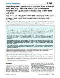
FIG. 3. Localization of the ECS in human fallopian tube. Images in the left column (A, E, I, M) are negative controls, described in the Subjects and Methods section. Samples are from the follicular phase. The top row represents FAAH-positive tissue sections of follicular phase (Fol; n ⫽ 10) (B), luteal phase (Lut; n ⫽ 10) (C), and ectopic pregnancy (Ect; n ⫽ 10) (D) samples. The second row depicts NAPE-PLD-positive slides in the follicular phase (F), luteal phase (G), and ectopic pregnancy (H). The third row shows typical representative images of CB1-positive samples from the follicular phase (J), luteal phase (K), and ectopic pregnancy (L), whereas the bottom row shows representative images of CB2 samples in the follicular phase (N), luteal phase (O), and ectopic pregnancy (P). There was FAAH and NAPE-PLD immunostaining in both the tubal epithelium (Ep) and stroma (St), with minimal to no immunostaining in the stroma for CB1 and CB2.
epithelium when compared with the rest of the epithelium (Fig. 3). Immunoreactivity was also demonstrated in the stroma for both proteins, although at a lower intensity than in the tubal epithelium. In the case of ectopic pregnancy, stromal staining for FAAH appeared reduced when compared with either phase of the menstrual cycle. At the dilutions used, cytoplasmic but no nuclear staining was demonstrated. No staining was observed with the negative controls (Fig. 3). Immunoreactivity for both CB1 and CB2 was also observed in the fallopian tube epithelium, with stronger intensity of staining along the luminal border of the epithelium (Fig. 3). Expression was primarily in the epithelial cytoplasm, with only minimal or absent staining demonstrated in the stromal cells. No staining was observed with the negative controls (Fig. 3). Histomorphometric analysis of ECS expression in fallopian tube epithelium There was a significant reduction in FAAH immunoreactivity in the tubal epithelium of the ectopic pregnancy Downloaded from https://academic.oup.com/jcem/article-abstract/97/8/2827/2823396 by guest on 09 April 2018
group compared with those from the luteal and follicular phase controls, with median (IQR) histoscore values of 2.36 (1.89 –2.52) for the follicular phase, 2.55 (2.07– 2.69) for the luteal phase, and 1.58 (0.55–2.21) for the ectopic pregnancy group (P ⬍ 0.05) (Fig. 4). There were no statistically significant differences (P ⬎ 0.05) in the histoscore values for NAPE-PLD expression, although there was a tendency toward higher scores in the ectopic pregnancy group [2.43 (2.26 –2.78)] and the follicular phase [2.46 (2.10 –2.57)] samples when compared with the luteal [2.17 (1.39 –2.44)] phase samples (Fig. 4). CB1 immunostaining was significantly different in both the follicular and ectopic pregnancy groups compared with the luteal phase controls, with histoscore values of 0.77 (0.67–1.03) for the follicular phase, 1.67 (1.37–2.01) for the luteal phase, and 0.22 (0.10 – 0.48) for the ectopic pregnancy group (P ⬍ 0.05) (Fig. 4). Unlike the immunostaining for CB1, there was no significant difference between all three groups with regard to CB2 expression. The histoscore values were 1.02 (0.32–2.44) for fol-
J Clin Endocrinol Metab, August 2012, 97(8):2827–2835
jcem.endojournals.org
2833
treated with the FAAH inhibitor URB597 (cyclohexyl carbamic acid 3⬘-carbamoyl-biphenyl-3-yl ester) and FAAH knockout mice showed evidence of oviductal embryo retention compared with untreated wild-type mice (6). Our data demonstrate a significant reduction in FAAH mRNA in tubes from ectopic pregnancy compared with controls. Although the Western blot analysis results were not statistically significant, there was a trend toward lower expression levels of this protein. The smaller numbers in the Western blot experiments and thus the potential differences observed may be related to the methodologies used (i.e. the lack of significance). The fact that histoscores are also reduced in the tubal epithelium supports the conclusion that FAAH expression is reduced in women with ectopic pregnancy. The observation that NAPE-PLD does not appear to be down-regulated in ectopic FIG. 4. Immunohistochemical scores (H-score) for FAAH (A), NAPE-PLD (B), CB1 (C), pregnancy is a significant finding because the and CB2 (D) in tubal epithelia of follicular phase (n ⫽ 10), luteal phase (n ⫽ 10), and ectopic pregnancy (n ⫽ 10) samples. Data are presented as median (IQR). Data with overall enzyme expressions observed would redifferent letters are significantly different (P ⬍ 0.05) from each other, with sult in high local levels of anandamide predisposcomparisons made using Kruskal-Wallis one-way ANOVA followed by Dunn’s ad hoc ing the embryo to an unfavorable environment posttest analysis. for normal development. licular phase, 1.59 (0.96 –2.06) for luteal phase, and 1.47 Previously, Horne et al. (14) demonstrated the presence (0.32–2.01) for ectopic pregnancy samples (Fig. 4). of CB1 protein and attenuation of its corresponding mRNA in fallopian tubes. Our data are consistent with these findings and in addition have shown immunohistoDiscussion chemically that CB1 levels are also reduced in ectopic pregnancy. In contrast, CB2 expression did not seem to be High AEA levels are associated with a plethora of adverse differentially regulated in ectopic pregnancy, which genevents in early pregnancy (6, 14, 29 –33), and what was erally concurs with animal studies suggesting that CB1 more significant from our study was the finding of higher and not CB2 dysfunction is involved in oviductal arrest of AEA levels in the tissues of women with ectopic pregnancy embryos (30). In the study of Wang et al. (6), the adverse compared with the luteal phase controls. Higher levels of OEA and PEA were also found in women with ectopic effects of FAAH deficiency were significantly improved by pregnancy, although the differences observed were not pharmacological silencing of CB1, suggesting that the delstatistically significant, but through the so-called “entou- eterious effects of high anandamide levels in the oviduct rage effect” (8 –11) these increased OEA and PEA levels are mediated via CB1. It is tempting to suggest that our could have a role in potentiating the actions of AEA in the observed reduction in CB1 expression in women with eclocal milieu. A previously unreported and interesting ob- topic pregnancy may be a compensatory mechanism to servation was the finding of FAAH and NAPE-PLD pro- limit the potentially adverse effects of endocannabinoids teins in human fallopian tubes and, in particular, their on early embryo development and transport. However, a expression in tubal epithelium. FAAH and NAPE-PLD are primary CB1 lesion could also lead to a compensatory considered the “gatekeepers” of endocannabinoid levels, increase in endocannabinoids and a decrease in FAAH and their activity and expression determine local concen- expression, especially because a small study has suggested trations of these ligands (34), with the idea that levels are that there may be differential distribution of the 1359G/A tightly regulated in early pregnancy with increasing ex- (rs1049353) polymorphism of the CB1 gene in women pression of FAAH in the embryo as development proceeds, with ectopic pregnancy compared with normal pregnant presumably to enhance its capacity to degrade AEA (7). controls (14). It is difficult to extrapolate these data to the FAAH dysfunction has been linked to high AEA levels and changes that may occur at the implantation site, but given embryo retention in mice (6) because wild-type mice the technical difficulty in obtaining significant tissue from Downloaded from https://academic.oup.com/jcem/article-abstract/97/8/2827/2823396 by guest on 09 April 2018
2834
Gebeh et al.
Endocannabanoids in Ectopic Pregnancy
the implantation site for protein expression analyses, it will perhaps remain a difficult area to evaluate with certainty. Nonetheless, our findings implicate CB1 and FAAH dysregulation in tubal pregnancy, while acknowledging the limitation of the controls used. There is a suggestion that the levels of endocannabinoid(s) are regulated in fallopian tubes such that a gradient exists to facilitate normal tubal transport and development (6, 7). The data presented here support this because a trend toward higher levels of endocannabinoids in the isthmus compared with the ampulla was observed, although this difference was not statistically significant. The reason may be that a true “endocannabinoid gradient” does not exist in human fallopian tubes or that our sample size was too small to detect any significant differences. Surprisingly, there was a trend toward higher levels in the fimbria compared with the ampulla for reasons that are not clear. The majority of tube samples were obtained from women in the luteal phase of the menstrual cycle, a time at which we have previously demonstrated high plasma anandamide levels and possibly in the peritoneal space, postovulation, because follicular fluid also contains high anandamide levels (35, 36). One possibility, therefore, for a trend toward higher levels in the luteal phase fimbria may be the higher postovulatory AEA levels. One limitation of this aspect of our study was our inability to confirm the presence of the cannabinoid receptor proteins using Western blotting methods due mainly to technical reasons with antibody specificity. What is perhaps more important is how these changes affect the “endocannabinoid tone” in the local milieu of the developing embryo rather than the absolute changes in protein expression. Another limitation of our study is the use of nonpregnant luteal phase samples as controls. However, obtaining fallopian tubes from normal pregnant women is unethical and rare in contemporary practice, and coupled with the absence of a validated ex vivo primary fallopian tube epithelium culture system, the use of these samples for ectopic pregnancy research is likely to remain common practice. Moreover, the fact that it is in this phase of the menstrual cycle that the initial stages of embryo development and tubal transport occur makes it a reasonable trade-off between what is “ideal” and what is “practical” while accepting the limitation. To the best of our knowledge, this is the first study to demonstrate the presence of the entire ECS in human fallopian tubes and at the same time quantify endocannabinoid levels in these tissues. Despite the relatively small numbers, our results suggest an apparent dysfunction of the ECS in ectopic pregnancy, with the implication that modulating this system could potentially provide a benchmark for further improvements in ectopic pregnancy care. Downloaded from https://academic.oup.com/jcem/article-abstract/97/8/2827/2823396 by guest on 09 April 2018
J Clin Endocrinol Metab, August 2012, 97(8):2827–2835
Whether the changes described here are a cause of, or are secondary to, ectopic pregnancy is difficult to ascertain, and experiments to prove this would be challenging to conduct in humans. One available option would be to evaluate whether or not high levels of endocannabinoids modulate key tubal functions such as cilia beat frequency or tubal smooth muscle contraction, especially because CB1 and FAAH knockout mice had infertility due to physical trapping of the embryos in their oviducts (6, 30).
Acknowledgments The authors thank Sarah-Jane Mason-Birks for assistance in patient recruitment and Shashi Rana, Angie Gills, and Karen Kulbici for general and specific laboratory support. The authors give special thanks to the volunteers and theater staff at University Hospitals of Leicester, United Kingdom, without whom this study would not have been possible. Address all correspondence and requests for reprints to: Dr. Anthony H. Taylor, University of Leicester, Reproductive Sciences, Department of Cancer Studies and Molecular Medicine, Clinical Sciences Building, Leicester Royal Infirmary, P.O. Box 65, Leicester LE2 7LX, United Kingdom. E-mail:
[email protected]; or Prof. Justin C. Konje, University of Leicester, Reproductive Sciences, Department of Cancer Studies and Molecular Medicine, Clinical Sciences Building, Leicester Royal Infirmary, P.O. Box 65, Leicester LE2 7LX, United Kingdom. E-mail:
[email protected]. This work was funded by the University Hospitals of Leicester National Health Service Trust and J.C.K.’s courses income. Disclosure Summary: The authors declare no conflicts of interest.
References 1. Norwitz ER, Schust DJ, Fisher SJ 2001 Implantation and the survival of early pregnancy. N Engl J Med 345:1400 –1408 2. Wilcox AJ, Weinberg CR, O’Connor JF, Baird DD, Schlatterer JP, Canfield RE, Armstrong EG, Nisula BC 1988 Incidence of early loss of pregnancy. N Engl J Med 319:189 –194 3. Pisarska MD, Carson SA 1999 Incidence and risk factors for ectopic pregnancy. Clin Obstet Gynecol 42:2– 8; quiz 55–56 4. Gebeh AK, Willets JM, Marczylo TH, Lam PMW, Taylor AH, Konje JC 2010 Molecular mechanisms of tubal pregnancy. Expert Rev Obstet Gynecol 5:727–739 5. Bambang K, Karasu T, Gebeh AK, Taylor AH, Marczylo TH, Lam PMW, Willets JM, Konje JC 2010 From fertilisation to implantation in mammalian pregnancy- modulation of early human reproduction by the endocannabinoid system. Pharmaceuticals 3:2910 –2929 6. Wang H, Xie H, Guo Y, Zhang H, Takahashi T, Kingsley PJ, Marnett LJ, Das SK, Cravatt BF, Dey SK 2006 Fatty acid amide hydrolase deficiency limits early pregnancy events. J Clin Invest 116:2122– 2131 7. Schuel H 2006 Tuning the oviduct to the anandamide tone. J Clin Invest 116:2087–2090 8. Jonsson KO, Vandevoorde S, Lambert DM, Tiger G, Fowler CJ 2001 Effects of homologues and analogues of palmitoylethanol-
J Clin Endocrinol Metab, August 2012, 97(8):2827–2835
9.
10.
11.
12.
13.
14.
15.
16.
17.
18.
19. 20.
21.
22.
amide upon the inactivation of the endocannabinoid anandamide. Br J Pharmacol 133:1263–1275 Rodríguez de Fonseca F, Del Arco I, Bermudez-Silva FJ, Bilbao A, Cippitelli A, Navarro M 2005 The endocannabinoid system: physiology and pharmacology. Alcohol Alcohol 40:2–14 Lambert DM, Vandevoorde S, Jonsson KO, Fowler CJ 2002 The palmitoylethanolamide family: a new class of anti-inflammatory agents? Curr Med Chem 9:663– 674 García Mdel C, Adler-Graschinsky E, Celuch SM 2009 Enhancement of the hypotensive effects of intrathecally injected endocannabinoids by the entourage compound palmitoylethanolamide. Eur J Pharmacol 610:75– 80 Trabucco E, Acone G, Marenna A, Pierantoni R, Cacciola G, Chioccarelli T, Mackie K, Fasano S, Colacurci N, Meccariello R, Cobellis G, Cobellis L 2009 Endocannabinoid system in first trimester placenta: low FAAH and high CB1 expression characterize spontaneous miscarriage. Placenta 30:516 –522 Chamley LW, Bhalla A, Stone PR, Liddell H, O’Carroll S, Kearn C, Glass M 2008 Nuclear localisation of the endocannabinoid metabolizing enzyme fatty acid amide hydrolase (FAAH) in invasive trophoblasts and an association with recurrent miscarriage. Placenta 29:970 –975 Horne AW, Phillips 3rd JA, Kane N, Lourenco PC, McDonald SE, Williams AR, Simon C, Dey SK, Critchley HO 2008 CB1 expression is attenuated in Fallopian tube and decidua of women with ectopic pregnancy. PLoS One 3:e3969 Habayeb OM, Taylor AH, Bell SC, Taylor DJ, Konje JC 2008 Expression of the endocannabinoid system in human first trimester placenta and its role in trophoblast proliferation. Endocrinology 149:5052–5060 Marczylo TH, Lam PM, Amoako AA, Konje JC 2010 Anandamide levels in human female reproductive tissues: solid-phase extraction and measurement by ultraperformance liquid chromatography tandem mass spectrometry. Anal Biochem 400:155–162 Lam PM, Marczylo TH, Konje JC 2010 Simultaneous measurement of three N-acylethanolamides in human bio-matrices using ultra performance liquid chromatography-tandem mass spectrometry. Anal Bioanal Chem 398:2089 –2097 Livak KJ, Schmittgen TD 2001 Analysis of relative gene expression data using real-time quantitative PCR and the 2⫺⌬⌬C(T) method. Methods 25:402– 408 Schmittgen TD, Livak KJ 2008 Analyzing real-time PCR data by the comparative C(T) method. Nat Protoc 3:1101–1108 Gebeh AK, Marczylo EL, Amoako AA, Willets JM, Konje JC 2012 Variation in stability of endogenous reference genes in fallopian tubes and endometrium from healthy and ectopic pregnant women. Int J Mol Sci 13:2810 –2826 Giang DK, Cravatt BF 1997 Molecular characterization of human and mouse fatty acid amide hydrolases. Proc Natl Acad Sci USA 94:2238 –2242 Okamoto Y, Morishita J, Tsuboi K, Tonai T, Ueda N 2004 Mo-
Downloaded from https://academic.oup.com/jcem/article-abstract/97/8/2827/2823396 by guest on 09 April 2018
jcem.endojournals.org
23.
24.
25.
26.
27.
28.
29.
30.
31.
32.
33.
34.
35.
36.
2835
lecular characterization of a phospholipase D generating anandamide and its congeners. J Biol Chem 279:5298 –5305 Willets J, Kelly E 2001 Desensitization of endogenously expressed ␦-opioid receptors: no evidence for involvement of G protein-coupled receptor kinase 2. Eur J Pharmacol 431:133–141 Bradford MM 1976 A rapid and sensitive method for the quantitation of microgram quantities of protein utilizing the principle of protein-dye binding. Anal Biochem 72:248 –254 Brighton PJ, McDonald J, Taylor AH, Challiss RA, Lambert DG, Konje JC, Willets JM 2009 Characterization of anandamide-stimulated cannabinoid receptor signaling in human ULTR myometrial smooth muscle cells. Mol Endocrinol 23:1415–1427 Nassar A, Cohen C, Agersborg SS, Zhou W, Lynch KA, Heyman ER, Olson A, Lange H, Siddiqui MT 2011 A new immunohistochemical ER/PR image analysis system: a multisite performance study. Appl Immunohistochem Mol Morphol 19:195–202 Taylor AH, Finney M, Lam PM, Konje JC 2011 Modulation of the endocannabinoid system in viable and non-viable first trimester pregnancies by pregnancy-related hormones. Reprod Biol Endocrinol 9:152 Taylor AH, Abbas MS, Habiba MA, Konje JC 2010 Histomorphometric evaluation of cannabinoid receptor and anandamide modulating enzyme expression in the human endometrium through the menstrual cycle. Histochem Cell Biol 133:557–565 Schmid PC, Paria BC, Krebsbach RJ, Schmid HH, Dey SK 1997 Changes in anandamide levels in mouse uterus are associated with uterine receptivity for embryo implantation. Proc Natl Acad Sci USA 94:4188 – 4192 Wang H, Guo Y, Wang D, Kingsley PJ, Marnett LJ, Das SK, DuBois RN, Dey SK 2004 Aberrant cannabinoid signaling impairs oviductal transport of embryos. Nat Med 10:1074 –1080 Maccarrone M, Bisogno T, Valensise H, Lazzarin N, Fezza F, Manna C, Di Marzo V, Finazzi-Agro` A 2002 Low fatty acid amide hydrolase and high anandamide levels are associated with failure to achieve an ongoing pregnancy after IVF and embryo transfer. Mol Hum Reprod 8:188 –195 Maccarrone M, Valensise H, Bari M, Lazzarin N, Romanini C, Finazzi-Agro` A 2000 Relation between decreased anandamide hydrolase concentrations in human lymphocytes and miscarriage. Lancet 355:1326 –1329 Paria BC, Song H, Wang X, Schmid PC, Krebsbach RJ, Schmid HH, Bonner TI, Zimmer A, Dey SK 2001 Dysregulated cannabinoid signaling disrupts uterine receptivity for embryo implantation. J Biol Chem 276:20523–20528 Fezza F, De Simone C, Amadio D, Maccarrone M 2008 Fatty acid amide hydrolase: a gate-keeper of the endocannabinoid system. Subcell Biochem 49:101–132 El-Talatini MR, Taylor AH, Elson JC, Brown L, Davidson AC, Konje JC 2009 Localisation and function of the endocannabinoid system in the human ovary. PLoS One 4:e4579 El-Talatini MR, Taylor AH, Konje JC 2009 Fluctuation in anandamide levels from ovulation to early pregnancy in in-vitro fertilization-embryo transfer women, and its hormonal regulation. Hum Reprod 24:1989 –1998