Effect of alkalizing agents on the processing of the P-amyloid precursor protein. Gesine Schrader-Fischer, Paolo A. Paganetti ^. Accepted 20 Drcemher 1995.
BRAIN RESEARCH ELSEVIER
Brain Reaearch 716 (1996) 91-100
Research report
Effect of alkalizing agents on the processing of the P-amyloid precursor protein Gesine Schrader-Fischer, Accepted
Paolo A. Paganetti ^
20 Drcemher
1995
Abstract
We investigated the processing pathway of the amyloid precursor protein (APP) to the secretion of PA4 under the treatment of ammonium chloride (NH,Cl), bafilomycin Al (bafA I), or chloroquine. all three agents thought to raise the pH in acidic compartments. HEK-293 cells expressing wild-type APP (APPwt) and APP carrying the Swedish double mutation (APPswe) were affected in a different manner: while cells expressing
APPswe decreased the secretion
of PA4 after treatment with bafA1 and NH,Cl, cells expressing APPwt of alternative PA4-related peptides. Within cells APP of a C-terminal fragment of APP generated by P-secretase was completely inhibited. Thus, BafAl and PA4 by inhibiting ,!3-secretase. Treatment with chloroquine did not alter PA4 secretion but. strikingly. intracellular PA4. The effect of reduced APP endocytosis was studied by expressing APP molecules (APPwt. 4, APPswe. A). Truncation of APP reduced PA4 secretion from APPwt but not from APPswe.
compensated the drug-induced decrease in PA4 by an increased generation accumulated, while the formation NH,Cl reduced the secretion of resulted in an accumulation of lacking the cytoplasmic domain
BafAl
and NH,Cl treatment inhibited the formation of /3A4 in cells expressing APPswe. 3 but not APPwt.A. With these constructs, accumulation of intracellular PA4 was observed. Since alkalizing agents still affected endocytosis-defi-
chloroquine had no effect and no cient APP containing the Swedish an acidic compartment along the also in rhe endosomal/ly5osomal
double mutation, we suggest that the formation of PA4 from this mutated APP takes place mainly in constitutive secretory pathway. Much in contrast to this. PA4 generation from APPwt appears to occur
compartment.
1. Introduction Alzheimer’s disease (AD) is characterized by the progressive cerebral accumulation of P-amyloid protein ( PA4). PA4 is a 39-43 amino acid peptide secreted from cells following proteolytic cleavage of a 100- 140 kDa glycoprotein, the amyloid precursor protein (APP). The primary structure of APP closely resembles a cell-surface receptor with a signal sequence, a large extracellular Nterminal region, a single transmembrane domain, and a small cytoplasmic C-terminal tail ([22]; for reviews [20,33]). PA4 is a product of normal cellular metabolism in vivo [34,35] and in vitro [ 1.5.34.351. Generation of the 4 kDa fragment PA4 involves at least two proteolytic cleavages: one at the N-terminus by an enzyme designated P-secretase and one at the C-terminus by an enzyme called
’ Corresponding author. Fax: (41) (3 1) 377.6936. 0006-8993/Yhj$l5.00 0 lY96 Elsevier Science PIZ SOOO6-8993(96)00002-9
B.V. All rights rrssrved
y-secretase [33]. The enzymes involved in these proteolytic events are still unidentified. Alternatively, proteolytic cleavage within the /3A4-region at position 16 by another unidentified enzyme, designated cu-secretase. leads to the production of a larger soluble fragment comprising most of the APP extracellular domain (sAPPcu) [I l] and to the secretion of a non-amyloidogenic 3 kDa fragment, designated P3 [2,15]. It is important to understand the mechanisms by which APP is cleaved to PA4 because abnormal APP processing may
be
involved
in
the
pathogenesis
of
AD.
Genetic
studies have revealed several missense mutations in the APP gene which are linked to pedigrees with early-onset AD (for a review see [33]). It has been shown that one of these mutations. the Swedish double mutation. leads to a 5-S-fold oversecretion of PA4 in vitro [3,5]. a finding reproduced also with primary skin fibroblast cultures obtained from Swedish mutation carriers [6,21]. There are still some open questions about the cellular mechanisms that lead to the secretion of PA4. In a recent
report we have shown that PA4 cleaved from an APP molecule carrying the Swedish mutation can be detected intracellularly [26] and intracellular PA4 was found also in untransfected cells [ 12,411. The generation of PA4 is completely inhibited by brefeldin A indicating that full maturation of APP is necessary for PA4 formation [ 17,261. Treatment with leupeptin, which can inhibit lysosomal thiol proteases, has no significant effect on PA4 production [2.17,35], and PA4 has not been found in isolated lysosomes [ 171. Application of agents that alter intracellular pH, such as ammonium chloride (NH,CI) or chloroquine, led to controversial effects. In some cases NH,Cl [10,12,17135] and chloroquine [10,12,35] led to substantial decreases in PA4 production suggesting the importance of an acidic intracellular compartment for its generation. Other authors could not find any effects of these agents on PA4 secretion [2]. Recent reports have shown that treatment with bafilomycin Al (bafA1). a specific inhibitor of the vacuolar Hf-ATPase, leads to a strong inhibition of PA4 secretion in HEK-293 cells. This is accompanied by a selective inhibition of p-secretase activity [ 18,231. While cells expressing APP carrying the Swedish mutation (APPswe) decrease the secretion of PA4 after bafA1 treatment, cells expressing wild-type APP (APPwt) are able to compensate the inhibition of PA4 formation by the enhanced secretion of alternative PA4-related peptides [ 181. We were interested in the direct comparison of three different alkalizing agents in regards to their effects on APP processing. For this, we expressed cDNAs for fulllength APP molecules (APPwt, APPswe) in human cell lines and treated them with NH,Cl, bafA1, or chloroquine. NH,Cl and chloroquine are weak bases and are thought to raise the pH of acidic compartments. NH,Cl has been shown to alkalize endosomes [ 131, chloroquine accumulates in lysosomes where it raises the pH [9]. It is not clear how these agents affect the Golgi apparatus or secretory vesicles. BafAl, a specific inhibitor of the vacuolar HiATPase [l], has been shown to affect different compartments in the cell. It can inhibit the acidification of endosomes and lysosomes [39.44] as well as of the trans-Golgi network and possibly also of secretory vesicles [43]. All these data indicate that these agents act at least in part in the endosomal/lysosomal compartment. Hence, we aimed to characterize the role of endocytosis and of the acidic endosomal/lysosomal compartments in the formation of PA4. The cytosolic domain of APP contains putative targeting signals, i.e. the signal for endocytosis [8,16,25]. Deletion of the cytoplasmic domain was shown to dramatically decrease the internalization of APP [24]. We generated truncated APP (APPwt. d, APPswe. d) and treated the cells with alkalizing agents. Our data revealed that PA4 generation from APPswe occurs mainly along the constitutive secretory pathway, in contrast APPwt is processed to ,!3A4 also in the endocytic pathway. Moreover, while NH,Cl and bafA1 inhibited PA4 secretion, chloroquine
had no effects on j3A4 secretion but induced accumulation of intracellular /3A4.
2. Materials
and methods
2.1. Plasmid construction The cDNA for APP695wt was inserted into a plasmid expression vector downstream of the cytomegalovirus promoter. The Swedish double mutation was obtained by replacing a 26 bp &oRI/BglII fragment with annealed oligonucleotides GATCTCTGAAGTGAATCTGGATGCAG and AATTCTGCATCCAGATTCACTTCAGA as described [5]. The truncated APP molecules APPwt.d and APPswe. d were obtained by site-directed mutagenesis (Bio-Rad Kit II) using the oligonucleotide CTTGGTCATGCAGCGCTAGTAACAGTAGGAC. Two stop codons (in bold face) truncated APP at the cytosolic side of the transmembrane domain, All APP cDNA constructs contained additional 12 amino acids VPEQKLISEEDL after the signal peptide using the oligonucleotides CCGAGCAAAAGCTTATTAGTGAAGAAGACCTGGTAC and CAGGTCTTCTTCACTAATAGCTTTTGCTCGGGTAC which were annealed and cloned into an endogenous KpnI site to create the c-myc epitope [30]. 2.2. Antiseru The hybridoma cells producing the mouse monoclonal antibody (mAb) against the c-nzq’c tag were obtained from ATCC (mAb 9E10, #1729). mAb 9E10 recognizes APP and secreted APP (s4PPtot). The rabbit polyclonal antiserum (As) NT1 1 and the mAb p 1 were raised against a synthetic peptide corresponding to amino acids l-40 of the PA4 sequence. This peptide was injected without coupling. As NT1 1 recognizes efficiently PA4 and P3 (as an example see Fig. 1). mAb p 1 reacts with PA4. fulllength APP and sAPPcu. As APPC was raised against the C-terminal end of APP (amino acids 676-695) and recognizes APP and all C-terminal fragments thereof ([29], for an example see Fig. 2). As 16 was raised against a synthetic peptide corresponding to the first 16 amino acids of PA4. This antigen was coupled to keyhole limpet hemocyanin using glutaraldehyde prior to immunization. As 16 recognizes the C-terminus of sAPPa (not shown). 2.3. Cell lines HEK-293 cells (transformed human primary embryonal kidney cells, ATCC CRL 1573) were co-transfected with a plasmid carrying the neomycin resistance (pNeo; Pharmacia) using calcium phosphate precipitation [4]. Stably transfected clones were selected with 0.75 mg/ml active drug of the neomycin analogue geneticin (G418: Gibco BRL). Cell lines were screened for APP expression histo-
93
logically and by Western blots using mAb 9ElO. Cells were routinely cultured in DMEM (Gibco), 10% FCS (Gibco). 1% penicillin/streptomycin (Gibco) and 0.25 mg/ml G418.
kdal
A.
2.4. Pulse-chase experiments
id: t
For the pulse-chase experiments 1.6 to 2.0 X 10” cells were plated on pOly-D-1ySine coated 6 cm dishes. After 24 h cells were starved in DME without cysteine/methionine (Sigma), 2% di a1yzed FCS (Gibco) for 45 min. Cells were pulse-labeled with 0.1 mCi/ml [ “S]Cys/Met (NEN DuPont, Expre ‘“S’“S Labeling Mix) in Cys/Met free DME, 2% dialyzed FCS, 20 mM HEPES, pH 7.4. and chased in DMEM, 2% FCS, 1 mM Cys/Met. If indicated we applied during the chase 10 mM ammonium chloride (1 M stock solution in water), 100 nM bafilomycin Al (100 PM stock in DMSO) or 100 PM chloroquine (10 mM stock in water). All drugs were from Sigma. Cell lysates and cell supernatants were prepared for immunoprecipitation as described [28] using 5 pg mAb 9ElOormAbP1,l ~lAsAPPC.8~lAsNTl1orAsl6 and protein A sepharose (Pharmacia). Immunoprecipitates were separated on Ttis/Tricine SDS-PAGE [32] (APP, sAPPtot, sAPPcr) or Tris/Bicine SDS-PAGE [42] (Cterminal fragments, PA4, P3). After incubation in Amplify (Amersham) gels were exposed to BioMax films (Kodak). Quantifications were performed by directly counting the dried gels using an InstantImager (Canberra Packard).
3. Results 3.1. Processing of ,full-length APPMv ulknlizing agents
and
Control 8,l
100
PA4
100
sAPPa P3
100 100
B. -6
PA4+
.*
APPswe
APPwt
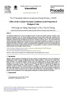
Fig. I. Effect of alkalizing agents on the secretion of PA4 and P3. HEK-293 cell lines stably expressing APPwt or APPswe were pulsed for 40 min and chased for 4 h in the absence (control) or presence of NH,CI. bafA1, or chloroquine. Conditioned media wre collected. proteins immunoprecipitated with As .UTI I (A) or mAb p I (B), and separated on a 13% Tris/Bicine SDS-PAGE. The position of molecular mass markers is shown on the right of the autoradiographs. Synthetic /3A3,-,,, and P3 ( PA4,i m-1o)loaded on separated lanes (not shown) migrated as indicated on the left of the autoradiographs. P3 was immunoprecipttated by As NTII but not by mAb PI.
After the chase, conditioned media and cell lysates were analyzed by immunoprecipitation with several antibodies against APP. lmmunoprecipitates were separated on SDSPAGE and quantified. Table 1 summarizes the effects of these agents on the processing products of APPwt and
agents on APPwt or APPswe processing
APPwt
4PP sAPPtot
PA4 + P3 +
APPswe under
HEK-293 cells stably transfected with cDNAs for APPwt or APPswe were pulse-labeled and chased for 4 h in the absence or presence of NH,Cl, bafA1, or chloroquine.
Table I Effect of alkaliring
-19
APPswe NH,CI
bafA 1
3Ok
8 *
120*
5 j
69+
5 *’
I23 i I8 80 + I2
Chloroquine
iY+ll *( 254 f II _ _ l57k
8 I’
172+ 19 r l67k 1s * *
46k
Y **
259151 ^ I48 If- 23 203 f I I * _ 186* I4 *
Control 9= I IO0 100 100 IO0
NH,CI 30*
IO ’
III *II 43*
4 _’
I41 + I7 160 k 36
baf.4 1
Chloroquine
S2& 6 *’ 243 f 24 * * 50+ 3 *373 + 88 * 376 + 50 ’
40*7 206 k 27 *
l45&I1
**
284 k 54 ’ 286 i 55 ^
HEK-293 cell lines expressing APPwt or APPswe were pulsed for 40 min and chased for 4 h in the absence (control) or presence of NH,CI. baf.41, or chloroquine. After 4 h chase, APP was immunoprecipitated from cell lysates with anti-c-mw mAb 9ElO (APP). APP processing products were immunoprecipitated from conditioned media with mAb 9EI 0 (,APPtot). mAb p I ( PA4. aAPPcu), As I6 (sAPPu) or Aa NTI I ( PAI, P3). Each value is the mean k S.E.M. of 4-6 independent determinations. For each determination. the values of the controls (no agents) were arbitrarily set as 100% but for cell associated APP, in this care 100% corresponds to the amount of APP at the beginning of the chase. For each construct one representative clone was selected. Significance compared to controls was calculated \vith the Student’s two-sample r-test (APP) resp. the one-sample r-test (sAPPtot. PA4, sAPPa, P3): * P < 0.05, - / P < O.OI.
G. Scht-u&r-Fischer,
94
P.A. Puganetti / Bruin Re.srurch 716 (1996) 91L 100
APPwt min
0
30
60
90
APPswe 120
240
0
=
30
60
90
120
240
kdal
: :g
APPX
-72
control
-47 -30
cssz CC83
bafA1
/( 1 :'Z.
; _
$$a
chloroquine
4:s
.!!:.
i
-
- 19 -15
C99‘
-
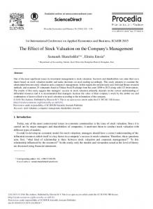
+C83+
Fig. 2. Effect of alkalizing agents on the C-terminal fragments of APP. Cells expressing APPwt or APPswe were pulse-labeled for 20 min for the indicated times. Cells were lysed, proteins immunoprecipitated with As APPC and separated on a 10% Tris/Bicine SDS-PAGE. molecular mass markers is shown on the right of the autoradiographs. As APPC efficiently immunoprecipitated full-length APP. and fragments of APP C99 and C83. The position of these proteins on the gels is indicated in the center between the autoradiographs. C99 was expressing APPswe but not APPwt; all agents blocked efficiently C99 generation in APPswe cells.
reduction of APP cleavage to soluble fragments released in the culture medium. Secreted APP derived from (Y- and /3-secretase (sAPPtot) was analyzed with an antibody
APPswe. NH,Cl, bafA1, and chloroquine led to an accumulation of full-length APP in the cell lysates. This may result from either a reduction of APP degradation or a A.
APPwt:
10 1 9
B.
C83
10
and then chased The position of both C-terminal detected in cells
APPswe:
C.
C99
APPswe:
C83
1
0 0
30
60
90
120
150
190
210
240
min
0
30
60
90
120
150
190
210
240
min
0
30
60
90
120
150
180
210
240
min
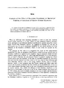
Fig. 3. Quantification of the effect of alkali7ing agents on the C-terminal fragments of APP in a pulse-chase experiment. The values were obtained from the experiment shown in Fig. 2 using the fragments C83 of APPwt cells (A), C99 of APPswe cells (B) and C83 of APPswe cells (C). The percentage of C99 resp. CX3 was normalized on the amount of intracellular APP detectable after 30 min of chase in consideration of the Cys/Met content of the fragments. The chase was perfomed without additions in the control (0). or in the presence of NH,CI ( n ). bafA I ( A ), chloroquine (0).
G. Schradrr-Fi&rr.
P.A. Pagunetti/ Brain Research 716 (19961 91-100
95
Table 2 Effect of truncation of the cytosolic tail of APP on processing
sAPPtot Total PA4 3.X kDa PA-l (‘?I of total PA4) sAPPa P3
APPh t
APPwt. d
APPswe
APPswe. d
100 f 12 lOOk 9
306 F 102 * 47* 342 * 290 *
105i 6 553 * 65 _ 4
247* 448 f 6+ 506 * 397 *
100 + 6 lOOk 9
56 * 10 2 29 - ^ 22 * *
85k 27*
9 4 =*
14 41 I 25 79
*** ’ . **
After an overnight pulse conditioned media were collected and analyzed as described in the legend of Table I. A normalization was necessary to account for a small variance in APP expression between the different stable HEK-293 cell lines. For this, all mean values were normalized with the amount of APP expressed during a 10 min pulse. Values for sAPPtot and sAPPa were obtained with one representative clone in triplicate, all other values with 4 independent clones in triplicate. Mean values f S.E.M. are given in percent of the respective amounts detected with APPwt cells: * P < 0.05. * * P < 0.01 (Student’s two-sample r-test).
against the N-terminus of APP. An increase in sAPPtot was observed with bafA1 and chloroquine with both constructs. NH,Cl treatment showed less stronger effects. The secretion of PA4 depended very much on the alkalizing agent and construct used. NH,Cl treatment led to a decrease in /?A4 secretion in cells expressing APPwt (69 * 5% of the control) and APPswe (43 k 4%). The effect of chloroquine was not significantly different from the control for APPwt but with APPswe a small increase in PA4 secretion was observed. Much in contrast to this. the PA4 secretion was increased in bafA1 treated cells expressing APPwt (1.57 + 8%). but markedly inhibited with APPswe (40 + 3%). By radiosequencing, Haass et al. [ 181 reported that the increase of PA4 secretion upon bafA1 treatment of APPwt cells was due to the enhanced production of /3A4-related peptides with alternative N-termini at Ile-6, Val-3, and Phe4 and a concomitant inhibition of PA4 starting at Aspl. By analyzing immunoprecipitated PA4 on Tris/Bicine SDS-PAGE we were able to clearly separate these different PA4 peptides. Under bafA1 treatment, with both mAb p I and As NT1 1 we observed indeed a shift in the migration characteristics of PA4 compared to the control (Fig. 1). Interestingly, this was also observed with NH,Cl (Fig. 1) and the ionophore monensin (data not shown) but not with chloroquine (Fig. 1). In contrast to bafA1, NH,Cl led to an general decrease of all secreted PA4-related peptides. When PA4 was immunoprecipitated Table 3 Effect of alkaliring
from cells expressing APPswe. neither bafA1, nor NH,Cl or chloroquine led to alternative /3A4-related peptides (Fig. 1). When the processing of APP by a-secretase was investigated, no inhibition was observed in the secretion of P3 and sAPPa after treatment with NH,Cl with either constructs. Both bafA1 and chloroquine led to an almost 2-fold increased secretion of P3 and sAPPw in cells expressing APPwt and APPswe (Table 1). 3.2. Influence of alkalizing C-terminal APP ,frugments
We then proceeded to analyze how the alkalizing agents affected the intracellular C-terminal amyloidogenic and non-amyloidogenic fragments of APP at different chase times. Cell extracts were immunoprecipitated with As APPC. The As APPC. raised against the C-terminus of APP. recognizes full-length APP, a 9 kDa and a 12 kDa C-terminal fragment of APP. The 9 kDa fragment could be immunoprecipitated with As APPC but not with mAb j3 1. Since this latter recognizes an epitope within the first 16 amino acids of PA4, m4o(data not shown). the 9 kDa fragment lacks this domain and can be designated as C83, the product of cy-secretase cleavage. In contrast, the 12 kDa fragment was immunoprecipitated by mAb /3 1 and therefore it contains the entire PA4 sequence, and we
agents on APPwt. _1 or APPswe. 3 processing APPW. J
APP sAPPtot PA4 3.8 kDa PA4 sAPPol P3
agents on the intracellular
APPswe. LI
Control
NH,Cl
II i I 100 100 100 100 100
18& 99* 94+ 106 + 106 + 981
bafA 1 1” 2 1 16 15 8
34* 95* 117z 106 k 105 * 98k
6” 8 x 18 13 17
Chloroquine
Control
NH,Cl
bafA 1
Chloroquine
36* 95* 95* 83k 93+ 92 f
II i I 100 100 100 100 100
19f2 ** lOOk8 62f6 ” 94 f 6 93 i- 6 116k9
33* 3 j^ 12X*26 43i 4” 99 * 32 91* 8 102i 11
35+13 * 107 + 24 1 IO f 12 101 * 23 84k 6 111 i 12
12 * 5 6 13 12 20
The values were obtained as described in the legend of Table 1; one representative cell line expressing APPwt.J or APPswe. J was chosen. The values of the control experiments were arbitrarily set as 100%. APP values are given for each clone as percent of APP at the beginning of the chase. Each value is the mean k S.E.M. of 4-6 independent determinations. Significance compared to controls was calculated with the Student’s two-sample t-test (APP) resp. the one-sample t-test (sAPPtot, PA4, sAPPa. P3); ’ P < 0.05. * _ P < 0.01.
96
G. Schrudrr-Fischer,
P.A. Puganetti/Bmin
believe that this 12 kDa fragment is identical to CYY. the C-terminal fragment of APP produced by the p-secretase. In lysates of cells expressing APPwt. C83 was detected already at the beginning of the chase, reached its maximum at 90 min and slowly decreased during the rest of the chase. Under treatment with NH,Cl. bafA1 or chloroquine, C83 accumulated steadily within the cells up to 4 h chase (Figs. 2 and 3A). In cells expressing APPswe, the time course of C83 formation was indistinguishable to that observed in APPwt cells and again treatment with NH,Cl, bafA1, or chloroquine led to the accumulation of C83 in the cells (Figs. 2 and 3C). We did not detect the C-termnal fragment C99 in the lysates of cells expressing APPwt under any of our experimental conditions (Fig. 2). In APPswe cells. C99 reached its maximum after 30 min and then gradually disappeared before the end of the chase. Treatment with NH,Cl, bafA1, or chloroquine blocked almost completely the generation of C99, only small amounts were detected at the end of the chase (Figs. 2 and 3B). These data indicate that p-secretase but not a-secretase activity was inhibited by alkalizing agents. The accumulation of C83 after treatment with different alkalizing agents may be due to a decrease in lysosomal degradation. 3.3. Alkali@ agents affect the processing of APP lacking the cytoplasmic domain NH,Cl, bafA1, or chloroquine were shown to raise the pH in endosomes, lysosomes and at least for bafA1 to act also along the constitutive pathway in mildly acidic intracellular compartments such as the Golgi apparatus or secretory vesicles. To explore the role of endocytosis in PA4 formation. we constructed cDNAs for APP molecules that lack the cytoplasmic tail and therefore the endocytosis signal (APPwt.d and APPswe.d). Firstly, we compared the amount of secreted APP processing products from cells expressing either full-length APP or truncated APP in an overnight pulse (Table 2). Truncation of APPwt did not lead to a change in the amount of secreted PA4 peptides (102 + 10%). Using the recently developed Tris/Bicine SDS-PAGE [42], we were able to observe that PA4 migrated as a doublet both in cells expressing APPwt.d and APPswe.d but not full-length APP molecules. The upper band co-migrated with PA4, m4,1at an apparent molecular mass of 4.0 kDa, the smaller band at approximately 3.8 kDa. This 3.8 kDa peptide was immunoprecipitated with As NT1 1 but not with mAb p 1 (Fig. 4), indicating that some of the first 16 amino acids of PA4 were lacking. The 3.8 kDa peptide probably corresponds to a PA4 peptide starting at Arg.5 [17]. The resolution of the PA4 doublet was such to allow exact quantification. With cells expressing APPwt. d the 3.8 kDa peptide amounted to 47 k 2% of the total PA4 peptides. The approximately 5-fold increase in PA4 secretion observed with the introduction of the Swedish mutation (553 + 65%) was also found with APPswe.d (448 f 47%). With this construct, the 3.8 kDa
Rraeurch
716 (1996j
91-100
kdal
-47 - 30 - 19 -15 -6
PA4 + P3 +
-3
B.
-6 -3
APPwt.A
APPswe.A
Fig. 3. Effect of alkalizing agents on the secretion of PA4 and P3 in cells expressing APP molecules lackin, - the cytosolic domain. Stably transfected cell lines expressing APPwt.d or APPswe.J were pulse-labeled for 40 min and chased for 4 h in the absence (control) or presence of NH,CI, bafAl, or chloroyuine. Conditioned media were collected, proteins immunoprecipitated with As NTI I (A) or mAb @I (B), and separated on a 13% Tris/Bicine SDS-PAGE. The position of molecular mass markers PA4, m4,jand P3 ( /3A4,7m1,1)is as indicated in the legend of Fig. I.
peptide amounted to only 6 2 1% of the total PA4 peptides or to about 60% of the 3.8 kDa peptide secreted from APPwt cells. Thus, we confirmed the published qualitative data indicating that the cytoplasmic tail is not absolutely necessary for the formation and release of PA4 [7,17,24]. In line with the data of Koo and Squazzo [24] demonstrating that APP lacking the cytosolic tail was not endocytosed. our data suggest that in cells expressing APPwt about 50% of the amount of secreted PA4 was generated along the constitutive secretory pathway. Crucially, in APPswe cells as much as 94% of PA4 was secreted from an endocytosis deficient APP molecule. Our data also imply that the alkalizing drugs may affect PA4 generation from the intact APP molecules not only in the acidic endosomal/lysosomal compartment but also along the constitutive secretory pathway. The effect of NH,Cl. bafA1. or chloroquine was investigated in cells expressing APPwt. d or APPswe. il. These agents led to an accumulation of APP in the lysates of cells expressing APPwt.d or APPswe.d (Table 3). This accumulation was less pronounced with the truncated APP than with the full-length APP (Table 1). In contrast to APPwt, cells expressing APPwt.4 showed no significant decrease in PA4 formation after application of NH,Cl
When cells expressing APPwt or APPswe were analyzed after a 4 h chase, the amount of intracellular PA4 in APPswe cells was below the limit of detection. But again accumulation of intracellular PA4 was observed in chloroquine treated cells (data not shown). No intracellular PA4 was detected in cells expressing truncated APP under any of the experimental conditions chosen (Fig. 5B).
4. Discussion -19
B.
-15 -6
c pA4+
APPwt
-3
APPswe
Fig. 5. Accumulation of intracellular @A3 after treatment with chloroquine. Cells expressing full-length APP molecules (A) or APPwt. 3 resp. APPswe.d (B) were pulsed overnight in the absence (control) or presence of NH,Cl, bafA 1. or chloroquine. Protein from cell lysates was immunoprecipitated with mAb p 1 and separated on a 10% Tris/Bicine SDSPAGE. Only the lower parts of the autoradiographs are shown. The position of molecular mass markers is given on the right. The position of synthetic PA4, m40which was loaded on a separate lane is indicated in the center: intracellular PA4 was detected in cells expressing APPm,t or APPswe but not in cells expressing the truncated APP molecules, hence the position of PA4 in B is given only as a reference.
(Table 3, Fig. 4). Moreover, while bafAl treatment in cells expressing an intact APPwt molecules led to the generation of several PA4 peptides. we were not able to see such an effect with APPwt.A (Fig. 4). Chloroquine did not affect the secretion of PA4: this was consistent with the results obtained with APPwt. NH,CI and bafA1 caused a decrease in PA4 secretion in cells expressing APPswe.A (62 f 6% resp. 43 + 4%) similarly to what found in APPswe cells (Table 3. Fig. 4). All three agents had no effect on the secretion of sAPPtot, sAPPur, the 3.8 kDa peptide or P3 from APPwt. A or APPswe. A cells (Table 3). 3.4. Treatment +t’ith chlornquine led to the accumulations of intracellular PA4 In an overnight pulse cells expressing APPwt or APPswe were treated with NH,Cl. bafAl, or chloroquine. Proteins of cell lysates were immunoprecipitated with mAb p 1. A small amount of intracellular PA4 was detected in cells expressing APPswe in control conditions (Fig. 5A) as previously reported by us [26]. Application of NH,Cl or bafA1 did not change the amount of intracellular PA4. Most interestingly, treatment with chloroquine led to a clear accumulation of intracellular PA4. Although in cells expressing APPwt no intracellular PA4 was detected, chloroquine - and to a lesser extent also bafA1 - led to the appearance of intracellular PA4 (Fig. 5A).
We investigated the effect of a raise in intracellular pH on the processing of APP to PA4 in cells expressing either full-length APP molecules or APP molecules lacking the cytoplasmic domain. APP is efficiently internalized from the cell surface [I 61 and is found in clathrin-coated vesicles [27,31]. Deletion of the whole cytosolic domain or of the consensus sequence for coated-pit localization causes a severe impairment of APP internalization [24]. We show that when the cytoplasmic domain of APP is deleted by truncation (APPwt. A) in addition to PA4 we were also able to observe the secretion of a 3.8 kDa peptide (Fig. 4). PA4 co-migrated on Tris/Bicine SDS-PAGE with synthetic PA4, m-1oat an apparent molecular mass of 4 kDa (Fig. 4). The 3.8 kDa peptide was immunoprecipitated with As NT1 1 but not with mAb p 1 and was resolved from PA4 only using Tris/Bicine SDS-PAGE. Failure of mAb p 1 to immunoprecipitate the 3.8 kDa peptide is indicative for this peptide to be a PA4 species lacking part of the N-terminus. Thus, this peptide may correspond to PA4 starting at position Arg5 identified by radiosequencing [17]. Using our experimental conditions we quantified separately the level of secretion of both components of the doublet generated from APPwt.A. When cells expressed this construct, PA4 secretion was half as high if compared to that found with full-length APPwt. On the other hand, the loss of PA4 secretion was compensated by the generation of the 3.8 kDa peptide (47% of total secreted /?A4 peptides, Table 2; 38% in [19]). We conclude that about 50% of PA4 is generated during transport along the constitutive secretory pathway while the residual 50% are formed upon endocytosis (APPwt) or cleaved at other N-terminal sites to form alternative PA4 peptides (APPwt. A). Interestingly, truncation of APP carrying the Swedish mutation affected only marginally PA4 secretion while the 3.8 kDa peptide amounted to about 6% of the total secreted PA4 (Table 2). The fact that the deletion of the cytoplasmic tail in cells expressing APPswe.A did not inhibit the secretion of PA4 implies that in these cells the formation of PA4 occurs mainly along the constitutive secretory pathway. This may be explained by the fact that. in the case of APPswe. P-secretase cleavage at Asp1 of PA4 is so efficient that is completed early in the constitutive pathway before APP reaches the cell surface. psecretase cleavage at Asp 1 may also strongly predominates over alternative cleavage sites.
Next, we were interested to test the effect of agents known to perturb the intracellular pH. In pulse-chase experiments, we treated cells with bafA1. NH,Cl or chloroquine. In cells expressing APPwt, bafA1 led to a strong inhibition of PA4 secretion while inducing alternative PA4-related peptides migrating on Tris/Bicine SDS-PAGE at different apparent molecular masses than synthetic PA4, m4o (Fig. IB). This suggest that bafA I treatment inhibited fi-secretase cleavage at Asp1 but resulted in more availability of the APP substrate for alternative psecretase-like activities [ 181. In cells expressing APPswe the inhibition of PA4 secretion by bafA1 was absolute since no /3A4-related peptides were detected (Fig. 1). The Swedish double mutation which is localized directly at the P-secretase processing site enhances the cleavage of APP by the ‘classical’ p-secretase [7] and may as well inhibit the recognition by alternative proteases. BafAl also efficiently blocked the p-secretase processing of APP to the C-terminal amyloidogenic fragment C99 (Figs. 2 and 3B). Most of the data obtained with bafA1 were reproduced with NH,Cl. Interestingly, although alternative /?A4 peptides in cells expressing APPwt were also observed, these did not compensate the reduction of PA4 starting at Asp1 since their overall secretion reached only 72 f 4% of the control (Table 1). Chloroquine treatment resulted in a small increase in the PA4 concentration measured in the conditioned medium of cells expressing APPwt, but had no inhibitory effect on the secretion of PA4 in cells expressing APPswe (Table 1). Importantly, under the same experimental conditions used for bafA1 or NH,Cl, no alternative PA4 peptides were observed with chloroquine (Fig. 1). Thus, the action of this latter agent on APP processing was clear different than that of bafA1 or NH,Cl (see also below). Surprisingly, the formation of C99, the C-terminal fragment of APP produced by p-secretase. was also blocked by chloroquine (Figs. 2 and 3B). This may be the first experimental evidence for C99 not being the processing intermediate for /?A4 generation. Nevertheless, chloroquine induced a strong accumulation of intracellular PA4 in cells expressing the full-length but. crucially, not the truncated APP constructs (Fig. 5). Since truncated APP are not endocytosed [24], this may indicate that upon APP endocytosis. PA4 was generated in the endosomal/lysosomal pathway where chloroquine inhibited its degradation and resulted in the accumulation of intracellular PA4. Purification of late endosomes/lysosomes from APPtransfected cells that release substantial amounts of PA4 did not reveal the presence of PA4 peptides [ 171, although APP or amyloidogenic APP metabolites were found in these structures by several authors [14,16,36-381. It is possible that chloroquine may be a strong agent to raise the pH in the lumen of lysosomes to an extent enabling PA4 generation. Another possibility is that in addition to the alkalizing effect, bafA1 or NH,Cl may reduce the transport of APP to the lysosomes. Recently bafA1 was
shown to strongly inhibit the transport of the transferrin receptor from late endosomes to lysosomes [40]. Application of bafA1, NH,Cl or chloroquine led also to a strong accumulation of APP (Table 1) as well as of the C-terminal fragment Cl33 (Fig. 2) within cells indicating that these agents were actively inhibiting protein degradation. Truncated APP molecules were less dramatically accumulated within cells (Table 3). These molecules are not endocytosed [24] and may be transported by bulk flow along the constitutive secretory pathway without being sorted to degradative compartments. Accumulation of intracellular APP was probably not due to an impairment of the secretory pathway, since higher amounts of sAPPcv and P3 were secreted from cells expressing full-length APP molecules (Table 1). Compared to controls the increase in sAPPa and P3 secretion was 2-4-fold. Thus, in contrast to p-secretase. a-secretase was not inhibited by the alkalizing agents. Interestingly, the release of processing products derived from cu-secretase cleavage was not affected in cells expressing APPwt. d or APPswe. A (Table 3). Truncation of APP by itself increased the secretion of sAPPtot. sAPPa and P3 compared to the full-length APP (Table 2, see also [5,8.17,24]). In conclusion. we showed that endocytosis of APP is important but not absolutely required for PA4 secretion. Much in contrast to this, when APP contains the Swedish double mutation, PA4 formation occurs mainly along the constitutive secretory pathway. We also report that the action of chloroquine on APP processing and PA4 secretion is clearly different from that observed with bafA1 or NH,Cl. Our data also indicate that in the presence of chloroquine @A4 production may occur also in lysosomes, but that under physiological conditions these structures may rather degrade this peptide.
References [I] Bowman. E.J.. Siehers, A. and Altendorf. K., Bafilomycins: A class of inhibitors of membrane ATPases from microorganisms, animal cells, and plant cells, Proc,. Nut/. Acad. Sci. USA, 85 (1988) 7972-7916. [2] Busciglio, J., Gabuzda, D.H., Matsudaira. P. and Yankner, B.A.. Generation of P-amyloid in the secretory pathway in neuronal and nonneuronal cells. Proc. Nat/. AUK/. Sci. USA, 90 (I 993) 2092-3096. [3] Cai. X.-D.. Golde, T.E. and Younkin. S.G.. Release of excess amyloid fi protein from a mutant amyloid p protein precursor. Science,
259 (1993)
514-5
16.
[4] Chen. C. and Okayama. H., High-efficiency transformation of mammalian cell? by plasmid DNA, Mol. Cell Bid.. 7 (1987) 2745-2752. [5] Citron. M.. Olteradorf, T., Haass, C., McConlogue. L.. Hung, A.Y., Seubert, P., Vigo-Pelfrey. C.. Lieberburg. I. and Selkoe. D.J.. Mutation of the P-amyloid precursor protein in familial Alaheimer’s disease increases B-protein production, Nature,360 (1992) 672-674. [6] Citron, M.. Vigo-Pelfrey, C., Teplow, D.B.. Miller, C., Schenk. D., Johnston, J., Winblad, B.. Venizelos. N., Lannfelt, L. and Selkoe, D.J.. Excessive production of amyloid P-protein by peripheral cells of symptomatic and presymptomatlc patients carrying the Swedish familial Alzheimer disease mutation, Pmt. Natl. Acud. Sci. USA. 91 (1994) 11993-l 1997.
[7] Citron, M., Teplow,, D.B. and Selkoe. D.J.. Generation of amylwd P-protein from its precursor is sequence specific. iVuuron. 14 (1995) 66 I-670. [8] De Strooper, B., Umans, L.. Van Leuven. F. and Van den Berghe, H.. Study of the synthesis and secretion of normal and artificial mutants of murine amyloid precursor protein (APP): Cleavage of APP occurs in a late compartment of the default secretion pathway. J. Cell Bid., I? 1 ( 1993) 295-304. [91 De Dube. C.. De Barsy, T.. Poole. 8.. Trouet, A., Tulkzns, P. and Van Hoof. F., Lysosomotropic agents, Bmchrm. Pharmcml.. 23 (I 974) 2495-253 I. [lOI Dyrks. T., Dyrks. E., Miinning, U., Urmoneit, B.. Turner, J. and Beyreuther, K.. Generation of DA4 from the amyloid protein precursor and fragments thereof, FEBS Lm., 335 (1993) 89-93. [I l] Esch, F.S.. Keim, P.S.. Beattle. E.C., Blather, R.W., Culwell, A.R., Oltersdorf. T.. McClure, D. and Ward. P.J., Cleavage of amyloid p peptide during constitutive procewng of its precursor, Scierxe, 248 (I 990) I 122- I 124. [12] Fuller, S.J., Storey, E., Li. Q.-X.. Smith. A.I.. Beyreuther. K. and Masters. CL., Intracellular production of PA4 amyloid of AILheimer’a disease: Modulation by phosphoramidon and lack of coupling to the secretion of the amyloid precursor protein, Biorhrnzistr?;. 34 (1995)
X091-8098.
[l3] Gekle, M., Mildenberger, S., Freudinger, R. and Silbernagl, S.. Endosomal alkalization reduce5 J,,,,j, and K,,, of albumin receptormediated endocytoais in OK cells, 4m. J. PIimioi. Rrnnl. Fluid Efectrofge Physiol., 268 (1995) F899-FY06. [14] Golde, T.E., Estus, S., Younkin. L.H., Selkoe, D.J. and Younkin. S.G., Processing of the amyloid protein precursor to potentially amyloidogenic derivatives. Sc,irnc,u. 255 (1992) 72X-730. [IS] Haass, C., Schlossmacher. M.G., Hung. A.Y., Vigo-Pelfrey. C., Mellon. A.. OstdsLewaki, B.L., Lieberburg, I., Koo, E.H.. Schenk, D., Teplow, D.B. and Selkoe, D.J.. Amyloid @peptIde is produced by cultured cells during normal metabolism, Nature. 359 (1992) 322-325. [I61 Haass. C.. Koo, E.H., Mellon, A., Hung, A.Y. and Selkor, D.J., Targeting of cell-surface P-amyloid precursor protein to lysoromes: Alternative processing into amyloid-bearing fragments, A’atw-e, 357 (I 992) 500-503. [17] Haass, C.. Hung, A.Y.. Schlossmacher, M.G.. Teplow, D.B. and Selkoe. D.J.. P-amyloid peptide and a 3-kDa fragment are derived by distinct cellular mechanisms, .I. Bid. Chwr.. 268 (I 993) 3021~ 3024. [I81 Haass, C., Capell. A.. Citron, M.. Teplow. D.B. and Selkoe. D.J., The vacuolar H--ATPase inhibitor bafilomycin A I differentially affects proteolytic processing of mutant and wild-type 0.amyloid precursor protein, J. Bid. Chm.. 270 (I 995) 6 I X6-6 192. [I91 Haass. C.. Hung, A.Y.. Citron, M.. Teplou,, D.B. and Selkoe. D.J., P-amyloid, protein processing and AlLheimer‘c disease, Drug Xes., 45 (1995) 39X-402. [20] Hardy, J. and Allsop, D., Amyloid deposition ah the central event in the aetiology of Alaheimer’s disease. Twnils Phrwrwucol. Sci., 12 (1991) 3X3-388. [2ll Johnston, J.A., Cowburn. R.F.. Norgrcn, S., Wiehager, B.. Venice10s. N., Winblad, B., Vigo-Pelfrey, C.. Schenk, D., Lannfelt, L. and O’Neill. C., Increased P-amyloid release and levels of amyloid precursor protein (APP) in fibroblast cell lines ft-om family members with the Swedish Alzheimer’s disease APP670/67 I mutation, FEBS Lrtt..
354 (1994)
274-27X.
[22] Kang, J.. Lemaire, H.G.. Unterbeck, A., Salbaum, J.M., Masters. CL., Graeschik. K.H.. Multhaup, G.. Beyreuther, K. and MuellerHill. B., The precursor of Alzheimer’s disease amyloid A4 protein resembles a cell-surface receptor, Mrrur-r. 325 (1987) 733-736. [23] Knops. J., Suomensaari, S., Lee, M.. McConlogue, L., Seubert. P. and Sinha, S.. Cell-type and amyloid precursor protein-specific inhibition of A /3 release by bafilomycin Al. a selective inhibitor of vacuolar ATPases, .I. Bioi. Chem.. 270 ( 1995) 24 19-2422.
[241 Koo. E.H. and Squa/.Lo. S.L., Evidence that production and release of amyloid P-protcin involves the endocytic pathway. L Bid. Chem.. 269 (1994) 17386-17389. [25] Lai, A., Sisodia, S.S. and Trowbridge, IS., Characterization of sorting signals in the @-amyloid precursor protein cytoplasmic domain. J. Bid. Chem., 270 (1995) 3565-3573. [26] Martin. B.L., Schrader-Fischer. G.. Busciglio, J., Duke, M.. Paganetti. P. and Yankner, B.A., Intracellular accumulation of p amyloid in cells expressing the Swedish mutant amylold precursor protein, submitted. [27] Nordstedt. C., Caporaho. G.L.. Thyberg, J.. Gandy, S.E. and Greengard. P., identification of the Allheimer P/AI amyloid precursor protein in clathrin-coated vesicles purified from PC12 cells. J. Bid. Chern.. 26X (199.1) 608-612. [2X] Paganetti. P. and Scheller. R.H., Proteolytic processing of the Aplysia A peptide precursor in AtT-20 cells, Bruin Rr.\.. 633 (1994) 53-62. [29] Palacios. G.. Palacios. J.M., Mengod, G. and Frey. P., /?-amyloid precursor protein localization in the Golgi apparatus in neurons and oligodendrocytes. An immunocytochemical structural and ultrastructural study in normal and axotomized neurons, Mol. Brairz Res., 15 (1992) 19.5-206. [30] Price. K.M., Cuthbcrtson. AS., Varndell, I.M. and Sheppard. P.W., The production and characterization of monoclonal antibodies to myc, c-erbB-2 and EFG-receptor using a synthetic peptide approach. Del,. Bid. srm1., 7 1 (I 990) 23-3 I. [.ill Sapirrtein. VS.. Durrie, R.. Berg. M.J. and Marks. N.. Amyloid precursor protein is enriched in axolemma and periaxolemmal-myelin and clathrin-coated vesicles. J. Nwrosc?. Rex. ( 1994) 348-35X. [32] Schaegger, H. and van Jagow, G., Tricine-sodium dodecyl rulfatepolyacrylamide gel electrophorehis for the separation of proteins in the range from 1 to 100 kDa. 4n0/. Bioc,hrm.. I66 (19871, 36X-379. [33] Selkoe, D.J., Normal and abnormal biology of the P-amyloid precursor protein. Anrzu. Rat,. Neum.~ci., 17 ( 1994) 4X9-5 17. [%I Seubert, P.. Vigo-Pelfrcy. C.. Esch, F., Lee. M., Doucy, H.. Davis. D., Sinha, S., Schlossmacher, M., Whaley, J.. Swindlehurst. C.. McCormack. R., Wolfe& R.. Selkoe. D.J., Licberburg, I. and Schenk, D., Isolation and quantification of soluble Alzheimer’s P-pcptide from biological fluids, ,%ture. 359 (1992) 32.5-327. [35] Shoji. M.. Golde, T.E., Ghiso, J.. Cheung. T.T.. Estus, S., Shaffer, L.M. Cai. X.D., McKay. D.M., Tintner, R., Frangione. B. and Younkin. S.G., Production of the Alzheimer amyloid @ protein by normal proteolytic processing, Scirnrc, 248 (I 992) 126- 129. [36] Siman, R., Mistretta. S., Durkin, J.T., Savage. M.J., Loh, T., Trusko, S. and Scott. R.W., Processing of the P-amyloid precursor. Multiple proteaaea generate and degrade potentially amyloidogenic fragments. .I. Bid. Chum., 268 (1993) 16602-16609. [37] Tagawa, K.. YaLaki. M., Kinouchi, T., Maruyama, K., Sorimachi, H., Tsuchlya. T.. Suzuki, K. and Ishiura, S.. Amyloid precursor protein is found in lysosomeh, Ge~or~rolr~~~, 39 (suppl.) (1993) 24-29. [3X] Tsuzuki, K.. Fukatsu, R.. Takamaru. Y.. Fujii, N. and Takahata. N., Potentially amyloidogenic fragment of 50 kDa and intracellular processing of amyloid precursor protein in cells cultured under leupcptin, Bruirl Rrs., 659 (1994) 213-220. [39] Urnala. T.. Moriyamn. Y., Futai, M. and Mekada, E., The cytotoxic action of diphtheria toxin and its degradation in intact vero cells are inhibited by bafilomycin Al, a specific inhibitor of vacuolar-type H+-ATPase, .I. Hiol. Clzer~., 265 (1990) 21940-21945. [401 Van Weert. A.W.M.. Dunn, K.W.. Geuze. H.J., Maxfield, F.R., and Stoorvogel, W., Transport from late endosomes to lysosomes, but not sorting of integral membrane proteins in endoaomes, depend5 on the vacuolar proton pump. J. Ceil Bid. . I30 (I 995) 82 I -X34. [41] Wertkin, A.M.. Turner. R.S.. Pleasure. S.J., Golde. T.E.. Younkin, S.G., Trojanowski, J.Q., Lee, V.M.-Y.. Human neurons derived from a teratocarcinoma cell line express solely the 69S-amino acid amyloid precursor protein and produce intracellular P-amylold or A4 peptides, Proc. Ntrtf. Awd. Sri. USA. 90 ( 1993) 95 13-95 17.
100
G. Sclzrzrdrr-Fischer,
P.A. Pngnrwtti/
[42] Wiltfang, J., &old. N. and Nruhoft, V.. A new mulliphaslc butter system for sodium dodecyl sulfate-polyacrylamide gel electrophoresis of proteins and peptides with molecular masses 100000-1000. and their detection with picomolar sensitivity, Electrophore.~is. 12 (I 991) 352-366. [431 Xu, H. and Shields. D.. Prosomatostatin processing in permeabilized cells, J. Bid. Chem.. 36 (1994) 22X75-2288 I,
Brain Re.setrd~
716 (IO961 9/- 100
1441 Yoshlmori. T., Yamamoto, A.. Morlyama, Y.. Futai, M. and Ta%hiro. Y., Bafilomycin Al, a specific inhibltor of vacuolar-type H+._ ATPase, inhibits acldltlcation and protein degradation in lysosomes of cultured cells, J. Bid. Ckem., 266 ( 199 I) 17707- 177 12.