on hard disk for subsequent processing. ... larization (A20), and recovery time at several repolariza- .... ent study shows that the recovery times of the action.
0014-4754/96/060577-0651.50 + 0.20 9 Birkh/iuser Verlag Basel, 1996
577
Effect of cold exposure on electrophysiological properties of rat heart P. de Martino Rosaroll, P. Venditti, S. Di Meo* and T. De Leo
Department of General and Environmental Physiology, University of Naples, Via Mezzocannone 8, 1-80134 Naples (Italy), Fax 4-39 81 5526194 Received 7 February 1995; received after revision 20 June 1995; accepted 7 September 1995 Abstract. Male rats exposed to the cold (4 ~
for five or ten days exhibited modifications in their thyroid state, as documented by increases in serum thyroid hormone levels, to which differently graded modifications of heart weight/body weight ratio, heart rate, and resting metabolic rate were associated. The values of the above mentioned thyroid state indicators returned to those of the control when the animals, kept at cold for ten days, were re-exposed to room temperature (24 ~ for an additional 10 days. The configuration of action potentials, recorded in vitro at 26 ~ from fibres of anterior papillary muscles, was different in control rats of different age and was affected by prolonged cold exposure. In fact, the action potential duration (APD) increased after ten days of cold exposure. In the re-exposed group the APD was not different from that of the controls. Such a pattern was not significantly modified when the stimulation frequency increased from 1 Hz to 5 Hz. The above results suggest that in cold exposure, as in experimental hyperthyroidism, thyroid hormone might exert a cardiac chronotropic effect by modifying heart electrophysiological properties. Thus thyroid hormone should play a basic role during the exposure to cold environment, stimulating the body metabolism and increasing heart rate as a response to the requirement for greater tissue perfusion. Key words. Action potential; cold exposure; thyroid hormone; heart potentials.
Previous studies indicate that the thyroid hormone effects on heart rate, like those on respiration 1, are probably due to several mechanisms. Indeed, heart rate regulation by thyroid hormone involves a modulation of the myocardium sensitivity to neurotransmitters by modifications of the adrenergic 2 and muscarinic cholinergic receptors 3. On the other hand, researches on rabbit sinoatrial node cells4 and on ventricular preparations of several homeotherm species4 s have shown that changes in thyroid state can affect myocardial electrical properties. Hypothyroidism, induced by thyroidectomy, produces increased action potential duration (APD), while hyperthyroidism, elicited by triiodothyronine (T3) or thyroxine (T4) treatment, is associated with decreased APD. Studies on rats, dealing with the time course of changes in repolarization time, indicate a late effect of thyroidectomy and T3 treatment 7, in agreement with the finding that bradycardia, slowly induced by thyroidectomy, is removed by a prolonged T3 treatmenff. Naturally the question arises whether the aforementioned modifications of heart electrical activity, produced by experimentally modifying the thyroid state, would also be found in the case of a physiological modification of such a state. In previous studies we showed that in neonatal rats 1~ as well as in young and adult rats 11, age-related changes in serum T3 levels are
* Corresponding author.
associated with alterations of action potential configuration comparable with those found after thyroidectomy or T3 administration. Changes in serum thyroid hormone levels are also associated with environmental temperature changes 12,13. In effect, the products of two major hormonal axes, the sympathetic nervous and hypothalamic-pituitary-thyroid systems, are believed to have integral roles in mammalian cold tolerance 14. Whereas plasma catecholamines appear to act during acute stages of cold exposure, thyroid hormones may be more influential after more prolonged cold exposure 15. Thus, the increased serum T 3 level has to be regarded as the major factor responsible for the sustained phase of cold adaptation, characterized by increased metabolism, elicited as a response to the seasonal change in environment temperature. The increase in basic metabolic rate, observed in mammals during cold exposure, places more demand on the heart for greater tissue perfusion. The responses to such a demand, consisting of increases in coronary blood flow, total cardiac output, and work, require increased cardiac contractile performance and heart rate. Some evidence indicates that in cold exposed rats heart rate varies according to serum level of thyroid hormones 9. Furthermore, it has been proposed that the increased sensitivity of fl and the decreased sensitivity of m-receptor-mediated responses, found in the cardiovascular system of cold-acclimated rats 14,17, may be mediated by changes in the animals' thyroid state 14. These results suggest that the cardiac chronotropism modifica-
578
Experientia 52 (1996), Birkh/iuser Verlag, CH-4010 Basel/Switzerland
tions, which the rat undergoes during cold exposure, are induced by thyroid hormone through mechanisms analogous to those operating in experimental hyperthyroidism. The aim of the present study was to determine whether changes in myocardial electrophysiological properties are associated with the changes in the thyroid state induced by chronic cold exposure. Accordingly, the values of peculiar thyroid state indicators and the characteristics of ventricular transmembrane potentials were examined in rats both exposed to cold and re-exposed to room temperature.
Materials and methods Animals. Young male rats of a Wistar strain, supplied by Nossan (Correzzana, Italy), were used in the experiments. All animals were kept at a temperature of 24 _+ 1 ~ up to 40 days of age and then randomly divided into six groups. The rats of two groups, kept at 4 _+ 1 ~ for 5 or 10 days respectively, were killed at 45 (CE45) and 50 (CEs0) days of age. Another group, exposed to 4 ~ for 10 days and then to 24 _+ 1 ~ for an additional 10 days, was killed at 60 days of age (CE60). The other three groups, constituted by rats kept at a temperature of 24 + 1~ C for 5, 10, or 20 days, and designed respectively as C45, C50, and C6o, were used as controls for cold-exposed animals which were killed at the same age. All rats were subjected to the same conditions (one per cage, constant artificial circadian cycle of 12 h of light and 12 h of darkness) and fed on the same diet, a commercial rat chow purchased from Nossan, and water on an ad libitum basis. Experimental procedure. Soon after 12-hour overnight fast the animals were subjected to the measurement of resting metabolic rate (RMR) by an open circuit indirect calorimetry system (Columbus Instruments International Corp., Columbus, Ohio, USA). Electrocardiographic recordings were performed on the animals, anaesthetized with Ethrane (Abbot, Aprilia, Italy) as previously reported 9. Arterial blood samples were subsequently collected to determine serum levels of total (TT3) and free (FT3) triiodothyronine by enhanced luminescence assay (Amerlite TT3 and FT 3 assays, Kodak Clinical Diagnostics). Anaesthetized rats were killed by decapitation and the hearts rapidly removed and placed in cold oxygenated Krebs' solution (135 mM NaC1, 5 mM KC1, 1 mM MgC12, 2 mM CaC12, 13 mM NaHCO3, 1 mM NaH2PO4, 11 mM glucose, pH 7.4). The heart great vessels and valves were trimmed away. The ventricles and atria were cut open and rinsed free of blood. After the heart weight determination the anterior papillary muscles were excised. The muscles were mounted horizontally into an experimental chamber between two bipolar silver electrodes insulated with teflon up to their tips. They were perfused continuously, at a rate of 11
Research Articles
ml/min, with Krebs' solution gassed with 95% 0 2 - - 5% CO2. A thermostat circuit kept the solution temperature around the preparations at 26 + 1 ~ The preparations were stimulated at 0.1 Hz by 2 ms rectangular pulses 20% above threshold value for an equilibration period of approximately 1 h before taking measurements. During these measurements the muscles were stimulated at 1 Hz. Papillary muscles from some animals of each group were stimulated at 1 and 5 Hz. Transmembrane potentials were measured by cellular impalement with 3 M KCl-filled glass capillary microelectrodes with resistances ranging between 15 and 40 MYL A compliant Ag-AgC1 wire served to feed signals into a high-input impedance preamplifier with input capacity neutralization. An Ag-AgC1 wire placed in the bath served as a reference electrode. The action potential signals were displayed and monitored on an oscilloscope (Tektronix 502A) throughout the experiment. Each action potential signal was recorded on-line in digital form at 80 gs intervals on a IBM-compatible computer (TechnoComp, Villaricca, Italy) and stored on hard disk for subsequent processing. The transmembrane potentials were analyzed by a computer program purchased from TechnoComp for the following characteristics: resting membrane potential, depolarization time, action potential amplitude, area above 20% depolarization (A20), and recovery time at several repolarization degrees. As impalements in approximately 20 cells of each preparation were performed, mean values of the above parameters were calculated for each preparation, and the sample means were averaged together. Resulting values were used to supply traces of action potential characteristic of each group. The values were expressed as the m e a n _ standard error in the tables and are indicated by vertical bars in the figures. The significance of the differences between each treatment group and its control was determined by the unpaired Student's t-test. For each group, action potential repolarization times (RT9o) at 1 and 5 Hz were compared by the paired t-test. The values were considered significantly different if p < 0.05.
Results Thyroid state assessment. Thyroid state was documented by modifications in: i) the heart weight/body weight ratio, ii) the heart rate, iii) the resting metabolic rate, iv) the plasma levels of F T 3 and T T 3 (table 1). The modifications of body parameters, heart rate, RMR, and serum levels of thyroid hormone demonstrate that the rat thyroid state changes in the period preceding sexual maturation (about 60 days of age) according to previous report 11. To such changes, those resulting from the periods of exposure at different ambient temperatures are added. While the body weight was not significantly affected by cold exposure, the heart weight increased so
Research Articles
Experientia 52 (1996), Birkh/iuser Verlag, CH-4010 Basel/Switzerland
579
Table 1. Indicators of thyroid status in control and cold-exposed rats. Groups
C45
CE45 Cso CEso C60
CE60
Heart weight/ body weight (HW/BW) (mg/g)
Heart rate beats/min
3.66 • 3.93 • 2.65 • 3.61 • 2.68 • 2.74 •
455 • 482 • 418 • 506 • 360 • 392 •
0.43 0.10 0.06 0. I3" 0.09 0.09
18 8 27 15" 14 13
Resting metabolic rate (RMR) ml O2/min/100 g
Hormonal levels TT3 (ng/dl)
FT 3 (pg/dl)
1.92 • 2.41 • 1.66 • 2.35 • 1.66 • 1.80 •
71 • 94 • 65 • 98 • 60 • 61 •
565 • 623 • 400 • 615 • 397 • 460 •
0.05 0.10" 0.12 0.06* 0.05 0.2
7 8* 9 17" 3 5
53 36 34 30* 16 39
Given values are the mean • SE of eight different experiments. HW/BW = heart weight/body weight, RMR = resting metabolic rate. *significant (p < 0.05) versus control rats of the same age. Table 2. Electrical properties of anterior papillary muscle fibres. Variable
Groups CE4s
C4 5
Resting potential (mV) Action potential (mV) DT (ms) A2o (mV 9ms) RTs0 (ms) RT70 (ms) RTs0 (ms) RTg0 (ms)
73.9 • 87.8 • 2.2 • 870 • 13.1• 20.2• 27.4• 40.5•
2.0 6.2 0.4 103 1.7
70.4 • 86.9 • 2.1 • 925 • 13.8• 21.9 • 30.0 • 43.2 •
Cs0 0.6 1.6 0.1 37
0.8
68.8 • 87.9 • 2.6 • 1282 • 19.9• 30.9• 40.4• 55.1 •
0.5 1.3 0.4 111 1.5 1.2
CEs0
C60
70.8 • 1.7 83.6 • 1.0 2.0 • 0.2 758 • 95* 1.6• 18.6• 25.1 • 37.0 • 5.0"
74.1 • 89.4 • 2.5 • 1202 • 18.6• 28.7 • 39.4 • 56.7 •
CE60 1.7 2.7 0.3 81 1.7 2.0
71.1 • 1.5 89.8 • 4.0 2.3 • 0.3 1064 • 66 16.7_+1.0 25.1 • 1.1 35.3 • 2.1 54.5•
Given values are the mean • SE of eight different experiments. DT = depolarization time; A2o = integrated area above 20% depolarization. RTso, RT7o, RTso, RT9o = recovery time at 50, 70, 80, and 90% repolarization respectively. *significant (p < 0.05) versus control rats of the same age.
that both the CE4s and CEso rats exhibited a heart/body weight ratio increased in comparison to the respective controls. The heart rate, not significantly different from controls after cold exposure for 5 days, showed an increase after 10 days. The R M R , on the contrary, was already significantly increased after 5 days of cold exposure. As expected, cold exposure increased plasma T 3 levels, though F T 3 levels for CE4s rats were not significantly different from those of the controls. After reexposure to r o o m temperature, following 10 days of cold exposure, the values o f all parameters used as indicators o f thyroid state were not significantly different from control values. Action potentials in papillary muscle fibers. In the beginning of this series of experiments, measurements were made on anterior and posterior papillary muscles from 45-day-old control rats (C4s). The analysis of the results concerning the repolarization times o f the action potential showed that their high standard errors were due to differences between the two papillary muscles. In fact, for every animal, the action potentials recorded from anterior papillary muscle (RTgo = 40.5 + 4.3 ms) were shorter than those recorded from posterior muscle ( R T g o = 50.7 + 1.8 ms). Consequently, in subsequent experiments, as well as in those reported in our previous works < to, ll, 17, action potentials were exclusively recorded from anterior papillary muscles.
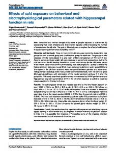
The analysis of the cold exposure effect on ventricular electrical activity in young rats was complicated because the responses were affected by modifications o f thyroid hormone levels, taking place both with age and cold exposure. Therefore, we used a group o f rats of the same age kept at a temperature o f 24 ~ as control for each group of cold-exposed animals. The time course o f the surface electrical activity in papillary muscle fibres shows that, in comparison to the respective controls, the action potential is shorter in 10 day cold-exposed rats, and no different in animals reexposed to ambient temperature (fig. 1). Evaluating the action potential characteristics, no significant difference was established in the resting membrane potential, action potential amplitude, and depolarization time obtained from control and cold-exposed preparations. The repolarization phase, and therefore the duration of the action potential is, on the contrary, affected by cold exposure. In fact, in 10 day cold-exposed rats the recovery times at different repolarization degrees are shorter than those of control rats (table 2). Ten days o f re-exposure at 24 ~ cause an almost complete restoration of action potential. In fact, the recovery time at 90% repolarization (RTg0) returns to 96% o f control (table 2). Effect of stimulus frequency on action potential duration. W e investigated the influence of stimulation frequency on A P D , by recording transmembrane potentials at 1 Hz
580
Experientia 52 (1996), Birkhfiuser Verlag, CH-4010 Basel/Switzerland 40
o m ~r
~
o
-40
-80
40-
C41
2-GO
Clio
ClIO
O.
-t,O 9
no -80 -
lO0
~.
.
150
o
100
8o
40-
CE4li
150
CEso
s
s
m iI
m
~
50
100
,
,
100
150
/ 100
, 150
0
-~.0
-80
APDoa
APDgo r
0
o
-8o .
APDIo
50
CEeo
-4o -
,
0
Time {ms}
~.
i~ -4.0
-80
APDoo
,
Time (ms}
o 84
E
-80
APDjo
,
Time (ms) 4.0
o
E
g.
APDto
0
Research Articles
i 150
r 100
Time (ms]
, 150
0
Time (ms}
GO
Time {ms}
Figure 1. Effect of cold exposure on action potentials recorded from papillary muscle fibres of rat. The action potential durations at 90% repolarization (APD90) are indicated by the arrows. The recordings were done as described in the text. The working frequency was 1 Hz.
+12
"~
+8
E 50
+4
I'-
,,-
o
C4 s
[~
CE4s
[]
Cso
[]
CEso
I~
Cjo
[~
CEeo
100
0 era
[]
~
~
ifL *
-4
Figure 2. Effect of stimulation frequency on action potentials recorded from papillary muscle fibres. Left: Change in recovery time at 90% repolarization after change of frequency from 1 to 5 Hz. Right: Recovery time at 90% repolarization at stimulation frequency of 5 Hz. *significant (p < 0.05) versus control rats of the same age. and 5 Hz in preparations from four animals of each group. In all preparations the repolarization phase is not essentialy affected by the increased stimulation frequency. In each group the RT90 changes are not significant, while the differences between RTg0 values in cold-exposed and control preparations, which are significant at 1 Hz, remain significant at 5 Hz (fig. 2). Discussion Cold exposure and thyroid state. The modifications in TT3 and FT3 serum levels, found in rats after five and
ten days of cold exposure and re-exposure to room temperature (table 1), are in agreement with previous researches which reported that TT3 and FT3 levels rapidly increased in cold-exposed animals, remained elevated during exposure and rapidly returned to control values in room temperature re-exposed animals 13. The cold-induced increase in TT3 and FT3 levels is accompanied by the appearance of a symptomatology peculiar to hyperthyroidism: weight loss, cardiac hypertrophy, tachycardia and high resting metabolism (table 1). The increases in resting metabolic rate and thyroid hormone level are strictly associated, while the resting
Research Articles
Experient!a 52 (1996), Birkh/iuser Verlag, CH-4010 Basel/Switzerland
heart rate increases in a more gradualway. This result is in agreement with a previous report, showing that heart rate change is a late effect of the thyroid state modification 9. Thyroid state and ventricular action potentials. The present study shows that the recovery times of the action potentials recorded in the anterior papillary muscles of controls are, at each age, shorter than those previously found TM. The differences between the results could be explained by the selection in animal farms of inbred strains with peculiar characteristics, Actually, the rats used in the present study exhibit values of the parameters indicators of thyroid state such as to be considered hyperthyroid in comparison with those used in previous works 7,1i. However, in these animals age-dependent modifications of thyroid state are also correlated with changes of electrophysiological parameters of anterior papillary muscle fibres. The decrease in T3 levels found between 45 and 60 days of age is associated with a decrease in heart rate and increases in action potential area and duration, conventionally measured by A2o and RT90 values. These results agree with most previous studies, showing that APD is shorter in hyperthyroid than in euthyroid ventricular preparations 5 7. Thus, the slow appearance of the tachycardia and the increase in repolarization time during cold exposure is in agreement with the previous findings indicating a late effect of thyroid hormone on the heart electrical activity7,9. However, the question arises whether results obtained by electrophysiological recordings at a stimulation frequency (1 Hz) well below those occurring in vivo (6.0-8.4 Hz) supply a realistic view of the effects of the cold-induced increase of thyroid hormone levels on electrical activity of the heart. The dependence on stimulation frequency of APD for the different groups could be such as to make the differences between the preparations from cold-exposed and control groups not significant. Actually, the measurements made at 1 Hz indicate that the cold exposure leads to a modification of basic electrophysiological properties of ventricular muscle fibres. Moreover, the recordings performed at 5 Hz show that in cold-exposed rats the decrease in ventricular action potential duration persists at stimulation frequencies closer to physiological ones (fig. 2). The ionic mechanisms responsible for the electrophysiological variations induced by changing the ambient temperature need to be investigated in the voltage-clamp setting. The mechanism by which the thyroid hormone operates on the regulation of myocardial electrophysiological properties also remains to be understood, even if it is thought that basically it can modulate specific ionic pathways by modifying either membrane-bound proteins or their lipid surrounding. Role of thyroid hormones in chronic cold-exposure. The results reported in this paper indicate that the modifications of heart electrical activity, found in animals made experimentally hyperthyroid, also take place in animals
581
which are in a state of functional hyperthyroidism. Furthermore, with previous reports, they help to clarify the role played by thyroid hormone in the adaptation process to a low environmental temperature. Chronic exposure to cold elicits a series of adaptive responses aimed at decreasing heat loss and increasing heat production. During the first period of cold exposure heat is produced by muscle shivering, whereas during chronic exposure heat is produced mostly by nonshivering thermogenic processes, which occur after an array of changes in metabolic activity at the level of ,the whole organism 18, It has now been recognised that thyroid hormones play an important role in chronic cold exposure. The sequence of events which, from the increased discharge frequency in peripheral temperature sensors ag, lead :to the increased serum levels of thyroid hormonesl2.13, includes: conduction of impulses to the Central nervous system along specific neural pathways 2~ Change in electrical activity of hypothalamic neurons 21, increased release of thyrotropin-releasing hormone mainly from the arcuate nucleus-median eminence area 22, and activation of TSH secretiona3 followed by increased thyroid activity24. In species possessing large quantities of brown adipose tissue (BAT), normal core temperature during cold exposure is maintained by the interaction of the thyroid axis and the SNS 14. Although the BAT is the principal source of thermoregulatory heat production in the rat, a contribution to the higher metabolic rate during the exposure to low temperatures is furnished by other tissues 25'26. In the liver the thermogenetic processes have been attributed to thyroid hormone-induced changes in both number of mitochondria per cell 13 and their oxidative capacity27. Studies on rats made experimentally hyperthyroid suggest that in the heart, in spite of a decrease in mitochondria per cell28, an increased oxidative capacity can be obtained by an increase in the mitochondrial content of cytochromes29. The changes of metabolic activity require suitable adjustments of cardiovascular system activity and in particular of the heart. As previously suggested 9, cold exposure-induced hyperthyroidism yields tachycardia by a modification of the adrenoceptor sensitivity and a direct action on pacemaker cells. It is possible that in cold-induced, as in experimental hyperthyroidism4 the heart rate increases owing to decreased duration of action potential of sinoatrial node cells. The heart rate increase requires a corresponding change in the duration of the action potential in atrial and in ventricular fibres in order to allow full electrical and mechanical recovery. In most species this result is obtained primarily by a rate-dependent mechanism explained by incomplete recovery of the plateau currents and an increase in the net outward currents 3~ in the rat, in agreement with previous results 7, a rate-independent mechanism, involving modifications of electrophysiological properties of ventricular muscle, is the dominant factor for the
582
Experientia 52 (1996), Birkh/iuser Verlag, CH-4010 Basel/Switzerland
m o d u l a t i o n of the action potential. T h u s thyroid hormone, t h r o u g h different biochemical a n d physiological mechanisms, plays a basic role in determining the integrated responses to modifications of a physiological or e n v i r o n m e n t a l stimulus, such as a m b i e n t temperature decrease. 1 Brand, M. D., and Murphy, M. P., Biol. Rev. 62 (1987) 141. 2 Williams, L. T., Lefkowitz, R. J., Watanabe, A. M., Hathaway, D. R., and Besh, H. R. Jr., J. biol. Chem. 252 (1977) 2787. 3 Sharma, V. K., and Banerjee, S. P., J. biol. Chem. 252 (1977) 7444. 4 Johnson, P. N., Freedberg, A. S., and Marshall, J. M., Cardiology 58 (1973) 273. 5 Sharp, N. A., Neel, S. V., and Parsons, R. L., J. molec, cell. Cardiol. 17 (1985) 119. 6 Binah, O., Arieli, R., Beck, R., Rosen, M. R., and Palti, Y., Am. J. Physiol. 252 (1987) H1265. 7 Di Meo, S., de Martino Rosaroll, P., and De Leo, T., Archs int. Physiol. Biochim. Binphys. 99 (1991) 377. 8 Di Meo, S., de Martino Rosaroll, P., Piro M. C., and De Leo, T., Comp. Biochem. Physiol. I05A (1993) 719. 9 Valente, M., De Santo, C., de Martino Rosaroll, P., Di Maio, V., Di Meo, S., and De Leo, T., Archs int. Physiol. Biochim. Biophys. 97 (1989) 431. 10 Di Meo, S., de Martino Rosaroll, P., Piro, M. C., and De Leo, T., Archs int. Physiol. Biochim. Biophys. 102 (1994) 129. 11 Di Meo, S., de Martino Rosaroll, P., Piro, M. C., and De Leo, T., Arch. int. Physiol. Biochim. Biophys. 100 (1992) 7. 12 Albright, E. C., Heninger, R. W., and Larson, F. C., in:
Research Articles
Current Topics in Thyroid Researches, p. 346. Eds C. Cassano and M. Andreoli. Academic Press, New York, London, 1965. 13 Goglia, F., Liverini, G., De Leo, T., and Barletta, A., Pfliigers Arch. 396 (1983) 49. 14 Fregly, M. J., Field, F. P., Katoich, M. J., and Barney, C. C., Federation Proc. 38 (1979) 2162. 15 Fregly, M. J., Pharmacol. Ther. 41 (1989) 142. 16 Harri, M. N. E., Melender, L., and Tirri, R., Experientia 30 (1974) 1041. 17 Di Meo, S., de Martino Rosaroll, P., Piro, M. C., and De Leo, T., Archs int. Physiol. Biochim. Biophys. 102 (1994) 153. 18 Jansky, L., Federation Proc. 25 (1963) 1297. 19 Zotterman, Y., in: Handbbok of Physiology. Neurophysiology. Sect. 1, vol. 1, p. 431. Ed J. Field. Am. Physiol. Soc., Washington 1959. 20 Landgren, S., Acta Physiol. Scand. 40 (1957) 202. 21 Hellon, R. F., Pfliigers Arch. 321 (1970) 56. 22 Hefco, E., Krulich, L., Illner, P., and Larsen, P. R., Endocrinology 97 (1975) 1185. 23 Bottari, P. M., Ciba Foundation Colloquia on Endocrinology 11 (1957) 52. 24 Straw, J. A., and Fregly, M. J., J. appl. Physiol. 23 (1967) 825. 25 Stoner, H. B., J. Physiol. 232 (1973) 285. 26 Jansky, L., Biol. Rev. 48 (1973) 85. 27 Liverini, G., Goglia, F., Lanni, A., Iossa, S., and Barletta, A., Comp. Biochem. Physiol. 97B (1990) 327. 28 de Martino Rosaroll., P., Di Maio, V., Valente, M., Di Meo, S., and De Leo, T., J. Endocrinol. Invest. 11 (1988) 559. 29 Di Meo, S., de Martino Rosaroll, P., and De Leo, T., Cell. Physiol. Biochem. 2 (1992) 283. 30 Carmeliet, E., J. Physiol. (Paris) 73 (1977) 903. 31 Boyett, M. R., and Fedida, D., J. Physiol. (London) 350 (1984) 361.