_______________________________________________________________________________________________________________________________________________________________
Research Article
_______________________________________________________________________________________________________________________________________________________________
Effect of filler addition on the bonding parameters of dentin bonding adhesives bonded to human dentin YONG-KEUN LEE, DDS, PHD, LILLIAM M. PINZON, DDS, MS, KATHY L. O’KEEFE, DDS, MS & JOHN M. POWERS, PHD ABSTRACT: Purpose: To determine the effect of filler addition on two total-etch, single component bonding systems on the bond strength, displacement at debonding, stiffness of debonding and energy absorbed to debonding of resin composites to human dentin. Methods: Two dentin bonding systems with no-filler (OS and SB) and filler-added (OSP and SBP) versions were studied. The dentin surfaces of human teeth were exposed with 600-grit SiC. TPH Spectrum A2 was used to bond to the dentin surfaces in the form of a truncated cone, 3 mm in diameter at the bonding surfaces and 5 mm in diameter at the base. Bonded specimens were stored in distilled water at 37°C for 24 hours. They were then debonded in tension with a universal testing machine at a cross-head speed of 0.5 mm/minute. Displacement at debonding, stiffness and energy to debonding were calculated based on the stress-displacement curve. Results: Bond strength, displacement at debonding and energy to debonding (measured and elastic) were influenced by the brand of the adhesive (OS/OSP vs. SB/SBP), but were not influenced by the filler addition based on two-way analysis of variance. Bond strength was in the range of 24.4-30.1 MPa, and there were significant differences between the bond strengths of OS and SB. Displacement and energy to debonding (measured and elastic) were different between the adhesives. Bond strength, bond stiffness and energy to debonding (measured) showed significant correlations. (Am J Dent 2006;19: 23-27). CLINICAL SIGNIFICANCE: Filler addition in dentin bonding adhesives did not change the bonding parameters such as bond strength, bond stiffness and energy to debonding.
: Dr. Yong-Keun Lee, Department of Dental Biomaterials Science, Dental Research Institute, College of Dentistry, Seoul National University, 28 Yeongeon-dong, Jongro-gu, Seoul, Korea. E- :
[email protected]
Introduction The clinical performance of present day adhesives has improved significantly, allowing adhesive restorations to be placed with a highly predictable level of clinical success. However, none of the modern systems appears yet to be able to guarantee hermetically sealed restorations with margins free of discoloration for a long time.1 Postoperative sensitivity, incomplete marginal seal, premature bond degradation, biocompatibility, and compromised bonding to abnormal substrates are still considered potential problems associated with dentin adhesives.2 Much attention has been directed to the determination of the optimal methods and materials for dentin conditioning and priming. Several bonding systems have recently included filled bonding resins. The addition of filler particles to the bonding resin should increase the viscosity of the bonding resin, and this increased viscosity would result in a thicker bonding resin layer.3 It has been suggested that thicker bonding resin layers create weaker joints because a thick layer would have a greater chance of containing flaws which would act as stress concentrators; a thicker layer is more likely to become deformed and fracture sooner; there would be more polymerization shrinkage; and there would be more fluid absorption in a thick layer.4 On the contrary, recent papers have supported the use of thicker bonding resin layers based on expectations of improved photopolymerization, reduced interfacial gap formation and better bonding,5 or strain energy absorption.6 Maximum dentin bond strength was obtained with a filler level of 10% in bonding resin and decreased with filler levels higher than 30%.7 Due to their filler content, filled adhesives may act as stress breakers.8 However, the effect of filler addition on bonding to dentin has
not been fully explained. Shear and tensile bond strength tests are commonly used to evaluate the integrity of the bond between the dental restorative material and tooth. However, conventional bond tests are highly dependent upon the specimen geometry. A reliable and valid method for the quantitative evaluation of dentin-bonded interface is needed to help predict, understand and assess clinical bonding failure.9 In conventional tests, bond strength is simply defined as the measured failure load divided by the original cross-sectional area. This presumes an approximately uniform stress distribution.10 However, shear and tensile bond strength tests do not always measure stresses at the dentin-composite interface because non-homogenous distribution of stress may cause cohesive fracture of dentin and resin composite.11 The dentin-composite interface should withstand stresses that develop initially during composite polymerization and later during clinical function. The interfacial microstructure and spatial distribution of the modulus of elasticity have a profound effect on load transfer at the dentin-composite interface.12 The elastic behavior of the dentin-composite interface, which could be represented by an interfacial stiffness parameter, is not completely understood. The reliability and validity of tensile and shear bond strength determinations of dentin-composite interfaces have been questioned. The fracture toughness (KIC) reflects the ability of a material to resist crack initiation and unstable propagation. When applied to an adhesive interface, it should account for both interfacial bond strength and inherent defects at or near the interface and should therefore be more appropriate for characterization of interface fracture resistance.13 The fracture toughness test was introduced as a clinically relevant method for assessing the fracture resistance
American Journal of Dentistry, Vol. 19, No. 1, February, 2006
24 Lee et al Table 1. Dentin bonding agents investigated.
__________________________________________________________________________________________________
Brand name (code)
Composition (wt.%)
__________________________________________________________________________________________________
One-Step (OS) One-Step Plus (OSP) Adper Single Bond (SB) Adper Single Bond Plus (SBP)
Acetone: 40-70, Bis-GMA, BPDM: 15-40, HEMA: 14-40 Acetone: 40-70, Bis-GMA, BPDM: 15-40, HEMA: 14-40, Fluoroaluminosilicate glass filler: 8 Ethanol: 30-40, Bis-GMA: 15-25, HEMA: 10-20, GDM: 5-15, CAI: 5-15, UM: 2-8, Water: 2-8 Ethanol: 25-35, Bis-GMA: 10-20, HEMA: 5-15, GDM: 5-10, CAI: 5-10, UM: 1-5, Water: 0.05), and no significant interaction between
American Journal of Dentistry, Vol. 19, No. 1, February, 2006
Filler addition and dentin bonding 25
Table 2. Bonding parameters of dentin bonding agent.
____________________________________________________________________________________________________________________________________________________________________
Code
Bond strength (MPa)
Displacement at debonding (mm)
Stiffness (N/mm3)
Energy to debonding (measured) (N/mm)
Energy to debonding (elastic) (N/mm)
30.1 (5.5) *a 28.6 (3.3) ac 24.5 (4.6) b 24.4 (8.6) bc
1.1 (0.2) a 1.0 (0.2) ac 0.9 (0.2) b 0.9 (0.2) bc
41.7 (5.3) a 41.1 (4.0) a 41.7 (3.5) a 39.9 (8.1) a
14.1 (4.1) a 12.7 (2.1) a 9.2 (3.0) b 9.8 (5.1) b
25.0 (8.4) a 22.2(10.0) ac 12.6 (1.1) b 16.7 (8.1) bc
____________________________________________________________________________________________________________________________________________________________________
OS OSP SB SBP
____________________________________________________________________________________________________________________________________________________________________ *
Standard deviations are in parentheses. Same superscript letter means the significantly same group based on Fisher’s PLSD interval (P< 0.05). Comparisons were performed between no-filler and filler-added versions of same brand (OS vs. OSP, SB vs. SBP), and between brand of adhesives within no-filler or filler-added versions (OS vs. SB and OSP vs. SBP). abc
Table 3. Correlations between bonding parameters.
____________________________________________________________________________________________________ a ____________________________________________________________________________________________________
Variable – 1
Variable – 2
r-value
Bond strength Displacement at debonding NS b Bond strength Stiffness 0.66 Energy to debonding (measured) Bond strength 0.90 Energy to debonding (elastic) Bond strength 0.48 Energy to debonding (measured) Stiffness NS Energy to debonding (elastic) Stiffness NS ____________________________________________________________________________________________________ a b
The correlation coefficients were significant at the significance level of 0.05. NS means no significant correlation (P> 0.05).
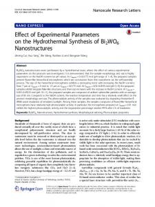
Fig. 2. Stress displacement curves for OS (above) and SBP (below).
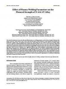
the two variables was observed. Energy to debonding (measured) was influenced by the brand of the adhesive but not by the filler addition, and there was no significant interaction between the two variables. Energy to debonding (elastic) was influenced by the brand of the adhesive but not by the filler addition, and there was no significant interaction between the two variables. Bond strength was in the range of 24.4-30.1 MPa, and there were significant differences between the bond strengths of OS and SB based on Fisher’s PLSD interval (Table 2). Displacement at debonding and energy to debonding (measured and elastic) of OS were higher than those of SB (P< 0.05), and energy to debonding (measured) of OSP was higher than those of SBP (P< 0.05). Stress-displacement curves for OS and SBP showed various curves (Fig. 2). There were significant correlations between bond strength, stiffness and energy to debonding (measured) (Table 3, Fig. 3).
Discussion The hypothesis regarding the addition of filler was accepted in both adhesives of OS and SB. There were no significant differences in all five bonding parameters between OS and OSP and between SB and SBP, even though in OSP, 8% filler was added, and 10-20% was added in SBP. Stress-displacement curves for each trial were different in the same adhesive, although bond strengths were similar (Fig. 2). Therefore, other bonding parameters in addition to the bond strength were evaluated in the present study. Depending on the bonding parameter, there were differences in values between adhesive pairs. It was reported that the use of the acetone-based systems resulted in a continuous and thick hybrid layer with reverse-
Fig. 3. Correlations between bonding parameters.
cone-shaped tags in close contact with the dentin tubule walls, and use of the water-based systems resulted in a thinner hybrid layer with some incompletely sealed dentin tubules.16 In the present study, OS and OSP were acetone-based and SB and SBP were ethanol-water-based systems. Bond strength of OS regardless of filler incorporation was 29.3 (4.4) MPa and that of SB was 24.5 (6.7) MPa, which were significantly different (P< 0.05). This result supports the previous report.16 The role of solvent and filler contents of bonding resin on microtensile bond strength to human dentin was evaluated by another investigator.17 Single Bond (SB), experimental Single Bond (with filler) (ExpSB), Prime & Bond NT (NT) and experimental Prime & Bond NT (without nanofiller) were studied. Bond strengths ranged from 58 MPa for ExpSB to 48 MPa for NT. The unfilled adhesives SB and ExpNT had bond strengths of 76 MPa and 39 MPa, respectively. Unfilled SB had significantly higher mean bond strength than the filled version. The ethanol-based adhesive SB had significantly higher mean bond strength than either the nonvolatile solvent-based or the acetone-based adhesives.17 In the present study, there was no
26 Lee et al significant difference in bond strength between unfilled and filled adhesives. Contrary to the previous study, acetone-based adhesives (OS and OSP) showed higher bond strengths than ethanol-water-based adhesives (SB and SBP) in the present study. This discrepancy may reflect the differences in composition of adhesives or testing methods. Large variations in bond strength determinations and lack of standardized test procedures have contributed to ambiguities in interpretation of bond strength data.13 Classical elastic theory is concerned with homogenous isotropic solids, or solids with known anisotropy. Therefore, there is a problem when experimental methods based on such theories are applied to natural materials which do not conform to the limitations of continua theory.18 Moreover the property of dentin is different depending on the region of dentin. The effect of dentin depth on the interfacial fracture toughness between dentin and composite was evaluated. Significant difference was observed between the superficial and deep dentin substrates.9 It was also confirmed that dentin pull-out during conventional bond testing was partly due to the biomechanics of the test and did not necessarily mean superior adhesive strength or even that the cohesive strength of the dentin was reduced.11 In the present study, superficial dentin was used as the bonding substrate. However, the coefficient of variation for bond strength was in the range of 11-35%, and those for other parameters were in the similar ranges except for energy to debonding (elastic) (9-49%). These high variations may reflect the anisotropy of dentin and difference in properties of individual tooth substrate. The elastic moduli of the successive layers across a dentincomposite bonding area were determined for dentin adhesive systems. A gradient of moduli of elasticity was observed from the rather stiff dentin over a more elastic resin-dentin interdiffusion zone. That gradient was more substantial in those systems that produced relatively thick adhesive resin layers. Elastic modulus of unaltered dentin (19.3 GPa) was significantly different from that of the interdiffusion zones, which was 4.9-9.7 GPa depending on the bonding agent. Elastic modulus of adhesive resin was 3.4 -4.8 GPa.6 Based on the above study, we can assume that deformation of a bonded interface will occur at the adhesive resin layer when external force is applied. However, the site of debonding is determined by strength and fracture mechanics of each layer. In the present study, debonding might have occurred within one or across combined layers. Bond strength and stiffness showed correlations, but the r value was not high (0.66). Strength values are often relied upon as indicators of structural performance for brittle dental materials. Strength, however, is more of a conditional than an inherent property, and strength data alone cannot be directly extrapolated to predict structural performance.19 Strength data are meaningful when placed into context by knowledge of material microstructure, processing history, testing methodology, testing environment and failure mechanism.19 In the present study, to determine more reliable bonding parameters instead of bond strength based on the tensile bond test, supplemental bonding parameters were evaluated. There were significant correlations between the parameters (Table 3). Although supplemental bonding parameters were calculated in this study and the meaning of these parameters was clear theoretically, practical
American Journal of Dentistry, Vol. 19, No. 1, February, 2006
meaning of these parameters should be studied further. The formation of a bonded interface is apt to produce microscopic flaws which could act as critical “stress risers” which promote interfacial failure.13 The initiation and propagation of such flaws under the mastication forces can be followed by fracture toughness or fracture energy.20 Gap-free intact interfaces were most frequently observed when a relatively thick layer of a separately polymerized and particlefilled adhesive resin was present. These observations provide evidence for an elastic bonding concept.6 Bond strength was significantly higher in all groups when the dentin bonding agent was painted on without being air thinned.5 It has been suggested that the dentin-composite joint should be reasonably flexible to accommodate polymerization shrinkage, and to minimize stress concentrations in bond during function,21 and this elastic bonding concept has recently been advanced as a method of achieving a superior seal.1 Significant differences in interfacial fracture toughness among dentin adhesives have been reported. Bond failures occurred at the interface between adhesive resin and the top of the hybrid layer with unfilled bonding resins with a film thickness of 30 µm. Filled adhesive (15% filled with 0.4 µm filler) formed thick films varying from 60-70 µm which failed cohesively; however, these adhesives sealed the dentin surface effectively despite moderate fracture toughness. Mean interfacial fracture toughness of SB was 0.84 (0.16) MPa/m1/2 and was 0.82 (0.12) MPa/m1/2 with OS.22 Energy to debonding observed in the present study is similar in concept to toughness in fracture. The value for OS was 14.1 N/mm and that for SB was 9.2 N/mm in the present study, which were not different (P> 0.05). The bond failure site might have been different as a result of incorporation of filler in the bonding resins. However, these differences were not reflected in the bonding parameters of the present study. Stress-resisting elastic buffers (thick bonding resin layers) underneath composite restorations may also better withstand shocks induced by occlusal loads, tooth flexure effects and thermal cycling during clinical function.6 The relative interfacial stiffness of dentin-composite interfaces was measured. The stiffness was determined from the initial slope of the forcedisplacement curve that was obtained from each fracture toughness test. The relative interfacial stiffness ranged from 1692 N/mm, and a significant positive linear correlation was found among the individual (r2=0.58) and the mean (r2=0.97) relative interfacial stiffness and KIC results.15 In the present study, the correlation coefficient between bond strength and stiffness was 0.66, which was lower than that in fracture toughness. This might suggest the inaccuracy of conventional tensile bonding test to determine the interfacial bond characteristics. Although the relative rankings of the average values for the dentin bonding agents were the same for both fracture toughness and tensile bond strength, there was no significant correlation between the individual KIC and TBS results within each test group (r2 < 0.5). It was therefore concluded that the fracture toughness test provided a valid method for characterization of the fracture resistance of the dentin-resin composite interface.13,23 Since adhesive joints produced by contemporary adhesives
American Journal of Dentistry, Vol. 19, No. 1, February, 2006
are brittle, future adhesive design may incorporate biomimetic intermediate-strength domains that can undergo stepwise reversible unfolding in response to varying functional stress levels before ultimate catastrophic failure of the adhesive joint occurs.2 These domains may also re-establish folded configurations on stress relaxation, making the adhesive both strong and tough.2 The practical meaning of stiffness and energy should be studied further with finite element analysis or comparison with results based on methods with a fracture mechanics approach. There were no significant differences in bonding parameters between no-filler and filler-added versions of acetone-based and ethanol-water-based adhesives. Bond strength, bond stiffness and energy to debonding (measured) showed significant correlations; however, the correlation coefficient varied by the parameters (r = 0.48 to 0.90). a. b. c. d. e. f.
Bisco, Inc., Schaumburg, IL, USA. 3M ESPE, St. Paul, MN, USA. Dentsply/Caulk, Milford, DE, USA. Vivadent, Schaan, Liechtenstein. Instron, Canton, MA, USA. SAS Institute, Cary, NC, USA.
Dr. Lee is Associate Professor, Department of Dental Biomaterials Science, College of Dentistry, Seoul National University, Seoul, Korea. Dr. Pinzon is Postdoctoral Scholar Fellow, Department of Preventive and Restorative Dental Science, University of California at San Francisco, San Francisco, CA, USA. Dr. O’Keefe is in private practice in Houston, TX, USA. Dr. Powers is Professor of Oral Biomaterials, Department of Restorative Dentistry and Biomaterials, Houston Biomaterials Research Center, The University of Texas Dental Branch at Houston, Houston, Texas, USA.
References 1. Van Meerbeek B, Perdigao J, Lambrechts P, Vanherle G. The clinical performance of adhesives. J Dent 1998; 26:1-20. 2. Tay FR, Pashley DH. Dental adhesives of the future. J Adhes Dent 2002; 4:91-103. 3. Van Meerbeek B, Conn LJ Jr, Duke ES, Eick JD, Robinson SJ, Guerrero D. Correlative transmission electron microscopy examination of nondemineralized and demineralized resin-dentin interfaces formed by two dentin
Filler addition and dentin bonding 27 adhesive systems. J Dent Res 1996; 75:879-888. 4. Retief DH. The principles of adhesion. J Dent Assoc S Afr 1970; 25:285-295. 5. Hilton TJ, Schwartz RS. The effect of air thinning on dentin adhesive bond strength. Oper Dent 1995; 20:133-137. 6. Van Meerbeek B, Willems G, Celis JP, Roos JR, Braem M, Lambrechts P, Vanherle G. Assessment by nano-indentation of the hardness and elasticity of the resin-dentin bonding area. J Dent Res 1993; 72:1434-1442. 7. Miyazaki M, Ando S, Hinoura K, Onose H, Moore BK. Influence of filler addition to bonding agents on shear bond strength to bovine dentin. Dent Mater 1995; 11:234-238. 8. Frankenberger R, Lopes M, Perdigao J, Ambrose WW, Rosa BT. The use of flowable composites as filled adhesives. Dent Mater 2002; 18:227-238. 9. Tam LE, Yim D. Effect of dentine depth on the fracture toughness of dentine-composite adhesive interfaces. J Dent 1997; 25:339-346. 10. Lin C-P, Chen R-S, Liu C-C, Lian S-S. Failure criteria of dentin-resin tensile bond test. A fracture mechanics approach. Scripta Materialia 1998; 38:115-121. 11. Versluis A, Tantbirojn D, Douglas WH. Why do shear bond tests pull out dentin? J Dent Res 1997; 76:1298-1307. 12. Misra A, Spencer P, Marangos O, Wang Y, Katz JL. Micromechanical analysis of dentin/adhesive interface by the finite element method. J Biomed Mater Res 2004; 70B:56-65. 13. Tam LE, Pilliar RM. Fracture toughness of dentin/resin-composite adhesive interfaces. J Dent Res 1993; 72:953-959. 14. Tam LE, Khoshand S, Pilliar RM. Fracture resistance of dentin-composite interfaces using different adhesive resin layers. J Dent 2001; 29:217-225. 15. Tam LE, Pilliar RM. The effect of interface stiffness on dentin-composite interfacial fracture resistance. J Dent 2000; 28:487-493. 16. Gregoire GL, Akon BA, Millas A. Interfacial micromorphological differences in hybrid layer formation between water- and solvent-based dentin bonding systems. J Prosthet Dent 2002; 87:633-641. 17. Nunes MF, Swift EJ, Perdigao J. Effects of adhesive composition on microtensile bond strength to human dentin. Am J Dent 2001; 14:340-343. 18. Braden M. Physics in dentistry. Phys Technol 1985; 16:58-62. 19. Kelly JR. Perspectives on strength. Dent Mater 1995; 11:103-110. 20. Toparli M, Aksoy T. Fracture toughness determination of composite resin and dentin/composite resin adhesive interfaces by laboratory testing and finite element models. Dent Mater 1998; 14:287-293. 21. Uno S, Finger WJ. Function of the hybrid zone as a stress-absorbing layer in resin-dentin bonding. Quintessence Int 1995; 26:733-738. 22. Walshaw PR, Tam LE, McComb D. Bond failure at dentin-composite interfaces with 'single-bottle' adhesives. J Dent 2003; 31:117-125. 23. Tam LE, Pilliar RM. Effects of dentin surface treatments on the fracture toughness and tensile bond strength of a dentin-composite adhesive interface. J Dent Res 1994; 73:1530-1538.