Journal of Medical and Biological Engineering, 23(4): 199-203
199
Effect of Hydroxyapatite Nano-Particle on Properties of Modified Tricalcium Silicate Bone Cements Ren-Jei Chung1
Chao-Yuan Lin1
Fong-In Chou3
Ping-Yu Shih2
Tsung-Shune Chin*,1,2
1
Department of Materials Science and Engineering, National Tsing Hua University, Hsinchu, 300, Taiwan, R.O.C. 2 Department of Materials Science and Engineering, National United University, Miaoli, 360, Taiwan, R.O.C. 3 Nuclear Science and Technology Development Center, National Tsing Hua University, Hsinchu, 300, Taiwan, R.O.C. Received 27 Oct 2003; Accepted 5 Dec 2003
Abstract Solid-state reactions were used to synthesize bone-cements based on zinc modified tricalcium silicate (C3S: Ca3SiO5). C3S is the main self-setting and the fastest hydration constituent in conventional cements. Zinc oxide was added to replace part of CaO in order to stabilize the C3S phase. 5 mole % ZnO addition (Zn-C3S) reveals the best compressive strength that reaches 166 MPa, compared with those without ZnO, 100MPa. In vitro direct-contact-tests using primary cultured osteoblast are persuasive of negligible cytotoxicity. Hydroxyapatite nano-particles prepared through hydrothermal methods were added as strengthener and resulted in the compressive strength increment of the Zn-C3S up to 175 MPa at an HA addition amount 7.5 wt%. The setting mechanism of C3S and Zn-C3S seems to be the same except varied gel properties that extend the fluidity of the slurry. These cements show remarkable self-setting properties and comparable strength with natural bones hence are potential in bony and dental restorations. Keywords: Bone-cement, Tricalcium Silicate, Self-setting, Zinc oxide, Hydroxyapatite, Nano-particles
Introduction Acrylic cements have been widely used in orthopedics and dentistry as fixation agents and fillers [1,2]. However, they have some disadvantages such as thermal necrosis of cells, shrinkage during polymerization, monomer releasing, and mechanical mismatch with the implant site, among others [3]. It has been known that glasses and derivatives based on the CaO-SiO2 system show excellent biocompatibility and form apatite layer on their surface in vivo as well as in vitro [4-5]. Tricalcium silicate (Ca3SiO5, C3S in short) is the main self-setting and the fastest hydration constituent in cements. C3S swells during setting and gets very high strength after being set [6]. From the phase diagram, it is manifested that C3S is a single phase at very high temperature, but will decompose into dicalcium silicate (C2S, Ca2SiO4) and calcium oxide (CaO) at temperatures below 1250 oC. Therefore, it’s difficult to obtain pure C3S phase by solid-state reaction. It is reported that some transition metals are able to stabilize the C3S phase [7]. Hydroxyapatite (HA) being the main inorganic content of human bone gets excellent biocompatibility and osteoconductivity, and accelerates the restoration and growth of *Corresponding author: Tsung-Shune Chin Tel: ++886-3-5742630; Fax: ++886-3-5719868 E-mail:
[email protected]
injured bone [8,9]. In this study, our aim was to synthesize quality improved C3S with high phase purity and mechanical properties, aiming to apply the aforesaid characteristics in bony and dental restorations. We chose zinc as a phase-stabilizing additive as well as making use of the special bio-effects of zinc. Zinc was excepted to substitute calcium in the crystal structure forming (Ca1-xZnx)3SiO5. The phase purity of developed materials was analyzed by XRD. Besides, hydroxyapatite nano-particle was added as a strengthener aiming also to benefit the biocompatibility. The setting time of developed cements were evaluated by the Gillmore needle method [10]. Mechanical properties were measured by an Instron machine. The pH value and in vitro biocompatibility were also tested.
Materials and Methods 1. Material preparation and analyses The precursory powders (CaCO3+ZnO):SiO2= 3:1 were mixed by ball milling the weighed constituents. Molar percentage ZnO to replace CaO was from 5 to 50%. After heating and soaking the mixtures at 1400 oC, they were quenched in liquid nitrogen. The products were ground to smaller than 37 µm (400mesh sieved). Powders were mixed with de-ionized water (in weight%) by firstly rapid shaking
J. Med. Biol. Eng., Vol. 23. No. 4 2003
200
(3000 rpm) and self-setting. The slurry was filled into a stainless mold (10mm in diameter), pressed by a pressure 8.3 kgf/cm2, incubated at 37oC under saturated steam for curing. 2. Preparation of hydroxyapatite nano-particles through a hydrothermal route Hydroxyapatite nano-particle was prepared by an autoclave hydrothermal process. Reagent grade Ca(H2PO4)2·H2O and Ca(OH)2 powders were mixed by a conventional ball-milling technique using zirconia balls immersed in ethanol for 1h. The molar ration of Ca(H2PO4)2·H2O to Ca(OH)2 was 3:7, which is based on the hydroxyapatite (HA) stoichiometric composition. After mixing, the slurry was dried by vacuum extractor. The HA powder was synthesized by heating mixtures of powders in 100~200 g batch with 2000 ml distilled water under 121 oC for 1~2 h in a conventional autoclave used for sterilization. The reaction product was filtered and washed several times with DI water for the complete removal of PO43- and Ca2+ ions, then frozen dried. 3. Cells and cell culture The primary cultural osteoblast was used for in vitro biocompatibility evaluation. Calvaria from Wistar rat (born in three days) were excised aseptically. Soft tissues on the surface were removed carefully. After rinsing the calvaria with PBS, the calvaria were minced and incubated in collagenase (1mg/ml PBS, Sigma Co.) at 37 OC for 10 min. The isolated osteoblasts were centrifuged for 5min at 1500 rpm and then cultured in DMEM. The medium was refreshed every two days. Each cement disc was gamma ray irradiation sterilized (10 kGy) before biocompatibility tests. 4. Characterization methods XRD patterns were obtained from samples using a wide-angle x-ray powder diffractometer (model D/max-IIB, Rigaku Co., Tokyo, Japan). X-ray radiation CuKα1 (λ = 1.5405Å) was set at 30 kV and 20 mA. Detailed peak features of the diffraction angles, 2θ, ranging from 24o to 38o were obtained by a θ-2θ scan rate of 0.4o per minute. The crystalline phases were identified using JCPDS files (HA: 24-0033 and Si: 03-0529). Setting properties were evaluated by the Gillmore needle method. When the needle can no longer penetrate the surface of cement it is meant that setting time for proper strength is reached. The compressive strength of the cylinder was measured using an Instron 4505 Universal testing instrument.
Results and Discussion In our past researches [11], through the synthetic procedures, C3S phase with substantial amount of free CaO can be obtained routinely for unmodified 3CaO-SiO2 compositions. And the amount of free CaO phase obviously decreases with increasing ZnO addition up to 7.5 mol%. The mechanical strength of the samples increases with curing time. After curing for 24 hours, the synthesized XRD-pure C3S shows a setting time of 56.3 min, a compressive strength around 100MPa. Among those ZnO modified products, the (Ca92.5Zn7.5)3SiO5
(a)
(b)
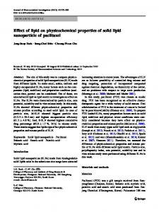
Figure 1. (a) Pure C3S sample shows no cytotoxicity; (b) Zn-C3S sample shows no cytotoxicity
samples have the least CaO impurity and show a compressive strength around 128MPa, yet with a worse handling property. The (Ca95Zn5)3SiO5 (Zn-C3S) samples show the best mechanical property with a setting time 54 min and the compressive strength reaches 166MPa. More ZnO addition leads to a lengthened setting time and a decreased compressive strength. Besides, the un-modified C3S slurry loses its fluidity in one minute, while that of the Zn-C3S slurry sample maintains fluidity until 3 minutes, which may be more suitable in clinical usage. Samples of pure C3S and Zn-C3S were selected for biocompatibility evaluation. After direct contact tests, the samples didn’t show obvious cytotoxicity (Figure 1). Because the calcium silicate bone cement was set with water, different weight ratios between water to cement powder lead to different working time and setting time. Figure 2 was the setting time of C3S and Zn-C3S cements with different amounts of water addition. It’s obviously that setting was positively dependent on water content. And when the water/powder weight ratio was 0.2, the C3S and Zn-C3S cements had the shortest setting time, however, the resulted solid got a loose structure. So the water/powder ratio was optimally set as 0.4 (200mg water versus 500mg powder) and applied for further studies. Figure 2 also tells that zinc addition extends the working time and setting time, and also provides better fluidity that was good for clinical usage. It is manifest that addition of zinc oxide varies the property of gel formed upon mixing with water as compared with that of un-modified C3S. The modified gel would extend the time to the formation
Zn-modified 3CaO-SiO2 with Hydroxyapatite Nano-Particle
201
600 550
140
Zn-C3S
131.8
120
450 400
99.2
100
350
116.1
C3S
94.1
MPa
300 250 200 150
C3S
100
80 54.9
60 40
35.3
49.7
31.7 31.2
50
34
20
0
0
0.2
0.4
0.6
0.8
1.0
5
10
15
20
Days
weight ratio of water to powder Figure 2. Setting time of C3S and Zn-C3S cements with different water addition
5 wt%
10 wt%
140
setting time (min)
120 100
ratio of water to powder is 2:5
80 60
7.5 wt%
40 20 0 0
20
40
60
80
HA content (wt%) (a)
Zn-C3S
140 130
setting time (min)
setting time (min)
500
120 110 100 90 80 70
C3S
60 50 40 0
2
4
6
8
10
HA content (wt%) (b) Figure 3. (a) Setting time of Zn-C3S with different amounts of HA addition; (b) Setting time of Zn-C3S and C3S with different amounts of HA addition.
Figure 4. Relationship between compressive strength and curing time of the cements
of cross-linking structure. Figure 3(a) shows the setting time of Zn-C3S with different amounts of HA addition. It’s obvious that setting time is prolonged upon HA addition less than 10 wt%, while decreased above 10 wt%. It’s suggested that when the HA is of small amount, the added HA will hinder the hydration of Zn-C3S that otherwise self-sets as usual. While in case of high HA concentration, the HA nano-particles tend to incorporate into the hydration mechanism leading to a loose hydrated structure and an uneven local setting. The setting time of C3S+HA cement shows a turning point at 5 wt% HA addition (Figure 3(b)). The difference between setting time of HA added Zn-C3S and C3S was due to fluidity as stated earlier. Figure 4 presents the relationship between compressive strength and curing time of the cements. After curing for twenty days, Zn-C3S has higher compressive than that of C3S. The mechanisms are elucidated due to the following steps and reasons: 1. Zn-C3S has better fluidity that is beneficial for early packing of the as hydrated components. 2. Zn2+ inserts into the lattice of final set mass. 3. From the analyses of fine structure of the final products, Zn-C3S shows more continuous and compact structure. There existed more boundaries and grains in final product of C3S cement. Besides, these two cements show similar time dependent curves of compressive strength during curing, it implies that the two cements have the same strengthening mechanism. Figure 5(a) shows the effects on compressive strength of C3S cements added with different HA: 5wt%, 7.5wt%, 10wt%, respectively. The results reveal that addition of HA decreases the final compressive strength. It’s due to the reason that added HA nano-particles might not be well separated and might react with the gel locally. Nevertheless the 7.5 wt% added one has relatively high strength than others. Figure 5(b) is same measurement upon the Zn-C3S cement. For HA added Zn-C3S, the compressive strength is raised, the best value as high as 175MPa is possible at an HA amount of 7.5 wt%. The reason
J. Med. Biol. Eng., Vol. 23. No. 4 2003
202
purity, lengthen the setting time and also increase the final compressive strength. The (Ca95Zn5)3SiO5 samples show the best mechanical property with a compressive strength 166MPa. In vitro cell culture tests showed that the studied samples show negligible cytotoxicity. Zn-C3S is concluded to be more suitable for clinical injecting. Besides, for Zn-C3S added with 7.5 wt% HA, its compressive strength is further improved and setting time is lengthened. Concerning clinical usages, Zn-C3S is more suitable and potential. The developed (C,Z)3S bone cements in this study show promising applications in bony and dental restorations.
110 100 90
MPa
80 70
7.5 wt% HA 10 wt% HA 5 wt% HA
60 50 40 30 20 0
10
20
30
Acknowledgments
40
Days
The authors are grateful for the sponsor of this research by the National Science Council of the Republic of China, under the grant NSC91-2216-E-239-001. The author R.J. Chung thanks for the partial funding supported from Lee Chau Jen Foundation, Biomedical Engineering Society of R.O.C. to present the paper in Bioceramics 15 (Australia).
(a) 200 180 160 140
MPa
120 100
5wt% HA-ZnC3S 7.5wt% HA-ZnC3S 10wt% HA-ZnC3S
80 60 40 20 0 -20 0
5
10
15
20
25
30
Day (b) Figure 5. (a) Effects on compressive strength of cured C3S solids by different amounts of HA addition: 5wt%, 7.5wt%, 10wt%, respectively; (b) Effects on compressive strength of cured Zn-C3S solids by different amounts of HA addition: 5wt%, 7.5wt%, 10wt%, respectively
is due to the good fluidity that helps dispersion of nano-particles, so that HA could be competent for strengthening. And the 7.5 wt% HA added one reaches highest strength in around 5 days that are faster than un-modified one.
HA the the the
Conclusions In this study, a series of ZnO modified 3CaO-SiO2 bone cements (C3S) was successfully deveoped. The results showed that addition of up to 7.5 mol% ZnO to replace CaO in the C3S composition would stabilize the C3S phase, increase the phase
References [1].
J.F. Cvar, G. Ryge, “Criteria for the clinical evaluation of dental restorative materials”, USPHS Publication. San Francisco: U.S. Government Printing Office. 244, 1997. [2]. H.R. Stanley, “Pulpal response to dental techniques and materials”, Dent. Clin. North. Am., 15, 115, 1971. [3]. W. Geurtsen, F. Lehmann, W. Spahl, G. Leyhausen, “Cytotoxicity of 35 dental resin composite monomers/additives in permanent 3T3 and three human primary fibroblast cultures”, J. Biomed. Mater. Res., 41(3):474-480, 1998. [4]. L.L. Hench, “Bioceramics: from conceot to clinical“, J. Am. Ceram. Soc., 74(7), 1487-1510, 1991. [5]. C. Ohtsuki, T. Kokubo and T. Yamamuro, “Mechanism of apatite formation on cao-sio2-p2o5 glasses in a simulated body-fluid”, J. Non-Cryst. Solids, 143(1): 84-92, 1992. [6]. K.L. Scrivener and A. Capmas: Calcium Aluminate Cements, 4th Edn.,(ed) P.C. Hemlett, Arnold, London. [7]. I. Fernandez Olmo, E. Chacon and A. Irabien, “Influence of lead, zinc, iron (III) and chromium (III) oxides on the setting time and strength development of Portland cement”, Cement Concrete Res., 31(8):1213-1219, 2001. [8]. K. de Groot, “Bioceramics of calcium phosphate”, CRC Press. Inc., Bocaa Ration, Florida, 1983. [9]. M.J. Dalby, L. Di Silvio, F. J. Harper and W, Bonfield, “Increasing hydroxyapatite incorporation into poly(methylmethacrylate) cement increases osteoblast adhesion and response”, Biomaterials, 23(2):569-576, 2002. [10]. G.C. Robert, L.W. Marcus, Restorative Dental Materials. Mosby-Year Book ch8:172-208, 1997. [11]. R.J. Chung, T.S. Chin, C.W. Huang, P.C. Lin, T.J. Hsu and F.I. Chou, Bone-Cements Based on Tricalcium Silicate, Key Engineering Materials, 240-2: 337-340, 2003.