JOURNAL OF BONE AND MINERAL RESEARCH Volume 18, Number 9, 2003 © 2003 American Society for Bone and Mineral Research
Effect of Hyper- and Microgravity on Collagen Post-Translational Controls of MC3T3-E1 Osteoblasts MITSURU SAITO, SHIGERU SOSHI, and KATSUYUKI FUJII ABSTRACT We attempted to study the effects of microgravity (by clinostat) and hypergravity (using centrifugation) on collagen metabolism using murine MC3T3-E1 osteoblasts, especially focusing on collagen cross-link formation. We found that altered gravitational load affected the post-translational modification of collagen, particularly the collagen maturation pathway, through altered expression of enzymes involved in cross-link formation. Introduction: Gravitational loading plays important roles in the stimulation of differentiated osteoblast function and in the maintenance of skeletal tissues, whereas microgravity seems to result in osteopenia caused by impaired osteoblast differentiation. The aim of our study was to clarify the effects of altered gravitational environments on collagen metabolism, particularly the relationship between post-translational collagen quality and enzymes involved in cross-link formation, using murine osteoblastic MC3T3-E1 cells. Materials and Methods: Cells were cultured under vector-averaged microgravity (1 ⫻ 10⫺3g) using a clinostat or under conventional centrifugation techniques to generate hypergravity (20g and 40g) for 72 h. We then examined the expression patterns of lysyl oxidase and the two lysyl hydroxylase isoforms telopeptidyl lysyl hydroxylase (TLH; procollagen-lysine, 2-oxyglutarate, 5-dioxigenase 2 [PLOD2]) and helical lysyl hydroxylase (HLH; [PLOD1]) by quantitative real time polymerase chain reaction (PCR) analysis. Quantitative analysis of reducible immature (dihydroxylysinonorleucine, hydroxylysinonorleucine, and lysinonorleucine) and nonreducible mature (pyridinoline and deoxypyridinoline) cross-links, and maturation rate analysis of immature to mature cross-links by conventional metabolic labeling using tritium lysine were also performed. Results: Hypergravity upregulated both TLH mRNA expression and enzyme activity compared with stationary cultures, whereas microgravity stimulated both HLH mRNA expression and enzyme activity. These results were consistent with increased relative occupancy rates of telopeptidyl hydroxylysine-derived cross-links and helical hydroxylysine-derived forms observed under hypergravity and microgravity, respectively. Hypergravity stimulated not only lysyl oxidase mRNA expression but also increased enzyme activity and the sum of immature and mature cross-links. Furthermore, the conversion rate of immature cross-links to mature compounds was markedly increased under hypergravity but decreased under microgravity. Conclusion: Altered gravitational loading may affect the post-translational modification of collagen through altered expression of enzymes involved in cross-link formation. These observations may be important in elucidating the mechanisms of osteopenia during space flight. J Bone Miner Res 2003;18:1695–1705 Key words:
microgravity, hypergravity, collagen, cross-links, lysine hydroxylation INTRODUCTION
ONE TISSUE ADAPTS both the quality and quantity of its constituents according to its mechanical environment.(1–5) In the absence of repetitive transient skeletal mechanical un-
B
The authors have no conflict of interest.
loading, such as experienced during spaceflight, osteopenia can occur as a result of diminished bone formation(6 – 8) and increased net resorption.(9) Morphological and functional studies have shown that osteoblast and osteoblast-like cells are highly sensitive to altered gravity. Microgravity, caused by space flight or simulated conditions such as those induced by the clinostat culture system, has been shown to exert profound
Department of Orthopaedic Surgery, Jikei University School of Medicine, Tokyo, Japan.
1695
1696
SAITO ET AL.
effects on important cellular processes such as proliferation(10) and differentiation.(11–13) These results suggest that decreased osteoblast function is the major contributing factor of spaceflight-induced osteopenia. In contrast, gravitational loading has an important stimulatory role in differentiated osteoblast function. Several studies have shown that hypergravity stimulates not only cellular differentiation, but also proliferation.(14 –16) Thus, hypergravity appears to exert stimulatory effects on bone formation. However, surprisingly little is known about the influence of microgravity and hypergravity on the metabolism of the most abundant bone matrix protein, type I collagen. In particular, collagen cross-linking is important for the proper biological and biomechanical function of collagen fibers,(17) and cross-link abnormalities are involved in connective tissue changes associated with disease and aging.(18 –20) Stereochemical and X-ray diffraction studies have revealed that differences in the molecular packing of collagen within fibrils are associated with differences in cross-link profiles.(21–23) Furthermore, proper mineralization seems to depend on the correct alignment of collagen molecules. For instance, the nucleation of calcium apatite crystals starts in gap regions, that is, in areas adjacent to cross-linking sites.(24) Thus, aberrant mineralization may also be caused by alterations in cross-linking patterns associated with changes in molecular packing. The sites of cross-linking in collagen molecules are mainly lysine (Lys) and hydroxylysine (Hyl) residues.(24 –27) The heterogeneity of type I collagen is due primarily to quantitative differences in the conversion of Lys to Hyl, with the enzymes responsible for the hydroxylation of the telopeptides and triple helices being telopeptidyl lysyl hydroxylase (TLH) and helical lysyl hydroxylase (HLH), respectively.(28) Cross-linking is initiated only after specific Lys or Hyl residues in the telopeptides are converted extracellularly by lysyl oxidase into the aldehydes allysine and hydroxyallysine, respectively.(24 –27) These aldehydes subsequently react with Lys or Hyl residues of the triple helix to give the characteristic di- and tri-functional cross-links.(29) The aim of this study was to examine the effects of microgravity (by clinostat culture) and hypergravity (using centrifugation) on collagen metabolism in osteoblastic MC3T3-E1 cells, particularly focusing on the correlation between the expression of the two lysyl hydroxylases, patterns of Lys hydroxylation, and collagen cross-linking.
MATERIALS AND METHODS Cell culture Murine MC3T3-E1 osteoblasts, purchased from RIKEN Cell Bank (Tsukuba, Japan), were maintained in alpha minimal essential medium (␣MEM; GIBCO, Life Technology, Gaithersburg, MD, USA) supplemented with 10% (vol/vol) fetal calf serum (HyClone Laboratories, Logan, UT, USA), 10 mM HEPES, 50 U/ml penicillin, 50 g/ml streptomycin, and 50 g/ml ascorbate (Sigma-Aldrich, St Louis, MO, USA) at 37°C in a humidified 95% air and 5% CO2 atmosphere. Cells (5 ⫻ 104 cells/cm2) were seeded into 25-cm2 culture flasks (Falcon; Becton Dickinson Labware, Franklin Lakes, NJ, USA) and were used for experiments after 14
days culture at the time of confluency. Before exposing cells to stimulated hypergravity or microgravity, flasks were filled completely with fresh medium to eliminate the presence of air bubbles to diminish the effects of turbulence and fluid shear forces during rotation culture.
Pulse labeling For analysis of collagen cross-link formation kinetics, confluent cell cultures were labeled with 250 Ci [3H] Lys (40 –100 mCi/mmol; Moravec Biochemical, Inc., Brea, CA, USA) per 25-cm2 flask for 24 h on culture day 13. Labeling was performed in medium lacking serum and Lys but supplemented with 50 g/ml ascorbate. The medium was then removed and replenished with fresh medium containing 10% fetal calf serum.
Hypergravity and microgravity Flasks were subjected to hypergravity (20g and 40g) by horizontal centrifugation at a constant rate in a swinging bucket rotor (TOMY Tech Inc., Tokyo, Japan). This gravity of centrifugation was selected after experiments using 5g, 10g, 20g, and 40g. It was found that 20g and 40g resulted in reproducible, detectable changes in collagen posttranslational modification compared with stationary controls. Four flasks were mounted on the sample stage of the centrifuge with left-right symmetry to maintain the balance of rotation. A three-dimensional clinostat was used to subject cells to simulated microgravity.(30,31) The clinostat is widely used to stimulate microgravity conditions on the ground. Its principle of action is based on gravity randomization by exposure to a vector-averaged gravity environment. In the threedimensional clinostat produced by MHI Co. (Kobe, Japan), samples are moved along a sphere by the cooperative rotation of two motors. The outer frame is rotated around a horizontal axis, with an inner supporting frame mounted on the outer frame. The rotational axis of the inner frame assembly and sample stage is perpendicular to the first horizontal axis. Rotational motions of the frame and the sample stage are driven by two servomotors, with the onset, rate, and duration of rotation of the motors controlled by computer. The rotational velocity of both frames was 60°/s. The three-dimensional clinostat allowed uniform generation of 1 ⫻ 10⫺3g stimulated microgravity on the sample stage and was located in a 37°C room. After 72 h of centrifugation or clino-rotation, MC3T3-E1 cell layers were washed three times with PBS and collected. In a preliminary study, we examined whether gravitational loading produced by clino-rotation and centrifugation for 72 h affected the temperature and pH of the conditioned medium. It was found that these parameters were identical to those of stationary culture medium.
Total cellular DNA content and alkaline phosphatase activity To estimate the effect of altered gravity on cell proliferation and differentiation, we measured both total cellular DNA content(32) and cellular alkaline phosphatase activity.(33) Briefly, cell layers were lysed in 0.05 M Tris-HCl,
COLLAGEN CROSS-LINKS IN HYPER- AND MICROGRAVITY TABLE 1. PRIMER SEQUENCES
AND
POLYMERASE CHAIN REACTION CONDITIONS USED
Gene COL1A1* Lysyl oxidase† HLH‡ TLH‡ GAPDH§
Primer sequence (5⬘–3⬘) Sense Antisense Sense Antisense Sense Antisense Sense Antisense Sense Antisense
GAACGGTCCACGATTGCATG GGCATGTTGCTAGGCACGAAG GGCGCCAGACAATCCAATGGGAG GCCTGGATGTAGTAGGGATCGGG TACCTTCACCGTCAACATAGCC TCCTTGAAGATGAGCGTTACCAT TACTATGCTCGCTCTGAAGATTACG GGACATTTTCATAACCACCCGATAT GGGTGGAGCCAAACGGGT GGAGTTGCTGTTGAAGCA
1697 FOR
AMPLIFICATION Size of amplification products (bp) 167 547 436 511 532
*,† Reported primer sequences in references 34 and 35 were used, respectively. ‡ Primers were constructed according to GenBank. § Primer pair was from Maxim Biotech, Inc., San Francisco, CA, USA.
pH 7.4, containing 1 mM MgCl2, and 0.2% NP-40 (SigmaAldrich). Total cellular DNA content was measured by adding the fluorochrome dye Hoechst 33258 (Pharmacia Biotech, Tokyo, Japan) to a final concentration of 10 g/ml. Fluorescence was determined by excitation at 350 nm and measurement of emission at 460 nm using a fluorescence spectrophotometer (Pharmacia Biotech). Herring sperm DNA was used as a standard. Alkaline phosphatase activity in cell lysates was determined by a colorimetric method using phenyl phosphate as substrate (Wako Pure Chemicals, Co., Osaka, Japan). Enzyme activity was expressed as micromoles of substrate used per minute per 1-ml sample per 1 g DNA at 37°C (IU/g DNA). Because microgravity may induce apoptosis in osteoblasts,(10) alkaline phosphatase (ALP) activity and collagen amount were normalized according to the amount of double-stranded DNA present after gravitational loading rather than according to cell number.
Total RNA isolation and reverse transcriptionpolymerase chain reaction Total RNA was extracted from harvested cell layers using TRIzol reagent (GIBCO-BRL, Grand Island, NY, USA) according to the supplied protocol. Isolated RNA was treated with 2.5 g RNase-free DNase (10 U; Worthington, Freehold, NJ, USA) for 30 minutes at room temperature in 10 l buffer containing 20 mM Tris-HCl (pH 8.3), 50 mM KCl, 2.5 mM MgCl2, and 40 U RNase inhibitor (Boerhinger Mannheim, Indianapolis, IN, USA). Reverse transcription-polymerase chain reaction (RTPCR) was carried out using a One-Step RT-PCR kit (QIAGEN Inc., Hilden, CA, USA), according to the manufacturer’s protocol. Aliquots of 1 g total RNA were reversetranscribed into cDNA at 50°C for 30 minutes in a thermal cycler. Sequences of each primer set used for amplification are detailed in Table 1. Primer sets specific for collagen ␣1 (COL1A1) and lysyl oxidase genes were derived from Matsumoto et al.(34) and Svinarich et al.,(35) respectively. Primers specific for glyceraldehyde-3-phosphate-dehydrogenase (GAPDH) were obtained from Maxim Biotech, Inc. (San Francisco, CA, USA) mRNA sequences of telopeptidyl Lys
hydroxylase (TLH, PLOD2, AF080572) and helical Lys hydroxylase (HLH, PLOD1, AF046782) were obtained from Genbank and used to design specific primers using primer design software (HYBsimulator 4.0; Advanced Gene Computing Technologies, Inc., San Francisco, CA, USA). Each PCR cycle included 1 minute denaturation at 95°C, 1 minute annealing at 58°C for COL1A1, 63°C for lysyl oxidase, or 61°C for HLH and TLH, and 1 minute extension at 72°C. The optimum cycle number that allowed amplification within the linear range to facilitate semiquantitative comparison between samples was found to be 30. Amplification products were run on 2.0% agarose gels and stained with ethidium bromide to visualize bands. To confirm the identity of the PCR products, products were cloned using a TA cloning kit (Invitrogen, San Diego, CA, USA) and nucleotide sequences analyzed. Real time PCR was performed to allow the relative quantification of lysyl oxidase, TLH, HLH, and GAPDH mRNA expression using the LyghtCycler system with SYBR Green I (QuantiTect SYBR Green RT-PCR kit; QIAGEN Inc.), according to the manufacturer’s protocol. Specific primers for each target gene were the same as described above (Table 1). Total RNA aliquots (1 g) were reversetranscribed into cDNA and RT reaction mixtures amplified for 45 cycles in a real time PCR cycler. Products were quantified using a standard curve that correlated each cycle number of linear-phase amplification with a value. Index mRNA levels were assessed using a threshold cycle (Ct) value. The relative expression of each target mRNA was computed from the target Ct values and the GAPDH Ct value using the standard curve method. To control for variability in amplification caused by differences in starting mRNA content, GAPDH was used as internal standard. Results are given as mean ⫾ SD of six independent experiments, normalized against the mean stationary control value.
Isolation of cell-matrix fraction containing insoluble collagen Cell layers were washed three times with ice-cold (PBS) and centrifuged at 3000g for 30 minutes at 4°C to separate
1698
the cell-matrix fraction from medium. Cell-matrix fractions were lysed with lysis buffer (0.1 M Tris-HCl, 0.125 M NaCl, 1% Triton X-100, 0.1% SDS, 1% deoxycholate; Sigma-Aldrich) and a cocktail of protease inhibitors including 0.5 mM phenylmethanesulfonylfluoride, 5 mM benzamidine, 2 mM pepstain A, and 1 mM leupeptin for 30 minutes at 4°C. Lysates were centrifuged at 3000g for 30 minutes at 4°C, and insoluble residues washed three times with cold distilled water and then lyophilized. This fraction was designated as the cell-matrix fraction. Most noncollagenous soluble proteins and procollagen were removed by this extraction and washing procedure. Total amount of collagen in cell-matrix fraction was calculated by the hydroxyproline (Hyp) assay using high performance liquid chromatography (HPLC), assuming that collagen weighed 7.5 the measured Hyp weight, with a molecular weight of 300,000.(18,36)
Lysine hydroxylation of collagen The level of Lys hydroxylation in the ␣1 and ␣2 chains of type I collagen, including or excluding telopeptides, was analyzed according to Uzawa et al.(37) with some modifications. To estimate the level of Lys hydroxylation in the collagen molecules, including both telopeptides and helical domains, cell-matrix fractions were extracted by 0.5N acetic acid for 2 days at 4°C with gentle agitation. Samples were then centrifuged at 20,000g for 30 minutes and supernatants were lyophilized. For the analysis of Lys hydroxylation in collagen molecules that excluded telopeptides, pepsin extractable collagen was isolated by digesting cell-matrix fractions with pepsin (Worthington) at an enzyme:substrate ratio of 1:10 in 0.5 M acetic acid (substrate content 10 mg/ml) at 5°C for 48 h.(38) After digestion, residues were removed by centrifugation at 20,000g for 30 minutes, NaCl was added to the supernatants to a final concentration of 0.7 M, and mixtures were stirred for 24 h. Precipitates were collected by centrifugation at 25,000g for 30 minutes, dialyzed against 0.5 N acetic acid, and lyophilized. Dried samples were subjected to 5% SDS-PAGE under nonreducing conditions, transferred onto Trans-Blot Transfer membranes (BioRad Laboratories, Inc.), and stained with 0.25% Coomassie Brilliant Blue (Wako). Visualized bands corresponding to the ␣1 and ␣2 chains of type I collagen were excised, hydrolyzed, and subjected to amino acid analysis using established methods.(18) Purified proteins were identified as ␣1 and ␣2 type I collagen chains by comparison with respective standards prepared from bovine skin type I collagen with and without pepsin treatment (KOKEN, Tokyo, Japan), with amino acid composition based on the known mouse type I collagen. The extent of Lys hydroxylation was expressed as the ratio of Hyl/(Lys ⫹ Hyl).(37,39)
Lysyl oxidase enzyme activity Lysyl oxidase enzyme activity was measured in cell layers and conditioned media using chick calvaria bone collagen labeled with [3H] Lys (250 Ci) (Moravec Biochemical, Inc., Brea, CA, USA) using standard methods.(40,41) Briefly, 0.5-ml aliquots of conditioned media and cell lysates (0.5 ml) extracted by 1 ml of lysis buffer (0.02 M boric
SAITO ET AL.
acid, 4 M urea, 0.15 M NaCl, pH 8.0) were prepared after gravitational loading. Samples were analyzed in quadruplicate in a final volume of 1.0 ml, containing 380,000 disintegrations per minute (dpm) tritiated collagen in the presence and absence of 5 ⫻ 10⫺4 M -aminopropionitrile. Reactions were incubated for 90 minutes at 37°C followed by distillation under vacuum. Units were defined as dpm and released over and above -aminopropionitrile only.
Lysyl hydroxylase enzyme activity Determination of TLH and HLH enzyme activities was performed according to the method of Miller,(42) using chick calvaria bone protocollagen labeled with [3H] Lys. Briefly, cell matrix fractions were homogenized in lysis buffer (0.2 M NaCl, 0.1 M glycine, 50 mM dithiothreitol, 0.1% [wt/vol] Triton-X, and 20 mM Tris-HCl, pH 7.5), and the homogenates were analyzed in quadruplicate in a final volume of 1.0 ml containing 220.000 dpm tritiated protocollagen. Samples were incubated for 2 h at 30°C, after which NaCl was added to a final concentration of 0.7 M. Precipitates were collected by centrifugation at 25,000g for 30 minutes. Precipitates with or without pepsin treatment were used to estimate the activity of the tritiated Hyl in the presence or absence of collagen molecule telopeptides, respectively. HLH enzyme activity was estimated by the tritiated Hyl activity in the pepsin extractable fraction, while TLH enzyme activity was assessed indirectly by the activity of tritiated Hyl in intact collagen molecules. Quantitative determination of enzyme activity as units of dpm of Hyl was carried out by high performance liquid chromatography (HPLC) after acid hydrolysis of the fractions according to established methods.(18)
Characterization of collagen cross-links The reduction of collagen with sodium borohydride (NaBH4; Sigma-Aldrich) and subsequent quantification of cross-links were carried out as previously described.(18) Briefly, cell-matrix fractions were suspended in potassium phosphate buffer, pH 7.6 (ionic strength ⫽ 0.15), and reduced at 37°C with nonradioactive NaBH4. The reduced samples were hydrolyzed in 6 N HCl at 110°C for 24 h. Hydrolysates were then analyzed for cross-links on a Shimadzu LC9 HPLC fitted with a cation exchange column (0.9 ⫻ 10 cm, Aa pack-Na; JASCO, Ltd., Tokyo, Japan) linked to an on-line fluorescence flow monitor (RF10AXL, Shimadzu, Shizuoka, Japan). Reducible cross-links (deHDHLNL, dehydro-dihydroxylysinonorleucine; deH-HLNL, dehydro- hydroxylysinonorleucine; deH-LNL, dehydrolysinonorleucine) were identified and quantified according to their reduced forms (DHLNL, HLNL, and LNL, respectively). Reducible cross-links and common amino acids, such as hydroxyproline, Hyl, and Lys, were detected with O-phthalaldehyde derivatization by the post-column method, whereas nonreducible cross-links, such as pyridinoline (Pyr) and deoxypyridinoline (Dpyr), were detected by natural fluorescence. Contents of each cross-link were expressed as mol/mol of collagen. Statistical analysis was carried out using one-way ANOVA followed by Fisher’s PLSD analysis, and statistical significance was set at p ⬍ 0.05.
COLLAGEN CROSS-LINKS IN HYPER- AND MICROGRAVITY
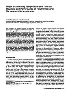
FIG. 1. Effect of hypergravity and microgravity on cell growth and alkaline phosphatase activity. MC3T3-E1 cells were exposed to 20g, 40g, or clino-rotation for 72 h, after which total cellular DNA content and the alkaline phosphatase activity were determined. Data represent mean ⫾ SD of six independent cultures. * and # represent significant increase (p ⬍ 0.01) and decrease (p ⬍ 0.01) vs. stationary culture, respectively.
RESULTS Cell growth and differentiation
1699
FIG. 2. Quantitative changes in collagen deposition in cell layers after exposure to hypergravity and microgravity for 72 h. Total collagen in the cell-matrix fractions was estimated by hydroxyproline assay using established HPLC methods. Collagen content is expressed as nanogram collagen extracted per 25-cm2 tissue culture dish and per microgram DNA. Data represent mean ⫾ SD of six independent cultures. * and # represent a significant increase (p ⬍ 0.01) and decrease (p ⬍ 0.01) vs. stationary culture, respectively.
We examined the effects of hypergravity and microgravity on cell proliferation and differentiation by measurement of total cellular DNA, with cellular ALP activity used as a marker of the mature osteoblastic phenotype.(43) As it was not feasible to get actual cell counts of loaded and unloaded cultures, total DNA level was used as the best representation of cellular proliferation. The promotion of cell proliferation by exposure to hypergravity was dependent on the magnitude of hypergravity, with 128% proliferation compared with stationary culture for cell grown at 20g and 142% proliferation at 40g. Similarly, hypergravity loading stimulated ALP activity up to 117% at 20g and 134% at 40g, while vector-averaged microgravity inhibited ALP activity down to 67% compared with stationary culture, without affecting cell growth (Fig. 1).
Collagen accumulation in cell layer The deposition of a collagenous matrix and COL1A1 expression were studied in MC3T3-E1 cells cultured in altered gravitational environments. Total collagen deposited was estimated by a hydroxyproline assay using established HPLC methods,(18) and results expressed as the amount deposited relative to DNA content of each dish in six separate experiments. Although hypergravity had no obvious effect of the deposition of collagen with respect to DNA content and COL1A1 expression, high accumulation of collagen was observed in the cell layers, with up to 1.16fold at 20g and 1.31-fold at 40g compared with stationary culture (Fig. 2). This effect was most likely because of the increased cell numbers. In contrast, microgravity resulted in significantly decreased in collagen matrix formation at both the protein and mRNA levels per cell, down to 75% and 92%, respectively, compared with stationary culture (Figs. 2 and 3).
FIG. 3. mRNA levels of COL1A1, lysyl oxidase, TLH, and HLH after exposure of MC3T3-E1 cells to hypergravity or microgravity for 72 h. Total RNA was extracted after altered gravitational loading and subjected to RT-PCR with primers listed in Table 1, as described in the Materials and Methods section. Amplification of GAPDH was used as an internal standard.
Lysyl oxidase and quantitative cross-link analysis While lysyl oxidase gene expression was slightly increased in hypergravity, microgravity had the opposite effect, as shown by semiquantitative RT-PCR and real time PCR analyses (Fig. 3; Table 2). However, because the differences in lysyl oxidase gene expression were not great, lysyl oxidase enzyme activity and cross-link content were
1700
SAITO ET AL. TABLE 2. RELATIVE mRNA LEVELS
AND
ENZYME ACTIVITIES
Relative mRNA levels (% of control)*
Stationary 20 g 40 g Simulated 0 g
FOR
LYSYL OXIDASE, TLH,
AND
HLH
Enzyme activity (dpm/total cellular DNA)†
Lysyl oxidase
TLH
HLH
Lysyl oxidase
Acid soluble collagen
Pepsin extracted collagen
100 ⫾ 4 144 ⫾ 12 222 ⫾ 10 48 ⫾ 8
100 ⫾ 8 233 ⫾ 22 198 ⫾ 25 105 ⫾ 14
100 ⫾ 5 87 ⫾ 22 103 ⫾ 28 297 ⫾ 19
4898 ⫾ 876 7698 ⫾ 703‡ 9039 ⫾ 908‡ 3298 ⫾ 687§
1948 ⫾ 49 2589 ⫾ 75‡ 2454 ⫾ 98‡ 1853 ⫾ 84
1456 ⫾ 34 1522 ⫾ 79 1564 ⫾ 85 1676 ⫾ 42‡
* Values are expressed as mean ⫾ SD of six independent experiment, normalized against the average of stationary control value. Quantitative real time PCR analyses were carried out as described in Materials and Methods section. † Values are expressed as mean ⫾ SD (tritiated Hyl) of six independent experiment, normalized by total amount of cellular DNA. Intact collagen including nonhelical and helical region of collagen and pepsin extractable collagen consisting of only helical region of collagen were used as substrates for determination of TLH and HLH enzyme activities, respectively, as described in Materials and Methods section. Symbols indicate a significantly higher (‡) or lower (§) mean than that of stationary culture ( p ⬍ 0.01).
determined by conventional methods. Culture at 20g and 40g stimulated lysyl oxidase enzyme activity compared with stationary culture, while microgravity suppressed enzyme activity (Table 2). The effect of altered gravitational stress on the extent of collagen cross-linking is shown in Fig. 4A. Culture at 20g and 40g increased the total amount of reducible (176% and 221%, respectively) and nonreducible (131% and 155%, respectively) cross-links compared with stationary culture in a loading magnitude-dependent manner. However, microgravity resulted in significantly decreased total cross-links (the sum of reducible and nonreducible cross-links) to 79% of stationary culture. The amount of reducible cross-links was particularly reduced by microgravity exposure to 68% of stationary culture, whereas nonreducible cross-links were diminished slightly to 94% compared with the control. Interestingly, the observed differences in total cross-links agreed with the lysyl oxidase enzyme activity results. To assess the effect of the altered gravitational stress on the kinetics of cross-link formation, we carried out pulsechase analysis using [3H] Lys. This experiment followed the metabolic fate of newly formed radiolabeled cross-links (results summarized in Table 3). The concentration of nonreducible cross-links and half-life of reducible cross-links observed during a 3-day period (between culture days 14 and 17) approximately doubled in stationary culture. Exposure to hypergravity decreased, in a load magnitudedependent manner, the tritium activity of reducible crosslinks, while the activity of nonreducible cross-links was elevated compared with stationary culture. Exposure to microgravity led to the opposite effect of tritium incorporation into cross-links. The biochemical maturation index of the collagen fibers, as estimated by the ratio of newly formed nonreducible to reducible cross-links, increased after culture at 20g and 40g (1.4- and 1.9-fold, respectively) compared with stationary culture, whereas exposure to microgravity diminished this index down to 50% of stationary culture.
Lysyl hydroxylases and Lys hydroxylation Steady-state levels of TLH and HLH mRNA were observed in MC3T3-E1 cells at culture days 14 and 17 under
each test condition (Fig. 3). Real time PCR analysis showed that expression of TLH mRNA after exposure to 20g and 40g increased only 2-fold compared with stationary culture despite no significant change in HLH mRNA expression. However, the opposite pattern of TLH mRNA expression was observed after exposure to microgravity (Table 2). Because changes in both TLH and HLH gene expression after exposure to altered gravitational environment were not significant, further analyses were performed by determining enzyme activities using conventional methods. HLH enzyme activity was estimated by the activity of tritiated Hyl in the pepsin extractable fraction, while TLH enzyme activity was assessed indirectly by the activity of tritiated Hyl in intact collagen molecules that included both nonhelical and helical regions. Tritiated Hyl activities in intact collagen molecules were greater in stationary culture than in cultures exposed to hypergravity, whereas microgravity increased tritiated Hyl activities in pepsin extractable collagen. This indicated that THL and HLH enzyme activities were stimulated in a similar fashion to that indicated by the trends in mRNA expression (Table 2). To estimate the level of Lys hydroxylation in ␣1 and ␣2 collagen I chains, in nonhelical domains and excluding these regions after pepsin digestion, cell-matrix fractions with or without pepsin treatment were subjected to SDSPAGE, and each chain was analyzed for amino acid composition (Table 4). The purity and identity of type I collagen were assessed by amino acid analysis of cell-matrix fractions (relative amounts of glycine in the range of 327–339 per 1000 amino acids and greater than 80 residues hydroxyproline per 1000 amino acids). SDS-PAGE analysis revealed no appreciable bands other than the ␣1 and ␣2 chains (data not shown). In the present study, acid soluble collagen and pepsin extractable collagen, considered as complete collagen and helical portions of the collagen molecules only, respectively, were analyzed for Lys hydroxylation. The solubilities of the cell-matrix fractions after acid and pepsin treatment were 30 –35% and 54 – 60% of cellmatrix fractions, respectively. However, our methods of biochemical analysis gave only an estimate of the soluble fractions after acid or pepsin treatment. For instance, it is possible that gravitational forces influenced the relative
COLLAGEN CROSS-LINKS IN HYPER- AND MICROGRAVITY
1701 TABLE 3. KINETICS
OF
CROSS-LINK FORMATION Days in culture
Tritium activity incorporated from [3H] lysine Total reducible cross-links (DHLNL ⫹ HLNL ⫹ LNL)
Stationary 20 g 40 g Simulated 0 g Stationary 20 g 40 g Simulated 0 g Stationary 20 g 40 g Simulated 0 g
Total nonreducible cross-links (Pyr ⫹ Dpyr)
Ratio of nonreducible to reducible cross-links (maturation index)
14
17 18.9 15.4 13.3 25.8 1.44 1.59 2.02 1.01 0.08 0.11 0.15 0.04
30.5
0.79
0.03
Values are the averages of three separate determination and represent the cpm for each cross-linked adduct relative to the cpm of hydroxylysine (normalized to 100) in each sample. Cells were labeled with [3H] lysine for 24 h on day 12 at the time of confluency. Labeling was in medium lacking serum and lysine and then cultures were continued in medium containing nonradioactive lysine until cell layers were harvested for analysis. DHLNL, dihydroxylysinonorleucine; HLNL, hydroxylysinonorleucine; LNL, lysinonorleucine; Pyr, pyridinoline; Dpyr, deoxypyridinoline.
TABLE 4. LYSINE HYDROXYLATION OF TYPE I COLLAGEN CROSS-LINKS IN CELL LAYER MATRIX Stationary
FIG. 4. (A) Effect of hypergravity and microgravity on total collagen cross-links. The total amount of cross-links in cell-matrix fractions was determined by adding the total reducible and nonreducible cross-link contents expressed as mol/mol of collagen. Data represent mean ⫾ SD of six independent cultures. * and # represent a significant increase (p ⬍ 0.01) and decrease (p ⬍ 0.01) vs. stationary culture, respectively. Proportion of (B) nonreducible and (C) reducible cross-link content with respect to total reducible and nonreducible cross-link contents, respectively.
Lysine hydroxylation (%)* Pepsin extractable collagen ␣1(I) ␣2(I) Acid soluble collagen ␣1(I) ␣2(I) Cross-links† DHLNL HLNL LNL Pyr Dpyr
20 g
40 g
AND
Simulated 0g
24.1 28.4
23.2 27.9
23.8 27.4
27.5 31.1
27.6 30.0
30.8 34.1
30.1 33.4
24.2 27.0
0.261 0.143 0.053 0.221 0.086
0.533 0.220 0.050 0.256 0.147
0.653 0.293 0.064 0.243 0.234
0.118 0.163 0.030 0.237 0.053
* Hyl/(Hyl ⫹ Lys) ⫻ 100 (%). † The content of cross-links is expressed as mol/mol of collagen.
proportions solubilized, such that there remains a variable “black box” in soluble fraction level. While hypergravity (20g and 40g) resulted in significantly increased Lys hydroxylation of intact nontriple helical domains of ␣1 and ␣2 collagen I chains compared with stationary culture, no significant differences in Lys hydroxylation levels in the triple-helical domains of ␣1 and ␣2 chains were observed between hypergravity-exposed cultures and stationary culture. In contrast, exposure to microgravity increased the triple-helical Hyl levels of both ␣1 and ␣2 collagen I chains compared with stationary culture, but dramatically decreased Lys hydroxylation in intact nontriple-helical ␣1 and ␣2 chain
domains. These differences in ␣1 and ␣2 chain Lys hydroxylation after exposure to altered gravity were associated with altered TLH and HLH enzyme activities, such that the extent of Lys hydroxylation in the triple-helical and nontriple-helical domains correlated with HLH and TLH enzyme activities, respectively.
Collagen cross-links Figures 4B and 4C show the proportion of reducible or nonreducible cross-links to total cross-links, while Table 4
1702
SAITO ET AL.
shows the content of each type of cross-link. Increases in both the DHLNL occupancy rate in reducible cross-links and the amount of pyridinium cross-links derived from telopeptidyl Hyl residues were observed after exposure to hypergravity compared with stationary culture. Interestingly, the occupancy rate of Dpyr, distributed in calcified tissue, showed marked increases in a load magnitudedependent manner. These findings were consistent with the levels of ␣1 and ␣2 collagen I chain Lys hydroxylation according to the amino acid composition analysis, such that Lys hydroxylation in telopeptides was very high after hypergravity culture compared with stationary culture. In contrast, microgravity resulted in a significantly increased HLNL and dramatically decreased DHLNL and pyridinium cross-links compared with stationary culture. This indicated that the Lys hydroxylation of triple-helical domains increased with the reduction of Lys hydroxylation in nontriple-helical domains. HLNL can result from both telopeptidyl Lys and Hyl. Based on LH enzyme activities and the level of Lys hydroxylation, the elevated occupancy rate of HLNL under microgravity conditions may be attributed to the elevated formation of HLNL derived from telopeptidyl Lys and helical Hyl. Thus, the proportions of cross-link types correlated with TLH and HLH enzyme activities and Lys hydroxylation in the nonhelical and helical domains of ␣1 and ␣2 collagen I chains (Tables 2 and 4; Figs. 4B and 4C).
DISCUSSION The process of mineralization by osteoblasts is stimulated by hypergravity(14 –16,44) but inhibited by microgravity.(45– 48) This is thought to be at least partly caused by altered regulation of the collagenous matrix formation and maturation process. Therefore, we examined whether hypergravity or microgravity directly affected osteoblast activity, collagen accumulation, or post-translational control of collagen using the murine MC3T3-E1 cell line.
Cell proliferation and differentiation To determine whether altered gravitational forces affected MC3T3-E1 cell proliferation and differentiation, we measured the total DNA content and ALP activity of cells grown for 72 h under centrifugation at 20g and 40g or under simulated microgravity using a clinostat. Standardized gravitational loading consistently led to a significant increase in both DNA amount and ALP activity, in a loading magnitude-dependent manner. These results were in agreement with the biochemical evidence that high-level hypergravity in excess of 10g enhanced MC3T3-E1 cell proliferation and differentiation.(44,49) However, the effects of highlevel hypergravity on differentiated functions and growth of MC3T3-E1 cells were in contrast to the effects of low-level hypergravity (less than 10g).(14) While the reason for this discrepancy is unknown, such bidirectional effects have been shown in cultures of osteoblasts exposed to stretching forces.(3) High-level strain produced increased cell proliferation and decreased ALP activity, while an opposite effect was observed at lower strain levels. Therefore, differences in osteoblastic response to different magnitudes of mechan-
ical stress may be a common phenomenon irrespective of the type of mechanical stress. Simulated microgravity inhibited cell differentiation, as estimated by cellular ALP activity, without affecting cell numbers. Carmeliet et al.(11) demonstrated that microgravity reduced the differentiation of human osteoblastic MG-63 cells because of decreased responses to systemic hormones and growth factors such as vitamin D and transforming growth factor 2 (TGF-2), but also found no significant difference in cell numbers between microgravity cultures and normal controls. Data have since accumulated that microgravity may induce apoptosis or reduce osteoblast growth through altered cytoskeletal organization.(10,50) In the present study, DNA content in cultures exposed to microgravity was slightly decreased compared with stationary control, but this difference was not significant. While it can be debated whether such data are attributable to the reported apoptotic phenomenon, it is clear that the role of apoptosis requires further study.
Collagen accumulation and maturation Because the synthesis and deposition of organic matrix plays an important role in the initial steps of mineralization by providing an organic scaffold for subsequent mineral deposition, new bone mass can only be acquired by increased matrix synthesis and maturation. Maximum procollagen biosynthesis by osteoblasts occurs during the proliferative stage, while maximum insoluble collagen accumulation takes place during the matrix accumulation and mineralization stages.(51) Insoluble collagen accumulation can depend on the number of cells synthesizing collagen, the level of collagen synthesis, extracellular processing steps of procollagen by procollagen C- or N-protease, and collagen cross-link formation.(41) In our experiments, crosslink formation appeared to play the most important role in insoluble collagen accumulation rather than amount of collagen synthesis. This finding may be because of the fact that cells were used 14 days postconfluency, which corresponds to the matrix accumulation stage rather than the mineral accumulation stage.(52) Calcium content in the cell-matrix fractions from our culture system was detected only at trace levels (data not shown). Our results agree with a previously report from Kuboki et al.(52) that examined time-dependent changes in calcium accumulation during long-term culture of MC3T3-E1 cells. Their study showed that calcium accumulation in the cell-matrix fraction began to rise from day 20 and increased consistently to peak values at day 50. Our results showed that under hypergravity conditions, the elevated accumulation of collagen compared with control cultures was loading magnitude-dependent, despite no apparent differences in the expression of COL1A1 or amount of collagen per microgram of cellular DNA. In contrast, microgravity resulted in significantly decreased collagen synthesis at the level of both mRNA, as normalized by GAPDH mRNA, and protein per microgram of cellular DNA. While expression of the lysyl oxidase genes was only slightly elevated under hypergravity and suppressed in microgravity, respectively, both lysyl oxidase enzyme activity and total amounts of cross-links were markedly increased under hypergravity, in a loading magnitude-dependent man-
COLLAGEN CROSS-LINKS IN HYPER- AND MICROGRAVITY
ner, but decreased under microgravity. Thus, our results indicated that insoluble collagen accumulation in MC3T3-E1 cells under altered gravitational conditions was not necessarily coincident with collagen synthesis. This suggests that other factors, such as cell numbers and crosslink formation, may play an important role in the control of insoluble collagen accumulation. However, the mechanism underlying the quantitative changes in cross-link formation under altered gravitational environments remains unclear. Little is known regarding the regulation of lysyl oxidase expression and activation in osteoblast and osteoblast-like cells. Boak et al.(53) reported that TGF-1 increased lysyl oxidase enzyme secretion and mRNA expression, while prostaglandin E2 reduced enzyme activity without affecting steady-state mRNA levels. Feres-Filho et al.(54) demonstrated that lysyl oxidase upregulation by TGF-1 in MC3T3-E1 cells occurred at both the pre- and posttranslational level. Interestingly, TGF- and prostaglandin E2 play crucial roles in the signal transduction pathway of mechanical load in skeletal tissues.(12,50,55) Thus, TGF-1 and prostaglandin E2 are candidates for regulatory factors of lysyl oxidase expression and activation in altered gravitational environments. To assess the effects of the altered gravitational stress on the kinetics of cross-link formation, pulse-chase experiments with [3H]-Lys were carried out that allowed us to analyze newly formed cross-links. We were surprised that not only lysyl oxidase expression and total amount of crosslinks, but also the conversion rate of reducible cross-links to mature compounds such as pyridinium cross-links, were markedly increased in hypergravity, in a load magnitudedependent manner, but were significantly decreased in microgravity. To examine whether these apparent alterations in conversion rate of cross-links were attributable merely to changes in collagen turnover, particularly the degradation rate of collagenous matrix, we measured the cross-link content in conditioned medium. As only trace amounts of cross-links were detected in the medium fraction, the effect of altered collagen turnover was excluded. It is generally thought that the conversion of reducible immature cross-links to nonreducible mature forms occurs through the nonenzymatic reaction of ketoimines with telopeptide aldehydes, in a time-dependent manner.(56 –58) In a previous study,(59) we characterized the age-related changes in biochemical characteristics of collagen from human weight-bearing/nonweight-bearing bone. We found that collagen maturation in weight-bearing bones occurred earlier than in nonweight-bearing bones after the beginning of bipedal gait in the growth phase. These results suggest that physiological load may accelerate collagen stabilization through the conversion of immature reducible cross-links to mature pyridinium compounds. Therefore, we speculate that unknown factors regulated by alterations in the mechanical environment may affect the cross-link maturation pathway. However, further studies into this hypothesis are required.
Lys hydroxylation and cross-links of collagen Two related routes for the formation of cross-links have been described based on whether the aldehyde-forming residue in the telopeptide is Lys (the allysine route) or Hyl (the
1703
hydroxyallysine route),(25,29) with each route resulting in chemically distinct cross-links. To explain the different pathways, it was proposed that two forms of Lys hydroxylase exist, one that specifically hydroxylates Lys in triple helical regions (HLH), and another capable of hydroxylating Lys in telopeptides (TLH).(28,60) Uzawa et al.(37) found that specific increases in telopeptidyl Lys hydroxylation in type I collagen caused by upregulated TLH gene expression were coincident with the onset of matrix mineralization. The low Pyr/Dpyr ratio is believed to be the result of specific molecular packing of collagen fibers, which gives rise to the unique molecular packing of type I collagen observed in bone.(34) Our analysis of the acid and pepsin extractable collagen showed that elevated telopeptidyl Lys hydroxylation seemed to coincide with TLH enzyme activity after exposure to hypergravity. Thus, elevated TLH enzyme activity was associated with the presence of telopeptidyl hydroxyallysine-derived cross-links such as DHLNL, Pyr, and Dpyr. In addition, low Pyr/Dpyr ratios caused by the relatively high levels of Dpyr compared with Pyr suggested that gravitational load promoted the synthesis of mature collagenous matrix as a scaffold for calcification. In contrast, microgravity had the opposite effects on cross-link formation. Microgravity led to high HLH enzyme activity that may explain the high Pyr/Dpyr cross-link ratio caused by overhydroxylation of Lys at the helical cross-linking sites in type I collagen. This trend toward increased loss of both collagen quality and quantity may result in the accelerated increase of bone fragility under conditions of microgravity. In summary, these findings provide evidence that altered gravitational load may affect the post-translational modification of collagen, particularly collagen maturation pathway through altered activities of enzymes involved in cross-link formation. These observations may also be important to elucidate the mechanism of osteopenia in space-flight.
ACKNOWLEDGMENTS We thank Dr Yuko Mikuni-Takagaki (Kanagawa Dental College), Dr Ryuichi Fujisawa (Hokkaido University School of Dentistry), and Dr Takaaki Tanaka (Department of Orthopaedic Surgery, National Higashi-Utsunomiya Hospital) for experimental discussions and technical support.
REFERENCES 1. Morey-Holton ER, Whalen RT, Arnaud SB, van der Meulen MC 1996 The skeleton and its adaptation to gravity. In: Fregly MJ, Blatteis CM (eds.) Handbook of Physiology: Environmental Physiology, vol. 1. Oxford University Press, New York, NY, USA, pp. 691–720. 2. Biewener AA, Bertran JEA 1993 Mechanical loading and bone growth in vivo. In: Hall BK (ed.) Bone Growth-B, vol. 7. CRC Press, Boca Raton, FL, USA, pp. 1–36. 3. Burger EH, Veldhuijzen JP 1993 Influence of mechanical factors on bone formation, resorption, and growth in vitro. In: Hall BK (ed.) Bone Growth-B, vol. 7. CRC Press, Boca Raton, FL, USA, pp. 37–56. 4. Jee WSS, Frost HM 1992 Skeletal adaptations during growth. Triangle 31:77– 88. 5. Lanyon LE 1992 Control of architecture by functional load bearing. J Bone Miner Res 7:S369 –S375.
1704 6. Morey ER, Baylink DJ 1978 Inhibition of bone formation during space flight. Science 201:1138 –1141. 7. Jee WSS, Wronski TJ, Morey ER, Kimmel DB 1983 Effects of spaceflight on trabecular bone in rats. Am J Physiol 244:R310 – R314. 8. Vico L, Alexandre C 1992 Microgravity and bone adaptation at the tissue level. J Bone Miner Res 7:S445–S447. 9. Vico L, Chappard D, Alexandre C, Palle S, Minaire P, Riffat G, Novikov VE, Backulum AV 1987 Effects of weightlessness on bone mass and osteoclast number in pregnant rats after a five-day spaceflight (COSMOS 1514). Bone 8:95–103. 10. Sarkar D, Nagaya T, Koga K, Nomura Y, Gruener R, Seo H 2000 Culture in vector-averaged gravity under clinostat rotation results in apoptosis of osteoblastic ROS 17/2.8 cells. J Bone Miner Res 15:489 – 498. 11. Carmeliet G, Nys G, Bouillon R 1997 Microgravity reduces the differentiation of human osteoblastic MG-63 cells. J Bone Miner Res 12:786 –793. 12. Westerlind KC, Turner RT 1995 The skeletal effects of spaceflight in growing rats: Tissue-specific alterations in mRNA levels for TGF-beta. J Bone Miner Res 10:843– 848. 13. Carmeliet G, Nys G, Stockmans I, Bouillon R 1998 Gene expression related to differentiation of osteoblastic cells is altered by microgravity. Bone 22:139S–143S. 14. Misawa M, Kozawa O, Tokuda H, Kawakubo A, Yoneda M, Oiso Y, Takatsuki K 1991 Effects of hypergravity on proliferation and differentiation of osteoblast-like cells. Bone Miner 14:15–25. 15. Guignandon A, Usson Y, Laroche N, Lafage-Proust MH, Sabido O, Alexandre C, Vico L 1997 Effects of intermittent or continuous gravitational stresses on cell-matrix adhesion: Quantitative analysis of focal contacts in osteoblastic ROS 17/2.8 cells. Exp Cell Res 236:66 –75. 16. Fitzgerald J, Hughes-Fulford M 1996 Gravitational loading of a simulated launch alters mRNA expression in osteoblasts. Exp Cell Res 228:168 –171. 17. Oxlund H, Brackman M, Ortoff G, Andreassen TT 1995 Reduced concentrations of collagen cross-links are associated with reduced strength of bone. Bone 17(Suppl):365S–371S. 18. Saito M, Marumo K, Fujii K, Ishioka N 1997 Single-column high-performance liquid chromatographic-fluorescence detection of immature, mature, and senescent cross-links of collagen. Anal Biochem 253:26 –32. 19. Bailey AJ, Wotton SF, Sims TJ, Thompson PW 1993 Biochemical changes in the collagen of human osteoporotic bone matrix. Connect Tissue Res 29:119 –132. 20. Masse PG, Rimnac CM, Yamauchi M, Coburn SP, Rucker RB, Howell DS, Boskey AL 1996 Pyridoxine deficiency affects biomechanical properties of chick tibial bone. Bone 18:567–574. 21. Mechanic GL, Katz EP, Henmi M, Noyes C, Yamauchi M 1987 Locus of a histidine-based, stable trifunctional, helix to helix collagen cross-link: Stereospecific collagen structure of type I skin fibrils. Biochemistry 26:3500 –3509. 22. Yamauchi M, Chandler GS, Tanzawa H, Katz EP 1996 Crosslinking and the molecular packing of corneal collagen. Biochem Biophys Res Commun 219:311–315. 23. Brinckmann J, Acil Y, Tronnier M, Notbohm H, Batge B, Schmeller W, Koch MH, Muller PK, Wolff HH 1996 Altered x-ray diffraction pattern is accompanied by a change in the mode of cross-link formation in lipodermatosclerosis. J Invest Dermatol 107:589 –592. 24. Yamauchi M, Katz EP 1993 The post-translational chemistry and molecular packing of mineralizing tendon collagens. Connect Tissue Res 29:81–98. 25. Eyre DR, Paz A, Gallop PM 1984 Cross-linking in collagen and elastin. Annu Rev Biochem 53:717–748. 26. Reiser K, McCrormick RJ, Rucker RB 1992 Enzymatic and nonenzymatic cross-linking of collagen and elastin. FASEB J 6:2439 – 2449. 27. Robins SP 1982 Analysis of the crosslinking components in collagen and elastin. Methods Biochem Anal 28:329 –379. 28. Bank RA, Robins SP, Wijmenga C, Breslau-Siderius LJ, Bardoel AF, van der Sluijs HA, Pruijs HE, TeKoppele JM 1999 Defective collagen crosslinking in bone, but not in ligament or cartilage, in Bruck syndrome: Indications for a bone-specific telopeptide lysyl hydroxylase on chromosome 17. Proc Natl Acad Sci USA 96: 1054 –1058.
SAITO ET AL. 29. Knott L, Bailey AJ 1998 Collagen cross-links in mineralizing tissues: A review of their chemistry, function, and clinical relevance. Bone 22:181–187. 30. Ichigi J, Asashima M 2001 Dome formation and tubule morphogenesis by Xenopus kidney A6 cell cultures exposed to microgravity simulated with a 3D-clinostat and to hypergravity. In Vitro Cell Dev Biol Anim 37:31– 44. 31. Hosen T, Kamisaka S, Buchen B, Sievers A, Yamashita M, Masuda Y 1996 Possible use of a 3-D clinostat to analyze plant growth processes under microgravity conditions. Adv Space Res 17:47– 53. 32. Labarca C, Paigen K 1980 A simple, rapid, and sensitive DNA assay procedure. Anal Biochem 102:344 –352. 33. Kind PRN, King EJ 1954 Estimation of Plasma phosphatase by determination of hydrolysed phenol with amino-antipyrine. J Clin Pathol 7:322–326. 34. Matsumoto T, Kawakami M, Kuribayashi K, Takanaka T, Minamide A, Tamaki T 1999 Effects of sintered bovine bone on cell proliferation, collagen synthesis, and osteoblastic expression in MC3T3–E1 osteoblast-like cells. J Orthop Res 17:586 –592. 35. Svinarich DM, Twomey TA, Macauley SP, Krebs CJ, Yang TP, Krawetz SA 1992 Characterization of the human lysyl oxidase gene locus. J Biol Chem 267:14382–14387. 36. Eyre DR, Koob TJ, Van Ness KP 1984 Quantification of hydroxypyridinium crosslinks in collagen by High-Performance Liquid Chromatography. Anal Biochem 137:380 –388. 37. Uzawa K, Grzesik WJ, Nishiura T, Kuznetsov SA, Robey PG, Brenner DA, Yamauchi M 1999 Differential expression of human lysyl hydroxylase genes, lysyl hydroxylation, and cross-linking of type I collagen during osteoblastic differentiation in vitro. J Bone Miner Res 14:1272–1280. 38. Rubin AL, Drake MP, Davison PE, Pfahl D, Speakman PT, Schmitt FO 1965 Effect of pepsin treatment on the interaction properties of tropocollagen macromolecules. Biochemistry 4:181– 190. 39. Lehmann HW, Bado M, Frohn C, Nerlich A, Rimek D, Notbohm H, Muller PK 1992 Lysyl hydroxylation in collagens from hyperplastic callus and embryonic bones. Biochem J 282:313–318. 40. Siegel RC 1974 Biosynthesis of collagen cross-links: Increased activity of purified lysyl oxidase with reconstituted collagen fibrils. Proc Nat Acad Sci USA 71:4826 – 4830. 41. Uzel MI, Shih SD, Gross H, Kessler E, Gerstenfeld LC, Trackman PC 2000 Molecular events that contribute to lysyl oxidase enzyme activity and insoluble collagen accumulation in osteosarcoma cell clones. J Bone Miner Res 15:1189 –1197. 42. Miller RL 1972 Rapid assay for lysyl-protocollagen hydroxylase activity. Anal Biochem 45:202–210. 43. Robinson RJ, Doty SB, Cooper RR 1973 Electron microscopy of mammalian bone. In: Zipkin I (ed.) Biological Mineralization. Academic Press, New York, NY, USA, pp. 257–296. 44. Kawashima K, Shibata R, Negishi R, Endo H 1998 Stimulative effect of high-level hypergravity on differentiated functions of osteoblast-like cells. Cell Struct Function 23:221–229. 45. Turner RT, Bell NH, Duvall P, Bobyn JD, Spector M, Holton EM, Baylink DJ 1985 Spaceflight results in formation of defective bone. Proc Soc Exp Biol Med 180:544 –549. 46. Vailas AC, Zernicke RF, Grindeland RE, Kaplansky A, Durnova GN, Li KC, Martinez DA 1990 Effect of spaceflight on rat humerus geometry, biomechanics, and biochemistry. FASEB J 4:47– 54. 47. Wronski TJ, Morey ER 1983 Effect of spaceflight on periosteal bone formation in rats. Am J Physiol 244:R305–R309. 48. Vico L, Chappard D, Palle S, Bakulin AV, Novicov VE, Alexandre C 1988 Trabecular bone remodeling after seven days of weightlessness exposure (BIOCOSMOS 1667). Am J Physiol 255:R243– R247. 49. Nakajima T 1991 Effect of hypergravity on migration proliferation and function of mouse osteoblastic cell line MC3T3–E1. J Stomatology Soc Japan 58:529 –544. 50. Hughes-Fulford M, Lewis ML 1996 Effect of microgravity on osteoblast growth activation. Exp Cell Res 224:103–109. 51. Gerstenfeld LC, Chipman SD, Kelly CM, Hodgens KJ, Lee DD, Landis WJ 1988 Collagen expression, ultrastructural assembly, and mineralization in cultures of chicken embryo osteoblasts. J Cell Biol 106:979 –989.
COLLAGEN CROSS-LINKS IN HYPER- AND MICROGRAVITY 52. Kuboki Y, Kudo A, Mizuno M, Kawamura M 1992 Timedependent changes of collagen cross-links and their precursors in the culture of osteogenic cells. Calcif Tissue Int 50:473– 480. 53. Boak AM, Roy R, Berk J, Taylor L, Polgar P, Goldstein RH, Kagan HM 1994 Regulation of lysyl oxidase expression in lung fibroblasts by transforming growth factor-1 and prostaglandin E2. Am J Respir Cell Mol Biol 11:751–755. 54. Feres-Filho EJ, Choi YJ, Han X, Takala TE, Trackman PC 1995 Pre- and post-translational regulation of lysyl oxidase by transforming growth factor-beta 1 in osteoblastic MC3T3–E1 cells. J Biol Chem 270:30797–30803. 55. Burger EH, Klein-Nulend J 1998 Microgravity and bone cell mechanosensitivity. Bone 22:127S–130S. 56. Eyre DR 1981 Cross-links maturation of bone collagen. Dev Biochem 22:51–55. 57. Eyre DR, Oguchi H 1980 Hydroxypyridinium cross-links of skeletal collagens: Their measurement, properties and proposed pathway of formation. Biochem Biophys Res Commun 92:403– 410. 58. Robins SP, Duncan A 1983 Cross-linking of collagen. Location of pyridinoline in bovine articular cartilage at two sites of molecule. Biochem J 215:175–182.
1705
59. Saito M 1999 Age-related changes in biochemical characteristics of collagen from human weight-bearing and non-weight-bearing bone. Tokyo Jikeikai Med J 114:327–337. 60. Royce PM, Barnes MJ 1985 Failure of highly purified lysyl hydroxylase to hydroxylate lysyl residues in the non-helical regions of collagen. Biochem J 230:475– 480.
Address reprint requests to: Mitsuru Saito, MD, PhD Department of Orthopaedic Surgery Jikei University School of Medicine 3-25-8 Nishi-Shinbashi Minato-ku Tokyo 105-8461, Japan E-mail:
[email protected] Received in original form November 27, 2002; in revised form February 13, 2003; accepted April 15, 2003.