Clinical Plasma Medicine 7–8 (2017) 1–8
Contents lists available at ScienceDirect
Clinical Plasma Medicine journal homepage: www.elsevier.com/locate/cpme
Original research article
Effect of N2/O2 composition on inactivation efficiency of Escherichia coli by discharge plasma at the gas-solution interface
MARK
Zhigang Kea, Prutchayawoot Thopanb, Greg Fridmanc, Vandana Millerc, Liangdeng Yub, ⁎ Alexander Fridmanc, Qing Huanga,d, a Key Laboratory of High Magnetic Field and Ion Beam Physical Biology, Institute of Technical Biology and Agriculture Engineering, Hefei Institutes of Physical Science, Chinese Academy of Sciences, Hefei 230031, China b Plasma and Beam Physics Research Facility, Department of Physics and Materials Science, Faculty of Science, Chiang Mai University, Chiang Mai 50200, Thailand c Drexel Plasma Institute, Drexel University, 200 Federal Street Suite 500, Camden, 08103 NJ, United States d School of Life Science, University of Science & Technology of China, Hefei 230026, China
A R T I C L E I N F O
A B S T R A C T
Keywords: Escherichia coli Discharge plasma Gas composition Bactericidal efficiency Nitrite
Bacterial inactivation by discharge plasma is affected by many factors including gas type. No uniform conclusion for the effect of working gas type on the inactivation efficiency has been reached until now. In this work, the gas type and composition (O2, N2 and air) on the inactivation efficiency of Escherichia coli by corona discharge plasma at the gas-solution interface produced using needle high-voltage electrode above the liquid were systemically investigated. It was found that air plasma had strong sterilization capability, followed by N2 plasma, while O2 plasma had the lowest sterilization capability. The reason for the gas type and composition to influence the sterilization capability was due to that they influenced the nitrite production in solution which further reacted with plasma-produced H2O2 in acidified medium to form a strong oxidant peroxynitrite. The produced peroxynitrite could damage both the membrane and DNA, especially the former, and consequently inactivate the bacteria lethally.
1. Introduction The term “plasma” in physics refers to an ionized gas which consists of electrons, ions, neutrals (fundamental and excited states), reactive oxygen/nitrogen species (ROS/RNS), and several kinds of electromagnetic radiation (infrared, visible and ultraviolet (UV) light) [1]. As the fourth state of matter, it occurs naturally and can be effectively generated in man-made systems via electric discharges. The man-made plasma provides opportunities for a wide range of applications in the fields of materials modification and pollutant treatment [2]. While in past some decades, plasma was yet rarely applied to the field of biology and medicine, a new multidisciplinary branch termed “plasma medicine” has emerged since the beginning of the 21st century [3]. Several valuable biomedical effects induced by discharge plasma have been reported, such as sterilization, blood coagulation, wound healing, cancer treatment and so on [4]. Among the plasma medicine areas, sterilization has been the research focus since Laroussi reported the capability of plasma to inactivate bacteria in 1996 [5]. Until now, numerous groups have reported promising results regarding the use of discharge plasma in bacterial inactivation in wastewater treatment [6],
medical sterilization [7–9], agricultural production [10], food safety management [11] and so on. One advantage of plasma used for sterilization is that it is a cocktail of various aforementioned active agents, all of which can potentially participate in the microbe inactivation process [3]. All experimental parameters which can influence the generation or action of these active agents on microbes can affect the bactericidal efficiency, such as mode of action (direct or indirect), voltage, gas composition, distance between the electrode and sample and so on [12]. Because the manmade plasma is produced via gas discharge, the gas type has great impact on the generation of active agents. Several studies have shown that gas composition has strong impact on the production and distribution of O atoms [13], hydroxyl radicals [14], nitric oxide [15] and so on. Therefore, it is expected that the gas composition also has significant effect on the bactericidal efficiency of plasma. However, up to now, a unified conclusion for the impact of gas composition on the bactericidal efficiency has not been reached. Some studies have found that the germicidal efficiency of O2 plasma was superior to air, N2, and argon (Ar) plasmas [16–18], while some other researchers have come to opposite conclusions [19,20]. Accordingly, no commonly agreed con-
⁎ Corresponding author at: Key Laboratory of High Magnetic Field and Ion Beam Physical Biology, Institute of Technical Biology and Agriculture Engineering, Hefei Institutes of Physical Science, Chinese Academy of Sciences, Hefei 230031, China. E-mail address:
[email protected] (Q. Huang).
http://dx.doi.org/10.1016/j.cpme.2017.05.001 Received 3 January 2017; Received in revised form 19 May 2017; Accepted 24 May 2017 2212-8166/ © 2017 Elsevier GmbH. All rights reserved.
Clinical Plasma Medicine 7–8 (2017) 1–8
Z. Ke et al.
clusion for which reactive agents are responsible for the difference in bactericidal effect has been drawn. Some researchers hold that in O2 plasma more short-life ROS such as hydroxyl radical, singlet oxygen, superoxide anion and so on are produced and they play the main role in the bacterial lethal effects [21–23]. Some other researchers deemed oxygen radicals and low pH contribute to the higher inactivation ability of air plasma [20]. In this study, we compared the inactivation behavior of Escherichia coli (E. coli) by corona discharge plasma at the gas-solution interface produced using needle high-voltage electrode above the liquid with shielding O2, N2, and air. The reactive species produced in solution, especially the final oxidation products of RNS (nitrite and nitrate), were detected and their correlation with bactericidal efficiency was found. By comparing the bactericidal efficiency of plasma with that of various active agents (nitrite, nitrate and H2O2), the main contributors to the difference in the bactericidal efficiency of different gas discharge plasma were determined. Additionally, membrane and DNA damage were assessed for comparing the changes of bacteria after air and O2 discharge plasma treatment, which are with the highest and lowest inactivation capability, respectively. The results shown in this work are helpful for unveiling the sterilization mechanism by plasma, and are highly relevant to future applications of discharge plasma in bacterial inactivation in solution. 2. Materials and methods
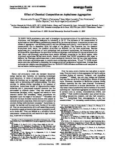
Fig. 1. Schematic diagram of discharge plasma device.
2.1. Chemicals
emission spectra of N2 discharge plasma with Lifbase software (Fig. S2). The working gas was introduced into the tube at side before plasma initiation for 2 min in order to exclude the air and it is exhausted via a pore at the cover. The gas fluxes were controlled by a flow meter. The high purity O2 and N2 were provided from high-pressure cylinders and the air was flowed by an air pump. After plasma treatment for certain time (20–240 s), 100 µL of the bacterial suspension after certain dilution was transferred and spread on a solid LB agar plate and then incubated at 37 °C overnight for CFU measurements. The temperature of the suspension after plasma treatment was in 32–42 °C which cannot kill the bacteria at all.
Tryptone and yeast extract for Luria-Bertani (LB) media preparation were obtained from OXOID. Sodium chloride (NaCl), sodium hydroxide (NaOH), potassium carbonate (K2CO3), disodium ethylenediaminete traacetic acid (EDTA), and phenol were purchased from Sinopharm Chemical Reagent Co., Ltd. NO donor diethylamine NONOate sodium salt hydrate, N-(1-Naphthyl)ethylenediamine dihydrochloride (NEDD) and sulfanilamide (SULF) were purchased from J & K Scientific Ltd. Propidium iodide (PI) with purity ≥ 94.0% was purchased from SigmaAldrich. SYBR green I (2-[N-(3-dimethylaminopropyl)-N-propylamino]−4-[2,3-dihydro-3-methyl-(benzo-1,3-thiazol-2-yl)-methylidene]−1-phenyl-quinolinium) was purchased from Invitrogen. All chemicals were used without further purification.
2.3. Determination of nitrite, nitrate, and H2O2 concentrations in water after plasma treatment
2.2. E. coli suspensions preparation, plasma treatment and colony-forming units (CFU) measurements
For determining nitrite, nitrate, and H2O2 concentrations, onemilliliter water was exposed to plasma treatment under the same conditions as with plasma treatment of bacterial suspension. The phenoldisulphonic acid method [24] was utilized to determine nitrate concentration with the acid prepared by dissolving 15 g of phenol in 135 mL concentrated H2SO4 and the solution was kept in water bath at 100 °C for 6 h. After plasma treatment, 0.8-mL plasma-treated water was added into 25-mL test tubes and then 50-µL H2O2 (30%) was added into the sample. The mixture was placed in boiling water for 10 min in order to convert nitrite into nitrate. Later, 0.8-mL K2CO3 (10 g/L) was added into the tube and the mixture was placed in a dry oven at 110 °C until dryness. 1-mL phenoldisulfonic acid was then added into the tube to dissolve the solid thoroughly and kept at room temperature for 10 min. Later, 1-mL EDTA (20 g/L) and 6-mL NaOH (10 M) were added into the tube and the detecting solution was titrated to the degree mark with distilled water. Absorbance at 420 nm of the mixture was determined on a UV–Vis spectrometer (SHIMADZU UV-2550) at ambient temperature. The detected nitrate content was the sum of nitrite and nitrate, and so it is defined as total nitrate as used in the following sections. The nitrite concentration in water after plasma treatment was determined with Griess reagents, N-(1-Naphthyl)ethylenediamine dihydrochloride (NEDD) and sulfanilamide (SULF) [25]. Because nitrite is easily oxidized to nitrate, its concentration was determined immedi-
One-hundred-microliter E. coli DH5 alpha stock at −20 °C was inoculated into 100-mL liquid LB (10% tryptone, 5% yeast extract and 10% NaCl) media and incubated at 37 °C overnight while shaking (180 rpm/min). After incubation, a working bacterial culture was prepared by diluting the overnight culture containing approximately 109 CFU/mL in distilled water by several times. One-milliliter working bacterial suspension was then transformed into a centrifugal tube and exposed to plasma treatment. The length and inner diameter of the tube is ca. 60 mm and 14 mm, respectively. The depth of the bacterial suspension is ca. 11 mm. The experimental apparatus is schematically shown in Fig. 1. Briefly, a needle-like anode with diameter < 1 mm which is made from stainless steel was inserted into the tube from the cover and placed ≈ 2–3 mm above the bacterial suspension. The upper end of the anode was connected with an AC power supply. The plasma was generated at the gas-solution interface. The peak voltage and current was about 9.7 kV and 2.8 mA, respectively, which were calculated from the voltage-current waveform (Fig. S1) and the instructions of the plasma power. The frequency was 9.6 kHz. The discharge power was about 12 W calculated from the area of Lissajous figure and the energy consumption was about 3600 J after treatment for 5 min. The gas temperature was about 950 K according to the rotational temperature simulation of OH (A-X) emission line in optical 2
Clinical Plasma Medicine 7–8 (2017) 1–8
Z. Ke et al.
ately following plasma treatment. Griess reagents, NEDD (0.1% w/v in H2O) and SULF (2% w/v in 5% HCl), were prepared prior to experiments as separate solutions. 50-µL plasma-treated water was added with 350-µL H2O, and then the sample was added with 100-µL NEDD solution and 100-µL SULF solution immediately. Absorbance of the solution at 540 nm was measured using a plate reader (Molecular Devices Spectra Max M2) following incubation at 37 °C for 45 min. Freshly prepared sodium nitrite (NaNO2) and sodium nitrate (NaNO3) were used for constructing the calibration curve for nitrite and nitrate, respectively. The H2O2 concentration in plasma-treated water was determined spectrophotometrically at 410 nm, after mixing with titanium sulfate in acidic condition [26]. Briefly, 0.8-mL plasma-treated water was added with 0.3-mL titanium sulfate (3 mM) and 0.3-mL H2SO4 (3 M), and then the mixture was incubated at room temperature for 30 min. 1.6-mL water was added into the detecting solution and absorbance of the mixture at 410 nm was measured on a UV–Vis spectrometer (SHIMADZU UV-2550). 2.4. Inactivation of E. coli by nitrite, nitrate, H2O2 and their combination in acidified medium E. coli suspensions were incubated with certain concentrations of nitrite, nitrate, H2O2 or their combination respectively in acidified solution adjusted by HNO3 at room temperature for 30 min, and then transferred and spread on solid LB agar plates after dilution. The inactivation efficiency was determined via CFU measurements after incubating at 37 °C overnight. The incubating time (30 min) was approximately equal to the time interval from the plasma treatment to spreading the bacterial suspension on LB agar plates. All the chemical concentrations and pH values were equal to that detected in H2O after air discharge plasma treatment for certain time.
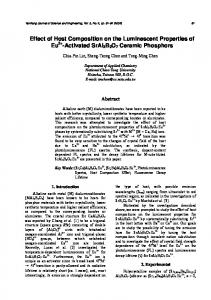
Fig. 2. Analysis of the effect of gas type on the inactivation efficiency of E. coli by discharge plasma. Upper: The photograph of plates spread with E. coli sample after O2, N2 and air plasma treatment for 20 s, respectively; Under: Dependence of inactivation ratio on O2, N2, and air plasma treatment time. Every experiment was repeated three times. The gas flow rate was 1.5 L/min. The bacterial density for plasma treatment was at about 104 CFU/mL.
efficiency of E. coli by discharge plasma at the gas-solution interface, bacterial suspension at about 104 CFU/mL was exposed to O2, N2, and air plasma treatment with a flow rate at 1.5 L/min and the inactivation effects were assessed by counting CFUs (Fig. 2). It showed that air as the working gas had 99% inactivation after 20 s plasma exposure, while for N2 plasma the inactivation was 50% and 82% after 20 s and 40 s treatment, respectively. In the case of O2 plasma, only 48% bacteria were inactivated after treatment for 40 s and even after 120 s treatment about 10% bacteria were still alive. Because air is mainly composed of N2 and O2, we also compared the bactericidal capability of air plasma with that of N2-O2 mixture (1.2 L/ min N2 + 0.3 L/min O2) plasma (Fig. S3). It can be seen from the figure that the inactivation efficiency of plasma in N2-O2 mixture is as high as that in air. These results indicate that the bacterial inactivation effects of discharge plasma strongly depend on the gas type – air the highest whereas oxygen the lowest. The reason for the high sterilization capability of air plasma is due to the synergy between N2 and O2 plasma.
2.5. Detection of membrane damage-PI uptake and SEM observations To test whether the plasma treatment caused membrane damage, the E. coli cells following plasma treatment were incubated with the DNA-binding probe PI at a final concentration of 20 mg/L for 10 min and then analyzed using a flow cytometer in FL3 channel. Additionally, the membrane damage was also detected with SEM observations. After plasma treatment for different time periods, the bacteria were collected by centrifugation at 3000 g for 5 min. Then, the bacteria were fixed by 4% glutaraldehyde precooled at 4 °C for 60 min, dehydrated with ethanol series, dried in air, coated with gold, and observed with a SEM (Sirion 200, FEI). 2.6. Detection of DNA damage To examine the intracellular DNA damage, double-strand DNA (dsDNA) concentration was investigated with SYBR green I [27]. Briefly, following plasma treatment, 0.8-mL E. coli suspension (107 CFU/mL) was mixed with 0.1-mL PBS (200 mM, pH 7) and 0.1-mL lysozyme at 1 mg/mL in 10 mM PBS (pH 7), and then the mixture was incubated at 37 °C for 4 h to break the cell envelope and release the intracellular DNA. Cells without plasma treatment were digested and used as the control. After digestion, the solutions were incubated with SYBR green I at working concentrations (1:20,000 dilution) at 37 °C for 15 min. The fluorescence spectra of the samples were detected at excitation wavelength of 485 nm on a fluorescence spectrometer (VARIAN Cary Eclipse).
3.2. Effect of gas type on nitrite and total nitrate formation in solution The high sterilization capability of N2-O2 plasma indicates that RNS which is composed of N and O may be crucial for the bacterial inactivation. Therefore, the final oxidation products of RNS, nitrite and nitrate, in water after plasma treatment with shielding different gas were measured (Fig. 3). The most nitrite and nitrate were formed for air plasma, nearly double that in solution after N2 plasma treatment for the same time. For example, the total nitrate concentration reached values of 962 µM and 400 µM after air and N2 discharge plasma treatment for 240 s, respectively. The nitrite concentration in water after air discharge plasma treatment for 30 s was 72 µM, which increased linearly to 197 µM with increasing treatment time to 120 s. For N2 discharge plasma, the nitrite concentration in water after treatment for 30 s was 29 µM and it increased to 126 µM after treatment for 120 s. With further increasing treatment time from 120 s to 240 s, the nitrite
3. Results 3.1. Effect of gas type on the inactivation efficiency To investigate the effects of shielding gas on the inactivation 3
Clinical Plasma Medicine 7–8 (2017) 1–8
Z. Ke et al.
Fig. 3. (a) Nitrite and (b) total nitrate concentration in water after O2, N2, and air discharge plasma treatment with flow rate at 1.5 L/min for different time. Every experiment was repeated three times.
completely, while sole H2O2 (legend 8), nitrate (legend 6) and their combination (legend 3) have no effect on the bacterial inactivation. This result illustrates that acidified nitrite is inevitable for the strong sterilization capability of air plasma. For further comparing the lethal effect of acidified nitrite and the combination of acidified nitrite and H2O2, nitrite alone and the mixture of nitrite and H2O2 at concentrations equal to that in water after air plasma treatment for 30, 60, and 120 s, respectively, were used to treat E. coli suspension at 107 CFU/mL. The pH in the medium was also equal to that in water after air plasma treatment for that time. After incubating for 30 min, the lethal effect was assessed with CFU measurements and the result is shown in Fig. 4. Acidified nitrite at 70 µM with pH 4.8 (equal to that in water after air plasma treatment for 30 s) can inactivate 16% bacteria, and this value is 24% with addition of H2O2 at 15 µM. If the nitrite concentration is increased to 120 µM with pH decreased to 4.4, the inactivation ratio is 43% and is increased to 63% with addition of H2O2 at 30 µM (equal to that in water after air plasma treatment for 60 s). If the concentrations of nitrite and H2O2 and pH value are equal to that in water after air plasma treatment for 120 s, both acidified nitrite alone and the combination of acidified nitrite and H2O2 can inactivate the bacteria completely (Fig. 4). The bacterial suspension was also subjected to air plasma treatment and it was found that the plasma-induced inactivation ratios (G and H in Fig. 4) were slightly higher than that induced by the “artificial” combination of acidified nitrite and H2O2 (B and D). The inactivation ratios after air plasma treatment for 30 and 60 s were 37% and 96%, respectively. The difference between the plasma effect and the acidified nitrite + H2O2 combination effect implies that the direct interaction of plasma also
concentration no longer increased dramatically for both air and N2 plasmas. For O2 discharge plasma, no nitrate and nitrite was detected. Optical emission spectroscopy (OES) measurements were also conducted to detect the excited nitrogen species generated in the gas phase during plasma treatment because they are the source of RNS in solution. For both N2 and air discharge plasma, NO and various nitrogen species emission lines were observed, and O emission line (777.2 nm) was observed for O2 discharge plasma (Fig. S4) [28]. The positive correlation between nitrite/nitrate concentration and bactericidal efficiency was also observed by changing the gas flow rate during plasma treatment. With increasing the N2 and air flow rates both the nitrite/total nitrate concentrations and the bactericidal capabilities decreased dramatically (Fig. S5 and Fig. S6). For O2 discharge plasma, both nitrite and nitrate cannot be generated even with the flow rate at 0.15 L/min (Fig. S5), and also the flow rate has no effect on the bactericidal efficiency (Fig. S6). Therefore, the results in Figs. 2–3 and Figs. S5–6 implied that RNS really contributes to the difference in the sterilization capability of different gas discharge plasma. For further confirming this speculation, NO donor (NONOate) which releases NO in solution was added into bacterial suspension and then the suspension was exposed to O2 plasma treatment, followed by CFU measurements. If RNS really contributes to the air plasma-induced bacterial inactivation, NONOate can enhance the bactericidal capability of O2 plasma because NO released from NONOate can react with the plasma-generated ROS to form various RNS [29]. The results in Fig. S7 which shows that NONOate greatly enhanced the sterilization ability of O2 plasma to the level of air plasma prove the speculation mentioned above.
3.3. The strong inactivation ability of air plasma is due to the combination of acidified nitrite with H2O2 Some studies have reported that combination of acidified nitrite and H2O2 made a great contribution to the antimicrobial activity of plasma activated water (PAW) [30,31]. In this work, H2O2 concentration and pH change in water after O2, N2 and air discharge plasma treatment were also measured (Fig. S8 and S9). In order to separate the contributions of nitrite, nitrate and H2O2 to the lethal effect of air plasma, acidified solutions (pH 3.5, by HNO3) prepared using nitrate (800 µM), nitrite (200 µM), H2O2 (200 µM) alone or a mixture were used to incubate E. coli at 107 CFU/mL for 30 min and their disinfection potentials were evaluated with CFU measurements (Fig. S10). The concentrations of these chemicals and pH used were equal to that in water treatment after air plasma treatment for 240 s in order to visualize the bacterial death induced by these chemicals clearly. It can be seen from the figure that the mixture of nitrate-nitrite-H2O2 (legend 2), nitrite-H2O2 (legend 4), nitrate-nitrite (legend 5) and nitrite alone (legend 7) in acidified medium can inactivate the bacteria
Fig. 4. Sterilization ability of acidified nitrite and the combination of acidified nitrite and H2O2. The concentration of nitrite and H2O2, and pH value are equal to that in water after air discharge plasma treatment with flow rate at 1.5 L/min for 30, 60, and 120 s, respectively. The unit for NO2- and H2O2 is µM.
4
Clinical Plasma Medicine 7–8 (2017) 1–8
Z. Ke et al.
played a role in the bacterial inactivation. But obviously, the combination of acidified nitrite and H2O2 played a major role in the bactericidal capability of air discharge plasma. To be noted, because the plasma acted on a small spot in the solution, in the region close to the plasma action, the nitrite and H2O2 concentrations could be higher than other zone near the action, and the pH value could be lower. Additionally, because part of generated nitrite and H2O2 reacted with each other during plasma treatment, the detected averaged concentrations of nitrite and H2O2 were actually lower than that with plasma in action. Furthermore, because there was no significant difference for H2O2 production from different sources of gas discharge plasma, we conclude that H2O2 was not a determinant for the difference in the bactericidal efficiency for different gas discharge plasmas. For example, after O2, N2 and air plasma treatment for 240 s, the H2O2 concentration in water is 389, 535, and 225 µM, respectively. To be noted, however, because part of generated H2O2 was consumed via the reaction with nitrite during plasma treatment, the actually produced H2O2 concentration was higher than the detected values in Fig. S8, especially for air plasma, but not for O2 plasma. Another reason for why H2O2 was not a determinant is that even in the absence of H2O2 the acidified nitrite already showed a strong sterilization capability (Figs. S10 and 4). Therefore, we confirmed that the difference in nitrite concentration in solution must be a major factor for the different bactericidal abilities measured from different plasma treatments. 3.4. Potential cellular targets of plasma In the elucidation of the mechanisms of plasma-mediated bacterial inactivation, it is important not only to identify the critical reactive agents responsible for mediating such effects but also to determine the cellular targets of plasma-driven effects. For simplifying the exploration of bacterial damage, air and O2 discharge plasma conditions with either high or low inactivation efficiency, respectively, were selected in the following research.
Fig. 5. The flow cytometry results of O2 (a) and air (b) plasma-treated E. coli for 60 s after incubating with PI. (c) The percent of permeated cells after air and O2 plasma treatment for different time periods.
3.4.2. DNA damage To identify whether air plasma induces more severe DNA damage than O2 plasma, the dsDNA quantities in E. coli after plasma treatment were assayed with SYBR green I, which showed an enormous increase in fluorescence at about 525 nm upon binding to dsDNA [27]. For E. coli after air plasma treatment, the fluorescence intensity of the dye at 525 nm after incubating with the digested cells decreased linearly with treatment time (Fig. 7). After treatment for 5 min, the value decreased to 36% compared to E. coli without plasma treatment. However, for E. coli after O2 plasma treatment, the fluorescence intensity was almost unchanged in the first 1 min and then decreased slowly with treatment time: it decreased only 12% after 5 min treatment. These results indicate that, compared with O2 plasma, air plasma induced more severe DNA damage.
3.4.1. Membrane destruction Membrane permeabilization of E. coli cells induced by O2 and air discharge plasma was demonstrated by PI uptake. PI is a good indicator of membrane integrity since it is not normally taken up by intact cells while becomes fluorescent when binding to nucleic acids of membranedamaged cells [32]. The determination of PI uptake in individual cells by flow cytometry revealed the proportion of cells with damaged membranes. The FSC-H subset and FSC-H subset-1 in Fig. 5a and b represent the intact and permeated cells after O2 and air discharge plasma treatment for 60 s, respectively. The proportion of membranepermeated cells was also plotted as a function of treatment time and the result is shown in Fig. 5c. For air plasma, the proportion of membranepermeated cells raised rapidly from 7.28% to 25.2% after treatment for 20 s, and it increased to 90% when the treatment time is 120 s. While for O2 plasma, the permeabilization proportion is 19.2% after 180 s treatment. The results clearly demonstrate that air plasma results in more serious membrane damage than O2 plasma. Furthermore, SEM observations, which can visually illustrate striking effects on cell morphology, were also used to illustrate the different effects of air and O2 plasma. It can be seen from Fig. 6 that the effects differed notably in these two gases. Before plasma treatment, the cell membrane of E. coli was smooth and the morphology was fairly complete. While after air plasma treatment for 5 min, the cell membranes were disrupted, even complete rupture of the membrane was observed in some bacteria. In some instances, the cells were damaged so significantly that they appeared as debris. In contrast, there were still many cells retaining their smooth surfaces even after O2 plasma treatment for 10 min. Because several steps of dehydration were needed for the preparation of SEM samples, the bacterial density was high (ca. 109 CFU/mL) in this experiment and so the plasma treatment time was extended to 10 min for visualizing the difference clearly.
4. Discussion Although all the plasma-produced active agents contribute to the bacterial inactivation processes, it is generally deemed that reactive chemical species including ROS and RNS are the key factors [12]. Because the category and content of the reactive species produced by plasma are strongly dependent on the working gas composition [33], the latter has a strong impact on the bacterial inactivation efficiency as shown in Fig. 2. Herein, it is found that nitrite concentration is the determinant for the difference in bactericidal ability of different gas discharge plasma. During plasma treatment, nitrite and nitrate generated in solution is from the dissolution of nitrogen oxides (NOx) formed in the gas phase. In the case of air discharge plasma, several reactive species including the atomic species (N, O) and the excited states are produced and they further react with each other to form NO in significant amounts by numerous elementary processes [34]. The produced NO can be further 5
Clinical Plasma Medicine 7–8 (2017) 1–8
Z. Ke et al.
Fig. 6. SEM observations of E. coli after air and O2 plasma treatment for different time.
O=NOO- form at pH7.4, while at pH6.2 up to 80% peroxynitrite will be in O=NOOH form [38]. In our case, the solution pH decreased dramatically during air and N2 plasma treatment, and so the peroxynitrite is mainly in the form of O=NOOH. The stability, reactivity, and capacity to permeate cell membranes of O=NOOH and O=NOO- are quite different. O=NOOH is much more reactive than O=NOO-. The former can undergo proton-catalyzed homolysis to yield NO2 and OH in ca. 30% yields, two strongly oxidizing/hydroxylating and nitrating species, respectively, while the latter is relatively stable [38]. Additionally, O=NOOH can cross through the cell membrane by simple diffusion with a rate significantly faster than the rates of its decomposition pathways while O=NOO- traverses membranes through anion channels [39]. Therefore, membrane offers no significant barrier to diffusion of O=NOOH within or between cells, although it is an important barrier between cell and the outer environment [39]. Because of its high reactivity and oxidation potential, peroxynitrite especially O=NOOH is highly bactericidal and can inactivate E. coli in direct proportion to its concentration [40]. It contributes to bacterial killing by interaction with diverse targets, such as protein modification, lipid oxidation, and DNA damage [41]. For example, early work suggested the existence of O=NOOH homolysis within the membrane
oxidized to NO2 and other NOx, and then dissolved into the solution to form nitrite and nitrate, accompanied by acidifications of the solution [31,35]. The pH reduction in solution is in accordance with the content of produced nitrite and nitrate. For the case of N2 discharge plasma, the required O for NO formation could originate from dissociation of water in the bulk liquid or at its surface, or from the O2 dissolved in the liquid [20]. Therefore, nitrite/nitrate are also detected in N2 plasma-treated water, although its concentration is nearly half of that in air plasmatreated water. But in the case of O2 plasma, no N2 could be gained except for negligible air diffusion from the surrounding and so nitrite and nitrate were not detected in solution. The mechanism for nitrite-induced bacterial inactivation is that nitrite reacts with H2O2 in acidified medium to generate a strong oxidant peroxynitrous acid (O=NOOH) or its conjugate base peroxynitrite (O=NOO-; pKa=6.8) (reaction 1 and 2) [36,37]:
NO−2 + H2 O2 + H+O = NOOH + H2 O ±H+
O = NOO−⟷O = NOOH
(1) (2)
The existing form of peroxynitrite (O=NOOH and O=NOO-) is strongly dependent on pH. For instance, ca. 80% peroxynitrite will be in
Fig. 7. (a) Fluorescence spectra of SYBR green I after incubating with digested E. coli with or without plasma treatment; (b) Fluorescence intensity of SYBR green I at 525 nm after incubating with digested E. coli after O2 or air plasma treatment for different time. Gas flow rate for plasma treatment: 1.5 L/min. Excitation: 485 nm; Excitation and emission slit: 5 nm.
6
Clinical Plasma Medicine 7–8 (2017) 1–8
Z. Ke et al.
Chinese Academy of Sciences (No. 2014289), and the Hundred Talents Program of the Chinese Academy of Sciences. The role of the funding body did not include any influence in the design of the study and collection, analysis, and interpretation of data and in writing the manuscript.
to yield NO2 and OH can induce lipid oxidation and nitration [42]. It has also been reported that peroxynitrite is a potent trigger of DNA damage including strand breakage and base modification [41]. Herein, both the extracellular (membrane) and intracellular (DNA) target was selected to investigate the oxidative damage induced by plasmagenerated peroxynitrite. Compared with O2 plasma, air plasma produced significantly more membrane permeated cells, and also more severe membrane damage, implying a key role of membrane damage for the bacterial inactivation by plasma-generated peroxynitrite. The dsDNA content in E. coli after air plasma treatment quantified with SYBR green I showed a reduction in a plasma dose-dependent pattern, while for O2 plasma treatment there was only a minor reduction, indicating that DNA is also a significant target of air plasma-generated peroxnitrite. For investigating the possible role of transient species in the different bactericidal activity of O2, N2 and air discharge plasma, hydroxyl radicals produced by the three gases discharge plasma were detected with coumarin-3-carboxylic acid (3-CCA). 3-CCA is a nonfluorescent product and can emit fluorescence at 450 nm after hydroxylation by hydroxyl radicals [43]. The fluorescence intensity of 3-CCA solution (2 mM) at 450 nm after O2, N2 and air plasma treatment is plotted as a function of treatment time and the result is shown in Fig. S11. It can be seen from the figure that the most hydroxyl radicals were produced by N2 plasma, followed by air plasma, and O2 plasma produced the least, which is however not in positive correlation with the bactericidal efficiency as shown in Fig. 2. Therefore, we conclude that although the transient species such as hydroxyl radicals play a role in the direct bacterial inactivation during plasma treatment, it is however not a main reason for the difference of bactericidal activity of different gas discharge plasmas. The results in Figs. S10 and 4 also show a possible role of acidified nitrite on plasma-induced bacterial inactivation. Several studies have shown the strong sterilization ability of acidified nitrite, which can form peroxynitrite and other nitrosating agents such as nitrous acid, nitric oxide, nitrogen dioxide and so on [44,45]. However, it is not known for us now how much contribution to the plasma-induced bacterial inactivation is from the acidified nitrite in the presence of H2O2, which needs further investigation in future.
Acknowledgments We thank Dr. Shen Jie and Dr. Cheng Cheng (Institute of Plasma Physics, Chinese Academy of Sciences, Hefei, China) for OES measurements and spectra analysis. Appendix A. Supporting information Supplementary data associated with this article can be found in the online version at http://dx.doi.org/10.1016/j.cpme.2017.05.001. References [1] Introduction to theoretical and applied plasma chemistry, in: A. Fridman (Eds), Plasma chemistry, Cambridge University Press, Cambridge, 2008, pp. 1–11. [2] P.K. Chu, X.P. Lu, Low Temperature Plasma Technology: Methods and Applications, CRC Press, Boca Raton, 2014. [3] Introduction to fundamental and applied aspects of plasma medicine, in: A. Fridman, G. Friedman (Eds), Plasma medicine, John Wiley & Sons, Ltd, West Sussex, 2013, p. 1–15. [4] Th. von Woedtke, S. Reuter, K. Masur, K.-D. Weltmann, Plasmas for medicine, Phys. Rep. 530 (2013) 291–320. [5] M. Laroussi, Sterilization of contaminated matter with an atmospheric pressure plasma, IEEE Trans. Plasma Sci. 24 (1996) 1188–1191. [6] H.S. Kim, Y.I. Cho, I.H. Hwang, H.L. Dong, D.J. Cho, A. Rabinovich, A. Fridman, Use of plasma gliding arc discharges on the inactivation of E. coli in water, Sep. Purif. Technol. 120 (2013) 423–428. [7] W. Heaselgrave, G. Shama, P.W. Andrew, M.G. Kong, Inactivation of Acanthamoeba spp. and other ocular pathogens by application of cold atmospheric gas plasma, Appl. Environ. Microbiol. 82 (2016) 3143–3148. [8] P.B. Flynn, S. Higginbotham, N.H. Alshraiedeh, S.P. Gorman, W.G. Graham, B.F. Gilmore, Bactericidal efficacy of atmospheric pressure non-thermal plasma (APNTP) against the ESKAPE pathogens, Int. J. Antimicrob. Agents 46 (2015) 101–107. [9] N.H. Alshraiedeh, S. Higginbotham, P.B. Flynn, M.Y. Alkawareek, M.M. Tunney, S.P. Gorman, W.G. Graham, B.F. Gilmore, Eradication and phenotypic tolerance of Burkholderia cenocepacia biofilms exposed to atmospheric pressure non-thermal plasma, Int. J. Antimicrob. Agents 47 (2016) 446–450. [10] L.K. Randeniya, G.J.J.B.D. Groot, Non-thermal plasma treatment of agricultural seeds for stimulation of germination, removal of surface contamination and other benefits: a review, Plasma Process. Polym. 12 (2015) 608–623. [11] V. Scholt, J. Pazlarova, H. Souskova, J. Khun, J. Julak, Nonthermal plasma-a tool for decontamination and disinfection, Biotechnol. Adv. 33 (2015) 1108–1119. [12] J. Guo, K. Huang, J.P. Wang, Bactericidal effect of various non-thermal plasma agents and the influence of experimental conditions in microbial inactivation: a review, Food Control 50 (2015) 482–490. [13] N. Knake, S. Reuter, K. Niemi, V. Schulzvon-von der Gathen, J. Winter, Absolute atomic oxygen density distributions in the effluent of a microscale atmospheric pressure plasma jet, J. Phys. D Appl. Phys. 41 (2008) 194006. [14] K. Mckay, D.X. Liu, M.Z. Rong, F. Iza, M.G. Kong, Generation and loss of reactive oxygen species in low-temperature atmospheric-pressure RF He+O2+H2O plasmas, J. Phys. D Appl. Phys. 45 (2012) 172001. [15] E. Wagenaars, T. Gans, D. O'Connell, K. Niemi, Two-photo absorption laser-induced fluorescence measurements of atomic nitrogen in a radio-frequency atmosphericpressure plasma jet, Plasma Sources Sci. Technol. 21 (2012) 042002. [16] Y. Ma, J.R. Chen, B. Yang, S.C. Pu, Q.S. Yu, A study of plasma inactivation effects on Desulfovibrio bastinii in liquid using dielectric barrier discharge, IEEE Trans. Plasma Sci. 42 (2014) 1607–1614. [17] K. Kadowaki, T. Sone, T. Kamikozawa, H. Takasu, S. Suzuki, Effect of water-surface discharge on the inactivation of Bacillus subtilis due to protein lysis and DNA damage, Biosci. Biotechnol. Biochem. 73 (2009) 1978–1983. [18] R.W. Zhou, X.H. Zhang, Z.H. Bi, Z.C. Zong, J.H. Niu, Y. Song, D.P. Liu, S.Z. Yang, Inactivation of Escherichia coli cells in aqueous solution by atmospheric-pressure N2, He, Air, and O2 microplasmas, Appl. Environ. Microbiol. 81 (2015) 5257–5265. [19] T. Takamatsu, A. Kawate, T. Oshita, H. Miyahara, A. Okino, G. Fridman, Investigation of reactive species in various gas plasmas treated liquid and sterilization effects, Int. Soc. Plasma Chem. 21 (2013) 370. [20] H. Jablonowski, M.A. Hänsch, M. Dünnbier, K. Wende, M.U. Hammer, K.D. Weltmann, S. Reuter, Tv Woedtke, Plasma jet's shielding gas impact on bacterial inactivation, Biointerphases 10 (2015) 029506. [21] J. Li, N. Sakai, M. Watanabe, E. Hotta, Study on plasma agent effect of a directcurrent atmospheric pressure oxygen-plasma jet on inactivation of E. coli using bacterial mutants, IEEE Trans. Plasma Sci. 41 (2013) 935–941. [22] Q. Zhang, P. Sun, H.Q. Feng, R.X. Wang, Y.D. Liang, W.D. Zhu, K.H. Becker,
5. Conclusion In conclusion, the gas type has a significant effect on the inactivation efficiency of E. coli by discharge plasma at the gas-solution interface, which is due to that it influences the nitrite content produced in solution. The produced nitrite further reacts with H2O2 in acidified medium to form a strong oxidant peroxynitrite. Among the three gases (O2, N2 and air) investigated in this study, the comparison showed that air plasma produced the maximum nitrite in solution and therefore had the highest inactivation ability, while O2 plasma could not produce nitrite and had the lowest inactivation ability. Both the cellular membrane and DNA are important targets of air plasma-produced peroxynitrite, in which the former is damaged more rapidly than the latter. Conflict of interest statement All authors disclose any financial and personal relationships with other people or organizations that could inappropriately influence their work. Funding sources This work was supported by the National Natural Science Foundation of China (No. 11405220, No. 11635013 and No. 11475217), the Natural Science Foundation of Anhui Province (No. 1608085MA20), the Youth Innovation Promotion Association of the 7
Clinical Plasma Medicine 7–8 (2017) 1–8
Z. Ke et al.
[23]
[24] [25] [26] [27]
[28]
[29]
[30]
[31]
[32] [33]
relevant solutions, Plasma Med. 3 (2013) 45–55. [34] M.A. Malik, Nitric oxide production by high voltage electrical discharges for medical uses: a review, Plasma Chem. Plasma Process. 36 (2016) 737–766. [35] Z. Machala, B. Tarabova, K. Hensel, E. Spetlikova, L. Sikurova, P. Lukes, Formation of ROS and RNS in water electro-sprayed through transient spark discharge in air and their bactericidal effects, Plasma Process. Polym. 10 (2013) 649–659. [36] Y. Kono, H. Shibata, K. Adachi, K. Tanaka, Lactate-dependent killing of Escherichia coli by nitrite plus hydrogen peroxide: a possible role of nitrogen dioxide, Arch. Biochem. Biophys. 311 (1994) 153–159. [37] W. Heaselgrave, P.W. Andrew, S. Kilvington, Acidified nitrite enhances hydrogen peroxide disinfection of Acanthamoeba, bacteria and fungi, J. Antimicrob. Chemother. 65 (2010) 1207–1214. [38] R. Radi, Peroxynitrite, a stealthy biological oxidant, J. Biol. Chem. 288 (2013) 26464–26472. [39] S.S. Marla, J. Lee, J.T. Groves, Peroxynitrite rapidly permeates phospholipid membranes, Proc. Natl. Acad. Sci. USA 94 (1997) 14243–14248. [40] L. Zhu, C. Gunn, J.S. Beckman, Bactericidal activity of peroxynitrite, Arch. Biochem. Biophys. 298 (1992) 452–457. [41] P. Ascenzi, A. di. Masi, C. Sciorati, E. Clementi, Peroxynitrite—An ugly biofactor? Biofactors 36 (2010) 264–273. [42] H. Rubbo, R. Radi, M. Trujillo, R. Telleri, B. Kalyanaraman, S. Barnes, M. Kirk, B.A. Freeman, Nitric oxide regulation of superoxide and peroxynitrite-dependent lipid peroxidation. Formation of novel nitrogen-containing oxidized lipid derivatives, J. Biol. Chem. 269 (1994) 26066–26075. [43] Y. Manevich, K.D. Heldt, J.E. Biaglow, Coumarin-3-carboxylic acid as detectot for hydroxyl radicals generated chemically and by gamma radiation, Radiat. Res. 148 (1997) 580–591. [44] R. Phillips, S. Kuijper, N. Benjamin, M. Wansbrough-Jones, M. Wilks, A.H.J. Kolk, In vitro killing of Mycobacterium ulcerans by acidified nitrite, Antimicrob. Agents Chemother. 48 (2004) 3130–3132. [45] J.G. Szabo, N.J. Adcock, E.W. Rice, Disinfection of Bacillus spores with acidified nitrite, Chemosphere 13 (2014) 171–174.
Assessment of the roles of various inactivation agents in an argon-based direct current atmospheric pressure cold plasma jet, J. Appl. Physi. 111 (2012) 123305. D.Z. Yang, L. Jia, W.C. Wang, S. Wang, P.C. Jiang, S. Zhang, Q.X. Yu, G.L. Chen, Atmospheric pressure gas–liquid diffuse nanosecond pulse discharge used for sterilization in sewage, Plasma Process. Polym. 11 (2014) 842–849. M.J. Taras, Phenoldisulfonic acid method of determining nitrate in water, Photom. Study Anal. Chem. 22 (1950) 1020–1022. K.M. Miranda, M.G. Espey, D.A. Wink, A rapid, simple spectrophotometric method for simultaneous detection of nitrate and nitrite, Nitric Oxide 5 (2001) 62–71. G.M. Eisenberg, Colorimetric determination of hydrogen peroxide, Ind. Eng. Chem. Anal. Ed. 15 (1943) 327–328. L. Han, S. Patil, D. Boehm, V. Milosavljević, P.J. Cullen, P. Bourkea, Mechanisms of inactivation by high-voltage atmospheric cold plasma differ for Escherichia coli and Staphylococcus aureus, Appl. Environ. Microbiol. 82 (2016) 450–458. J. Shen, Q. Sun, Z.L. Zhang, C. Cheng, Y. Lan, H. Zhang, Z.M. Xu, Y. Zhao, W.D. Xia, P.K. Chu, Characteristics of DC gas-liquid phase atmospheric-pressure plasma and bacteria inactivation mechanism, Plasma Process. Polym. 12 (2015) 252–259. D.A. Wink, J.B. Mitchell, Chemical biology of nitric oxide: insights into regulatory, cytotoxic, and cytoprotective mechanisms of nitric oxide, Free Radic. Biol. Med. 25 (1998) 434–456. G. Kamgang-Youbi, J.M. Herry, M.N. Bellon-Fontaine, J.L. Brisset, A. Doubla, M. Naïtali, Evidence of temporal postdischarge decontamination of bacteria by gliding electric discharges: application to Hafniaalvei, Appl. Environ. Microbiol. 73 (2007) 4791–4796. P. Lukes, E. Dolezalova, I. Sisrova, M. Clupek, Aqueous-phase chemistry and bactericidal effects from an air discharge plasma in contact with water: evidence for the formation of peroxynitrite through a pseudo-second-order post-discharge reaction of H2O2 and HNO2, Plasma Sources Sci. Technol. 23 (2014) 015019. P. Breeuwer, T. Abee, Assessment of viability of micro-organisms employing fluorescence techniques, Int. J. Food Microbiol. 55 (2000) 193–200. H. Tresp, M.U. Hammer, K.D. Weltmann, S. Reuter, Effects of atmosphere composition and liquid type on plasma-generated reactive species in biologically
8