Kuo WU*, CLAUDE WASTERLAINt, LEONARD SACHSt, AND PHILIP SIEKEVITZt§. *Department of ..... Peterson, D. W., Collins, J. F. & Bradford, H. F. (1983) Brain. Res. 275, 169-172. 45. ... Gilbert, M. E. (1988) Brain Res. 463, 90-99. 49. Yeh ...
Proc. Natl. Acad. Sci. USA Vol. 87, pp. 5298-5302, July 1990 Neurobiology
Effect of septal kindling on glutamate binding and calcium/ calmodulin-dependent phosphorylation in a postsynaptic density fraction isolated from rat cerebral cortex (N-methyl-D-aspartate acid receptors/y-aminobutyric acid receptors/synaptic cytology)
Kuo WU*, CLAUDE WASTERLAINt, LEONARD SACHSt, AND PHILIP SIEKEVITZt§ *Department of Neurology, Cornell University Medical College, New York, NY 10021; tDepartment of Neurology & Brain Research Institute, UCLA School of Medicine, Los Angeles, CA 90024; and *Rockefeller University, New York, NY 10021
Contributed by Philip Siekevitz, April 16, 1990
ABSTRACT Postsynaptic density (PSD) fractions were isolated from the cerebral cortices of control and kindled rats and assayed for glutamate and y-aminobutyric acid-binding capacities and for the Ca2+/calmodulin-dependent protein kinase. Glutamate binding was found to be increased by =50% in the PSDs isolated from kindled rats as compared to controls; this increase was almost completely from an increase in B..; Kd decreased only slightly. Studies with inhibitors indicate that the receptors involved were of the N-methyl-D-aspartate and quisqualate types. PSDs isolated from control and kindled rats did not differ in y-aminobutyric acid or flunitrazepam binding. The in vitro autophosphorylation of the Ca2+/calmodulindependent protein kinase was depressed by 45-76% in PSDs isolated from kindled rats as compared to controls, with little change in amount of the kinase. Therefore, we infer that (i) the kindled state is associated with an increase in glutamate activation of postsynaptic sites, allowing Ca21 to enter dendritic spines, (ii) a change has occurred in activity ofthe protein kinase, which is the major cerebral cortex PSD protein, and (iii) perhaps major alterations in the PSD are a concomitant to the long-lasting nature of the kindled state.
chemical changes in isolated PSDs, such as neurotransmitter binding and phosphorylations, were associated with septal kindling.
METHODS Kindling. Male Sprague-Dawley rats were stereotaxically implanted at the medial septal nuclei; the bipolar twisted stainless steel electrodes were located close to midline so that both hippocampi showed after-discharges. After 2 weeks of being handled daily, experimental animals received three daily stimulations (400 uA for 1 sec at 50 Hz) 3 hr apart for 5 days per week through the septal electrode. Kindling criteria included five stage-V seizures, three of them consecutive. Kindled rats were tested for 2 weeks before sacrifice by decapitation without anesthesia. Previous experiments have shown that rest periods of up to 2 months do not reduce either the kindled seizures or the change in protein phosphorylation associated with it (12). Cortices were dissected by free hand in Los Angeles, frozen on dry ice, and shipped to New York. Isolation ofPSD. PSDs were isolated from five to six pooled cortices, as described (21), and checked for purity by electron microscopy, as described (22). Electron microscopic examination of cerebral cortex slices was by the same method (22). Other Techniques. Phosphorylation was done by using 50 ,ug of PSD fraction and described methods (21, 23). Electrophoresis and autoradiography were performed as described (22, 23). Bands from dried gels were excised by cutting and soaked in Biofluor (4 ml per band); radioactivity was counted in a Beckman LS-180 scintillation counter. The bindings of L-[3H]glutamate (24) and of [3H]y-aminobutyric acid (GABA) and [3H]flunitrazepam (25) were assayed by described procedures. Samples were soaked in 4 ml of Hydrofluor each and were counted in a Beckman LS-180 scintillation counter. When used, calmodulin (CaM) was pretreated to remove Ca2" (21). Scatchard binding plots were obtained by the procedure of Rosenthal (26), and protein concentrations were estimated by the method of Lowry et al. (27) with bovine serum albumin as standard.
Kindling is the term coined by Goddard (1) to describe the progressive development, in response to initially subthreshold electrical stimulation of specific brain sites, of epileptic seizures that progressively spread and increase in intensity. Progression of the kindling phenomenon is characterized by increases in after-discharge duration and amplitude (2-5), a spread to secondary sites (5, 6), and decreases in threshold in distant cortical sites (2). Kindling shows remarkable temporal and spatial specificity as seen in many species from frog (7) to subhuman primates (8) and, once established, may persist without further stimulation for the life of the animal (9). After sufficient training, some animals develop spontaneous seizures (10). Strong pharmacological evidence indicates that this phenomenon is generated through stimulation of identifiable synaptic populations (11). Kindling by stimulation of the medial septal nuclei is associated with a decrease in the activity of the enzyme calmodulin kinase II in cortex and hippocampus (12, 13). However, calmodulin kinase II is a ubiquitous enzyme in brain. It is present in the presynaptic apparatus (14) where it may regulate transmitter biosynthesis by phosphorylation of 5-tyrosine and 5-tryptophan hydroxylases (15) and may regulate transmitter release via synapsin I phosphorylation (16). The enzymatic activity is also present in large quantities postsynaptically, particularly in postsynaptic densities (17), and calmodulin kinase II appears identical to the major postsynaptic density (PSD) protein (18-20). The goal of our study was to investigate whether any bio-
MATERIALS L-[2,3-3H]Glutamate (25 Ci/mmol; 1 Ci = 37 GBq), [y32P]ATP (3000 Ci/mmol) and [methyl-3H]flunitrazepam (77 Ci/mmol) were obtained from New England Nuclear, and [3H]GABA (54 Ci/mmol) was from Amersham. L-Glutamate, GABA, N-methyl-D-aspartate (NMDA), quisqualate, DLAbbreviations: NMDA, N-methyl-D-aspartate; GABA, -t-aminobutyric acid; DL-AP4, DL-2-amino-4-phosphonobutyrate; DL-AP5, DL-
The publication costs of this article were defrayed in part by page charge payment. This article must therefore be hereby marked "advertisement" in accordance with 18 U.S.C. §1734 solely to indicate this fact.
2-amino-5-phosphonovalerate; CaM, calmodulin; PSD, postsynaptic density. §To whom reprint requests should be addressed.
5298
Neurobiology: Wu et al.
Proc. Natl. Acad. Sci. USA 87 (1990)
2-amino-4-phosphonobutyrate (DL-AP4), DL-2-amino-5-phosphonovalerate (DL-AP5), and diazepam were all obtained from Sigma.
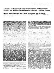
RESULTS Cytology and Chemistry. Cerebral cortex slices from control and kindled rat brains were examined by electron microscopy, and the number of synapses per field was counted at 10,000 magnification. In 17 fields from each group, number of synapses was 5.4 per field in the control and 5.6 per field in the kindled rat. Also, the two groups did not differ in length and thickness of the PSD or in number of synaptic vesicles in the axon terminals. The isolated PSD fraction from the control and kindled rat cortices were morphologically similar to each other and resembled those obtained from canine cerebral cortex (28) or from canine hippocampus (22). SDS gels of the PSD fractions gave identical profiles of the proteins of PSDs from control and kindled rats. Ca2+/CaM-Dependent Phosphorylation. Kindling has been shown to reduce phosphorylation of a 50-kDa protein in hippocampal synaptic membrane preparation (29) and of 50-kDa and 58- to 60-kDa proteins in septal-kindled rats by using a post hoc assay (12). These two proteins were further identified as the a and p subunits of a Ca2+/CaM-dependent protein kinase 11 (13). PSDs isolated from cerebral cortex gave similar results. Fig. 1 shows that the in vitro Ca2+/ CaM-stimulatable phosphorylation of the 51-kDa and the 60-kDa proteins were depressed in the isolated PSD fraction from cerebral cortex of kindled rats as compared to those from control animals. When these bands were excised and counted (Table 1), depression of the Ca2+/CaM-stimulated 32P-incorporation into 50-kDa and 60-kDa bands was from 40-45%; little change was seen in basal phosphorylation. In another experiment, when using PSDs prepared from another set of control and kindled animals, depression ranged from 64-76% (data not shown). Little difference was observed by gel electrophoresis in amounts of the 51- and 60-kDa proteins in PSDs from control and kindled rats (data not shown). Therefore, the marked change in autophosphorylation was not from the much smaller change in amounts of protein. Also noted in the figure is a decrease after kindling in the phosphorylations of two high-molecular-mass (180 and 200 kDa) K-CONTROL .~
~
KINDLED
--
~
6OkDa-51 kDa--
Ca2+ CaM
'._,
+
-
+
+
+
+
+
+
+
FIG. 1. Ca2+/CaM-dependent phosphorylation of PSD fractions from cerebral cortices of control and kindled adult rats. Fifty micrograms of PSD protein was used for each slot. Additions to the incubation mixture are indicated, where [Ca2+] equals 0.5 mM, and 1.5 ,g of CaM is the amount added.
5299
Table 1. Effect of septal kindling on Ca2+/CaM-dependent phosphorylation in PSD fractions Phosphorylation, cpm Control Kindled Addition(s) 51 kDa 60 kDa 51 kDa 60 kDa None (baseline) 77 36 72 33 Ca2+ 86 45 96 46 CaM 80 38 73 36 Ca2+/CaM 728 315 430 206 Ca2+/CaM minus baseline 358 651 173 279 The 51-kDa and 60-kDa bands of Fig. 1 were excised from the gel and counted.
PSD proteins; the same decrease after kindling was found earlier (30) for glycoproteins with similar molecular weights from hippocampal and cerebral cortex membranes. Binding of L-[3HJGlutainate. We next examined, by binding studies, the binding of some neurotransmitters that may be involved in the kindling phenomenon (cf. refs. 31-33). Table 2 shows that at two concentrations of glutamate the binding of this neurotransmitter to PSD fraction was increased by -50% in the kindled state as compared to control, whereas no change was seen in the binding ofGABA or flunitrazepam. Similar results were obtained by using PSDs obtained from another set of control and kindled rats. When we examined the type of glutamate receptor present in these PSD fractions, it was inferred from the data in Table 3 that our preparation contained a mixture of quisqualate and NMDA types. The inhibition by DL-AP5, but not by DL-AP4, indicated an NMDA type of receptor (34, 35), a situation similar to that previously seen with PSD fractions from canine cerebral cortex and hippocampus (22). Furthermore, no difference was seen in the degree of inhibition between the PSD fractions isolated from the cortex of control and kindled rats (Table 3). To account for the increase in the binding of glutamate in the PSDs from kindled rat, we did concentration curves of the binding, Scatchard plots, and calculated Bmax and Kd for each. Fig. 2 shows the data from a representative experiment, indicating a slight decrease in Kd and a large increase in Bmax in the PSD fractions from kindled rats as compared to control ones. This experiment was repeated two more times; Bmax and Kd from the control rats were 25.8, 23.9, and 22.8 pmol/mg, and 427, 456, and 443 nM, respectively, whereas Bmax and Kd for the kindled animals were 36.8, 36.8, and 33.9 pmol/mg and 369, 371, and 379 nM, respectively. Averaging these values showed that although the Kd of kindled preparations decreased by 16% compared to control, the Bmax of kindled preparations increased by 48%, indicating that the kindled state was associated with a large increase in the apparent number of glutamate-binding sites and only a small increase in their affinity for glutamate.
DISCUSSION Because evidence indicates that kindling involves a transsynaptic mechanism, we examined properties of PSDs. To obtain sufficient quantities of PSD material, we used cerebral cortex, which undergoes changes very similar to those described in hippocampus during kindling (5, 6, 12). We examined the receptors for glutamate and GABA because of the many reports of the involvement of those neurotransmitters in kindling (cf. refs. 36-39). Thus, in studies with hippocampal slices, stimulation resulted in an increase in glutamate binding (40) from an apparent increase in glutamate receptors (41). Cytological (42) and electrophysiological experiments (43-48) clearly indicated an involvement of NMDA-type glutamate receptors in kindling. A
5300
Proc. Natl. Acad. Sci. USA 87 (1990)
Neurobiology: Wu et al.
Table 2. Specific binding of L-[3H]glutamate, [3H]GABA, and [3H]flunitrazepam to PSD fractions isolated from cerebral cortices of kindled and control rat brains Specific binding, % control [3H]Flunitrazepam [3H]GABA L-[3H]glutamate 2 1 AM 2 nM 5 ,M 1M 50 nM 50 nM 100 100 100 100 100 100 Control 88 94 83 91 142 156 Kindled Cerebral cortex PSD fractions (25 ,ug per assay) were incubated with L-[3H]glutamate, [3H]GABA, and [3H]flunitrazepam at two concentrations each and processed for binding. Specific binding was obtained from the difference between total and nonspecific binding, the latter being defined as amount of label bound in the presence of 0.5 mM unlabeled L-glutamate for labeled L-glutamate, 100 ,M unlabeled GABA for labeled GABA, or 100 ALM diazepam for labeled flunitrazepam. Results are the averages of duplicate determinations that varied by