EXPERIMENTAL AND THERAPEUTIC MEDICINE 12: 2568-2576, 2016
2568
Effect of systemic injection of heterogenous and homogenous opioids on peripheral cellular immune response in rats with bone cancer pain: A comparative study JUN‑YING DU*, YI LIANG*, JUN‑FAN FANG, YONG‑LIANG JIANG, XIAO‑MEI SHAO, XIAO‑FEN HE and JIAN‑QIAO FANG Department of Neurobiology and Acupuncture Research, The Third Clinical Medical College, Zhejiang Chinese Medical University, Hangzhou, Zhejiang 310053, P.R. China Received May 7, 2015; Accepted June 23, 2016 DOI: 10.3892/etm.2016.3647 Abstract. Exogenous and endogenous opioids have been shown to modulate the immune system. Morphine‑induced immunosuppression has been investigated extensively. However, the immune‑regulating function of endogenous opioid peptides is unclear. The present study aimed to evaluate the difference in effects on cellular immune function between recombinant rat β ‑endorphin (β ‑EP; 50 µg/kg) and plant source morphine (10 mg/kg) via intraperitoneal injection treatment in a rat model of bone cancer pain. Walker 256 cells were injected into a tibial cavity injection to establish the bone cancer pain model. The paw withdrawal thresholds and body weights were measured prior to surgery, at 6 days after surgery, and following 1, 3,6 and 8 treatments. The spleen cells were harvested for detection of T cell proliferation, natural killer (NK) cell cytotoxicity, and the relative quantities of T cell subtypes (CD3+, CD4+ and CD8+ cells). Plasma levels of interleukin‑2 (IL‑2) were also determined. It was found that single or multiple treatments with β‑EP (a homogenous opioid peptide) and morphine (a heterogenous opioid) had good analgesic effects on bone cancer pain, while the analgesia provided by morphine was stronger than that of β‑EP. Treatment with β‑EP 3, 6 and 8 times increased the body weight gain in the rat model of bone cancer pain, while morphine treatment had on effect on it. With regard to immunomodulatory functions, β‑EP treatment increased T cell proliferation and NK cell cytotoxicity, and increased the relative quantities of T cell subtypes,
Correspondence to: Professor Jian‑Qiao Fang, Department of
Neurobiology and Acupuncture Research, The Third Clinical Medical College, Zhejiang Chinese Medical University, 548 Binwen Road, Binjiang, Hangzhou, Zhejiang 310053, P.R. China E‑mail:
[email protected] *
Contributed equally
Key words: bone cancer pain, cellular immune, heterogenous, homogenous, opioid peptide
but no effect on T cell secretion. However, morphine treatment decreased T cell proliferation and the levels of T cell subtypes. These data indicate that opioids from different sources have different effects on cellular immune function in vivo. A small dose of homogenous opioid peptide exhibited positive effects (analgesia and immune enhancement) on cancer pain. These results provide experimental evidence supporting the exploitation of human opioids for the treatment of cancer pain. Introduction In patients with cancer, pain is a common symptom and the major factor responsible for decreasing the quality of life (1,2). A number of studies concerning the prevalence of pain in cancer patients have shown that 24‑60% of patients undergoing active anticancer treatment (3,4) and 62‑86% of terminal cancer patients (5,6) suffer from burdensome pain symptoms. The unique pathophysiology of cancer pain causes it to exceed that of a combination of inflammatory and neuropathic pain (7). In addition, there is evidence suggesting that patients with chronic pain always exhibit immune suppression symptoms (8). It has been suggested that the levels of CD4+ T cells in the serum of patients with cancer pain are decreased (9). Therefore, cancer pain and immune suppression are two main symptoms in cancer patients. Opioids are used widely to treat acute pain following extensive surgery and many kinds of chronic pain, particularly cancer pain (10‑13). It is known that opioids not only result in analgesia but also modulate the immune system (14). Opioids include endogenous opioid peptides and exogenous opiates. There is growing evidence that acute and long‑term administration of exogenous opiates, especially morphine, which is a heterogenous opioid, mediates immunosuppression (15). However, the effects of endogenous opioids on the immune system remain a subject of debate, with some reports that endogenous opioids promote the immune function and others supporting the opposite view (15,16). Although numerous studies have observed the effects of opioid drugs on immune responses, the clinical relevance of these observations for heterogenous and homogenous opioids remains uncertain. Few studies have analyzed the association
DU et al: EFFECT PF SYSTEMIC INJECTION OF OPIOIDS ON CELLULAR IMMUNE FUNCTION
between opioids and the immune system in vivo. To address this, in the present study, Walker 256 cells were injected into a tibial cavity in rats to establish a bone cancer pain model. Recombinant rat β‑endorphin (β‑EP; 50 µg/kg) and plant‑derived morphine (10 mg/kg) were administered by intraperitoneal injection and the analgesic effects were compared. In addition, the effects of the opioids on cellular immune function, specifically T lymphocyte proliferation, natural killer (NK) cell cytotoxicity and the levels of T cell subgroups in the bone cancer pain models were examined, and the differences between the effects on cellular immune function were compared between the heterogenous and homogenous opioid treatment groups. The aim of this study was to provide scientific evidence useful in the development of human opioids to treat cancer pain. Materials and methods Animals. A total of 40 adult female Sprague‑Dawley (SD) rats weighing (150‑170 g; Shanghai SLAC Laboratory Animal Co., Ltd, Shanghai, China) and 10 female SD rats (weight, 70‑80 g; Shanghai SLAC Laboratory Animal Co., Ltd.) were raised in a 12‑h light/dark cycle with access to plentiful amounts of food and water. They were housed five per cage and were acclimatized for 1 week prior to behavioral studies. Efforts were made to minimize animal discomfort and reduce the numbers of animals used. The animal protocols were approved by the Animal Ethics Committee at Zhejiang Chinese Medical University (Hangzhou, China). Surgery. Walker 256 cells (1x10⁷; The Cell Bank of Type Culture Collection of Chinese Academy of Sciences, Shanghai, China) were administered by intraperitoneal injection into the abdominal cavity of juvenile rats (70‑80 g). After 7 days, ascites were generated in the peritoneal cavity and carcinoma cells were harvested through sterile syringes. The percentage of cellular activity was checked to ensure that it was >95%, as measured using a TC10™ Automated Cell Counter (Bio‑Rad Laboratories, Inc., Hercules, CA, USA). Female SD rats were anesthetized by the administration of 10% chloral hydrate (0.35 ml/100 g) intraperitoneally, and then placed in a supine position. The left leg of the rat was shaved and the skin sterilized with iodophor and 75% ethanol. A 1‑cm rostro‑caudal incision was then made in the skin in the upper half of the tibia. The tibia was carefully exposed with minimal damage to the muscle and blood vessels. A 21‑gauge needle was inserted at the site of intercondylar eminence at a 30‑45˚ angle and pierced 5 mm below the knee joint into the medullary cavity of the tibia. The needle was then removed and replaced with a 10‑µl syringe (Hamilton Co., Bonaduz, Switzerland) containing the carcinoma cells (3x10⁵) to be injected into the tibial cavity. The syringe was kept in position for 2 min prior to removal from the tibial cavity to prevent cells from leaking out along the injection hole. The injection site was quickly sealed using bone wax and the wound was closed with stitches. Penicillin (20,000 units, intramuscular injection) was given to avoid infection. Rats of the sham surgery group were injected with the same volume of phosphate‑buffered saline (PBS) into the tibial cavity, and the other protocols were the same as those used in the surgery group.
2569
Experimental groups. The rats were separated randomly into four groups: i) Sham surgery group (n=10); ii) surgery group (n=10); iii) morphine group (n=10); and iv) β‑EP group (n=10). Paw withdrawal thresholds (PWTs). The PWTs were observed at six time points: Baseline (prior to surgery), at 6 days after surgery and following 1, 3, 6, and 8 treatments (as described below). As in a previous study (17), rats were adapted to the new environment by being placed on a metal mesh table. A mechanical stimulus (force 0‑50 g over a 20 sec time period) was delivered to the plantar surface of the left hind paw using a Dynamic Plantar Aesthesiometer (37450; Ugo Basile, Monvalle, Italy). When the animal withdrew its hind paw, the mechanical stimulus was automatically stopped, and the force at which the animal withdrew its paw was recorded as the PWT. Withdrawal responses were taken from four consecutive trials with ≥3 min between trials and averaged. Administration of treatments. Immediately after finishing the measurement of PWTs on 6 day, rats in the morphine group were intraperitoneally injected once every other day with 10 mg/kg morphine hydrochloride injection (C81004‑2; Northeast Pharmaceutical Group Shenyang No. 1 Pharmaceutical Co., Ltd., Shenyang, China) and rats in the β‑EP group were injected intraperitoneally with 50 µg/kg β‑EP (H‑284; Bachem AG, Hauptstrasse, Switzerland) for 15 days, once every other day. Sham surgery and surgery groups did not received any treatment. Body weight measurements. The body weights of the rats were measured at baseline, at 6 days after surgery and following 1, 3, 6, and 8 treatments. The increase in body weight was calculated as follows: Body weight growth rate (%) = measured value/basal value x 100. Extraction of splenic monocytes. Rats were sacrificed by cervical dislocation after the last PWT had been measured. The dead rats were soaked into 75% alcohol, and then moved onto a super clean bench. The spleen was excised, soaked in RPMI‑1640 medium, HEPES [RPMI‑1640 supplemented with 10% fetal bovine serum (FBS), 10,000 U/ml penicillin G and 10,000 µg/ml streptomycin; Thermo Fisher Scientific, Inc., Waltham, MA, USA] for 20 min. The spleen was placed on a 200‑mesh stainless steel screen, cut into pieces and then ground, using PBS (sterile) to keep the tissue moist during the whole experiment. The obtained cell suspension was combined with 3‑5 volumes of red blood cell lysis buffer (Beyotime Institute of Biotechnology, Haimen, China), and mixed gently. After standing for 2 min, the suspension was centrifuged at room temperature for 10 min at 1,000 g, and the supernatant was discarded. The obtained cell suspension was combined with 5 volumes of PBS (sterile), mixed gently, then centrifuged at room temperature for 10 min at 1,000 x g, twice. Cells were suspended in RPMI‑1640 (10% FBS, 100 U/ml penicillin G and 100 µg/ml streptomycin) after cleaning, and the concentration of the cell suspension was adjusted to 1x10⁶/ml. T lymphocyte proliferation assays. Splenic monocytes were seeded into 96‑well plates at 2x10⁵ cells/well in triplicate. For the test samples, 20 µl concanavalin A (ConA; 10 µg/ml;
2570
EXPERIMENTAL AND THERAPEUTIC MEDICINE 12: 2568-2576, 2016
Sigma‑Aldrich, St. Louis, MO, USA) was added to each well. For the control, 20 µl RPMI‑1640 was added to three wells. The plates were incubated at 37˚C for 72 h. Cell viability was assessed using Cell Counting kit‑8 (CCK‑8; Beyotime Institute of Biotechnology) and according to the manufacturer's protocol. WTS‑8 (20 µl) was added to each well and plates were incubated at 37˚C for 4 h. The absorbance of each sample was measured at 450 nm using a microtiter plate reader (SpectraMax M4; Molecular Devices, LLC, Sunnyvale, CA, USA). The reference wavelength was >650 nm. The T lymphocyte proliferation function was determined using the following equation: T lymphocyte activity (%) = absorbance of test sample/absorbance of control x 100. NK cell cytotoxicity assays. YAC‑1, a mouse lymphoma cell line, was purchased from Shanghai Institutes for Biological Sciences, Chinese Academy of Sciences (No. TCM28; Shanghai, China). Cells were grown in suspension in a culture bottle (Corning Incorporated, Corning, NY, USA), with RPMI‑1640 medium, HEPES (RPMI‑1640 supplemented with 10% FBS, 100 U/ml penicillin G and 100 µg/ml streptomycin). Only cells in the exponential growth phase were used for cytotoxicity assays. The YAC‑1 cells were used as sensitive target cells for the evaluation of NK cell cytotoxicity in vitro (18). Determination of NK cell function was implemented using an enzymatic colorimetric technique involving lactate dehydrogenase (LDH) release (LDH‑cytotoxicity assay kit; BioVision, Inc., Milpitas, CA, USA). Splenic monocytes as effector cells were incubated in RPMI‑1640 medium, HEPES (RPMI‑1640 supplemented with 10% FBS, 100 U/ml penicillin G and 100 µg/ml streptomycin). Viability of effector and target cells was determined by the trypan blue dye exclusion test prior to the cytotoxicity test to confirm that the viability was >95%. In the test sample, the effector cells at a concentration of 1x10 6 in 100 µl culture medium were mixed with 100 µl YAC‑1 cells at a concentration of 2x10⁴, resulting in an effector cell:target cell ratio of 50:1. As the background control, 200 µl medium/well was added to triplicate wells (the background value was subtracted from all other values). As the low control, 1x10⁶ cells/well in 200 µl culture medium were added to triplicate wells. As the high control, 1x10⁶ cells/well in 200 µl culture medium containing 1% Triton X‑100 were added to triplicate wells. Each test sample and all controls were evaluated in triplicate in 96‑micro‑well plates, and incubated at 37˚C in a thermostatic incubator with 5% CO2 for 4 h. The micro‑well plates were centrifuged at 250 x g for 10 min, and the supernatant was isolated. The LDH reaction mixture was added and maintained for 30 min at room temperature with the absence of light. The absorbance was measured using the SpectraMax M4 reader at 490 nm and the percentage of cytotoxicity was determined using the following equation: Cytotoxicity (%) = [absorbance (test sample ‑ background control ‑ low control)]/[absorbance (high control ‑ low control)] x 100. Flow cytometry assay. Spleen cell suspensions were collected from the cell culture bottles, and were adjusted to a concentration of 1‑5x10⁶ cells/ml. Fluorescein isothiocyanate (FITC)‑conjugated anti‑rat CD3 monoclonal antibody (cat. no. E00051‑1631; eBioscience, Inc., San Diego, CA, USA),
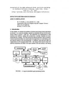
Figure 1. Effect of opioid treatment on the PWTs in an animal model of bone cancer pain. The analgesic effect of morphine on allodynia was stronger than that of β ‑EP. *P