Research Institute of Infectious Diseases, Fort Detrick, Frederick, Maryland 21701-501 I,. **Department of Virus ... WATTS ET AL. study was designed and ...
Am. J. Trap. Med. Hyg., 36(l), 1987, pp. 143-152 Copyright G 1987 by The American Society of Tropical Medicine and
EFFECT OF TEMPERATURE
ON THE VECTOR
OF AEDES AEGYPTI FOR DENGUE
EFFICIENCY
2 VIRUS
DOUGLAS M. WATTS,* DONALD S. BURKE,** BRUCE A. HARRISON,-/RICHARD E. WHITMIRE,* AND ANANDA NISALAKg *Departmentof Pathogenesisand Immunology, DiseaseAssessment Division, U.S. Army Medical ResearchInstitute of InfectiousDiseases,Fort Detrick, Frederick,Maryland 21701-501 I, **Department of Virus Diseases,Walter Reed Army Institute of Research,Washington,DC 20012, t Walter Reed BiosystematicsUnit, NHB Stop 165, SmithsonianInstitute, Washington,DC 20506, *United StatesArmy MedicaI ResearchUnit-Kenya, Box 401, APO New York 09675, and #Departmentof Virology, U.S. Army Medical Component,AFRIMS, Rajvithi Road, Bangkok 4, Thailand
Abstract. The effect of temperature on the ability of Aedesaegypti to transmit dengue (DEN) 2 virus to rhesus monkeys was assessedas a possible explanation for the seasonal variation in the incidence of dengue hemorrhagic fever in Bangkok, Thailand. In two laboratory experiments, a Bangkok strain of Ae. aegypti was allowed to feed upon viremic monkeys infected with DEN-2 virus. Blood-engorgedmosquitoes were separatedinto two groupsand retained at constant temperatures. Virus infection and transmission rates were determined for Ae. aegypti at intervals ranging from 4 to 7 days during a 25-day incubation period. Results of the first experiment for mosquitoes infected with a low dose of DEN-2 virus and maintained at 20,24,26, and 3O”C,indicated that the infection rate ranged from 25% to 75% depending on the incubation period. However, DEN-2 virus was transmitted to monkeys only by Ae. aegypti retained at 30°C for 25 days. In the second experiment, the infection rate for Ae. aegypti that ingested a higher viral dose,and incubated at 26, 30, 32, and 35°C ranged from 67% to 95%. DEN-2 virus was transmitted to monkeys only by mosquitoes maintained at L 30°C. The extrinsic incubation period was 12 days for mosquitoes at 3O”C, and was reduced to 7 days for mosquitoes incubated at 32°C and 35°C. These results imply that temperature-induced variations in the vector efficiency of Ae. aegyptimay be a significant determinant in the annual cyclic pattern of denguehemorrhagic fever epidemics in-Bangkok. Epidemicsof denguehemorrhagicfever (DHF) occur annually in Bangkok, Thailand, where Aedes aegypti (L) has been incriminated as the primary vector of the four dengue (DEN) virus serotypes.lg 2 While all serotypes have been associated with epidemics, DHF epidemics have been attributed most frequently to DEN-2 virus infections.3T 4 Case rates begin to increase during the latter half of the hot-dry season, March through May, and attain peak case rates during the rainy season,June to November. The incidence of DHF casessubsidesmarkedly during the cool-dry season,November to March.5 Initially, the associationof DHF epidemics with the rainy seasonwas attributed to an increase in the population density of Ae. aegypti.2 Data generated subsequently failed to support this observation and also revealed that an increase in the longevity of this mosquito was negatively corAccepted26 June 1986.
related with the increased incidence of DHF.6 However, observations on the seasonal feeding pattern ofAe. aegyptisuggestedthat annual DHF epidemics were more likely the result of increasedfrequency of feeding on humans during the hot-dry and rainy seasons.‘.* Also, Yasuno and Tonn’ alluded to the possibility that variation in case rates might reflect the influence of seasonaltemperature fluctuations on the extrinsic incubation period of DEN viruses in Ae. aegypti. Definitive data demonstrating that the extrinsic incubation period of DEN viruses was temperature-dependent have not been published, but this phenomenon has been documented for other virus-mosquito vector systems.9-16 An analysis of the incidence of DHF casesin relation to meteorologicvariables in Bangkok for the past two decadesprovided indirect evidence that temperature influenced the vector efficiency of Ae. aegypti for DEN viruses.17The present
143
144
WATTS
study was designedand conducted to assessthe ability of Ae. aegypti to transmit DEN-2 virus under environmental temperatures approximating those of the different seasonsin Bangkok, Thailand. MATERIALS
AND METHODS
ET AL.
obtained from eachmonkey; serum wascollected and storedat -20°C. Specificity and titer of each antiserum was determined by serial 2-fold dilutions tested against 80 to 100 PFU of each DEN virus serotype in a PRNT.19 Infection of mosquitoes
Mosquitoes were infected by allowing them to feed on viremic monkeys that had been inocuAdult rhesusmonkeys (Macaca mulatta) were lated via the saphenousvein with 0.5 ml of 105.0 obtained commercially and maintained in mos- PFU/ml of DEN-2 virus. Immediately after the quito-proof rooms according to standard labo- mosquitoes fed, blood was obtained from monratory procedures. Heparinized blood was ob- keys and centrifuged at 300 x g for 20 min. The tained from monkeys prior to use in the plasma component of each blood sample was experiments and centrifuged at 300 x g for 20 stored at -70°C until assayedfor virus at 1:5 or min. A 1:10 dilution of each plasma was tested serial log,, dilutions by direct and delayed plaque for hemagglutination inhibition (HI) antibody by assay in LLC-MK2 cells.*OIn addition, plasma employing 8 units of sucroseacetone-extracted obtained on day 28 post-inoculation was tested DEN-2 and Japaneseencephalitis (JE) virus-infor DEN-2 virus HI and neutralizing antibody fected, suckling mouse brain antigens.18A sim- as described previously. la, I9 Blood-engorged ilar dilution of heat-treated (56°C x 30 min) mosquitoes were incubated at temperatures applasma was assayedfor DEN- 1, 2, 3, and 4 and proximating the hot-dry, the rainy, and the coolJE virus antibody by plaque reduction neutral- dry seasonsfor Bangkok, Thailand. ization tests(PRNT), l9 with 50% endpoints, employing LLC-MK2 cells. Viral transmission Man keys
At 3- to 7-day intervals during the incubation period, DEN-2 virus transmission was attemptThe Ae. aegypti adults were F, progeny from ed at room temperature. A sample of mosquitoes eggsoviposited by adults collected as larvae in representingeach temperature was transferred to a low socioeconomic sector of Bangkok. Eggs a 0.5-l cylindrical carton, one end of which was were hatched, and larvae were reared to adults, enclosedby nylon netting. Each carton was taped according to standard procedures, at 25°C and securely to the shaven abdomen of a monkey to at 70% to 80% RH. All mosquitoes were 6- to allow mosquitoes to feed through the nylon netlo-days-old when initially used in each experi- ting. Immediately after feeding, mosquitoeswere ment. After blood engorgement, a continuous stored at -70°C until assayed for virus. Blood supply of 10% sucroseand an oviposition sub- was obtained from monkeys before the mosquistrate were provided. toes fed and again 28 days later. Serial 2-fold dilutions of plasma of each blood specimen were tested for DEN-2 virus HI antibody,18 and a 1: Virus 10 dilution of each plasma was assayedfor virusDEN-2 virus was isolated during 1978 from specific neutralizing antibody by PRNT.19 The the blood of a DHF patient hospitalized at the absence of DEN virus HI and neutralizing anChildren’s Hospital, Bangkok.The virus had been tibody in the first plasma specimen and the prespassagedtwice in LLC-MK2 cells and was iden- ence of antibody in the second was considered tified before and after passageby PRNT em- evidence of virus transmission. ploying DEN virus 1, 2, 3, and 4 monospecific antisera.19Each antiserum wasprepared by a sinViral infectivity assays gle intravenous injection of adult rhesus monkeys with 0.5 ml of approximately 1 x 105.0 The Ae. aegypti used in preliminary experiplaque forming units (PFU)/ml of each DEN viments were triturated individually in 1.0 ml of rus type. On day 28 post-inoculation, blood was medium RPM1 1640 containing 10% heat-treatMosquitoes
DENGUE
VIRUS
TRANSMISSION
ed fetal bovine serum, 500 pi/ml of streptomycin, and 500 U/ml of penicillin. Mosquito SUSpensions were clarified by centrifugation at 1,000 x g for 30 min at 4°C and tested for virus by direct and delayed plaque assay.2o The distribution of DEN-2 virus in a sample of individual Ae. aegypti employed in actual experiments was determined as follows. Mosquito headswereremoved and assayedindividually for virus by the direct fluorescent antibody technique (DFAT). 21,22Heads of uninfected and parenterally DEN-2 virus-infected Ae. aegypti were included as controls for theseassays.DEN virus antisera for the DFAT were obtained from convalescing DHF patients and conjugated with fluoresceinisothiocyanate.The thorax-abdomen of eachmosquito wasplacedin a drop of medium RPM1 1640 and the salivary glands were extracted with sterile insect minuten pins. Glands were disrupted by sonic energy, either as individual specimensin 0.2 ml or as pools of 5 pairs of glands in 0.5 ml of medium RPM1 1640, supplemented as described above for testing individual Ae. aegypti. Suspensionsprepared from individual glands were tested for virus in Toxorhynchitessplendens(Weidmann)22y23 and suspensionsderived from pooled glands were tested by the direct and delayed plaque assay.2oAssay of suspensionsin TX. splendenswas performed by inoculating each of 8 mosquitoes intrathoracically with aliquots of 0.85 ~1 per mosquito. These mosquitoes were then incubated for 14 daysat 32°C and then storedat - 70°C until virus assay. After extracting salivary glands from an individual Ae. aegypti, the corresponding thoraxabdomen components were triturated and suspensions were assayed for DEN-2 virus in LLC-MK2 cells, as described above. Undiluted suspensionsof eachthorax-abdomen were tested in the first and secondexperiments. Viral infectivity titers were also determined for additional whole mosquitoes from the second experiment by assayingserial 1O-fold dilutions of each mosquito suspension. The body component of individual Ae. aegypti was labeled such that the viral assayresultscould be analyzed for individual mosquitoes. The relation of DEN virus titer in mosquitoes to temperature was analyzed by Tukey’s Studentized Range (HSD) test.23Recoveryof DEN-2 virus from plasma of monkey blood specimens and from suspensionsderived from whole mos-
BY AEDES AEGYPTI
145
quitoes, thorax-abdomens, and salivary glands was confirmed by PRNT employing DEN-2 virus, monospecific antiserum.1q EnvironmentaI temperature Environmental temperature data recorded for Bangkok from 1958 through 1978 were received from the Ministry of Communications, Bangkok, Thailand. These recordswere used to prepare an annual temperatureprofile. I7Data were collected with a maximum-minimum thermometer and a hygrothermograph located at a meteorological station in Bangkok. Also, during 1978 and 1979 temperature was monitored continuously with a mechanical hygrothermographin an Ae. aegyptiinfested houselocated approximately 1 km from where this specieswas collected for viral transmission trials. RESULTS
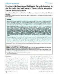
Environmental temperature The annual temperature profile for Bangkok, based on an analysis of 20 years of daily temperature recordings,is presentedin Figure 1. The mean temperature during the cool-dry season, November through February, ranged from 25.5 to 28.0°C, with a mean minimum of 21.0 to 25.O”Cand a mean maximum of 3 1.O to 33.5”C. The hot-dry and rainy season (March through October) mean temperature ranged from 28.0 to 30.0°C, and the mean minimum and mean maximum ranged from 25.0 to 26.0°C, and 32.O”C to 34.5*C, respectively. Year-to-year temperature variations were slightly greater during the cool-dry seasonsthan during the hot-dry and rainy seasons.A comparable cyclical pattern was recorded inside an Ae. aegypti-infested house where temperatures were from 1 to 2°C lower than the above. Rhesusmonkey modelfor infectingAedes aegypti with DEN-2 virus A preliminary experiment was conducted to determine the duration of DEN-2 viremia in rhesus monkeys and to ascertain the day post-infection that the viremia would produce maximum infection rates in Ae. aegypti. Mosquitoes fed upon eachof thesemonkeys on days 2 through 10 post-inoculation and were retained for 14 days
146
WATTS
ET AL.
TABLE1
Dengue2 virusviremiain rhesusmonkeys Monkey number
228
258
294
Days postinoculation*
Aedesaegyptr Viremia**
infection rate+
2 3
+ +
00 (O/5) 20 (l/5)
4 5 6 2
+ + + +
60 (3/5) 00 (O/5) 00 (O/5) 00 (O/5)
3 4 : 2 3 4
+ + + 0 + + +
00 (O/5) 80 (4i5) 00 (O/5) 00 (O/5) 20 (l/5) 60 (3/5) 20 (l/5)
5 6
+ +
00 (O/5) 00 (O/5)
l Viremia not detected on day 1 or after days 5 or 6 through day 10 post-inoculation. ** + = virus positive. 0 = virus negative. 7 Percent infected (No. pos./No. tested).
18 1I
4
JFMAMJJASOND MONTHS
FIGURE1. Average monthly temperaturefor Bangkok, 1958-l 978. Top line = maximum mean temperature with range, middle line = mean with range, and bottom line = minimum mean with range.
at 32°C. DEN-2 virus was recovered from the blood of each of 3 monkeys on days 2 through 5 or 6 following inoculation (Table 1). However, infection rates were highest for mosquitoes that fed upon monkeys on day 3 and/or 4 post-inoculation. In all subsequent experiments, mosquitoes were fed upon monkeys on day 4 postinfection. DEN virus HI or neutralizing antibody was not detected in plasma obtained from monkeys before inoculation with virus. On day 28 post-infection, HI antibody titers were 2 1:40, and virus-specific neutralizing antibody titers were > 1:10 for all monkeys. Dengue virus infection and transmission experiments In the first experiment, 300 Ae. aegypti ingestedblood from a DEN-2 virus-infected monkey. Fib-e separategroups of 60 fully blood-engorged mosquitoes were maintained in environmental chambers at 20, 24, 26, 28, and
30°C. At intervals ranging from 3-7 days during the 25-day incubation period, virus transmission trials were conducted by allowing the surviving mosquitoes (5-13,‘group) an opportunity to refeed as a group on an individual recipient monkey. Overall feeding rates for mosquitoes at all temperatures on incubation day 3 ranged from 42?&83%, and from 70%-100% during subsequent virus transmission trials. All mosquitoes maintained at 28°C died before incubation day 12 due to a malfunction of the incubator. An infective dose of DEN-2 virus was not transmitted to monkeys by mosquitoes maintained at 20, 24. and 26°C during the 25-day incubation period (Table 2). However, mosquitoes retained for 25 days at 30°C transmitted virus, as evidenced by an HI antibody titer of 1: 160 and a neutralizing antibody titer of 2 1:10 in plasma of the recipient monkey. Although a viremia was not demonstrable for the monkey usedto provide an infectious bloodmeal to the mosquitoes, it was infected, as indicated by its development of an HI antibody titer of 11:40 and DEN-2 virus neutralizing antibody titer of I 1:10. Data presentedin Table 2 show that mosquitoesbecame infected, and that the frequency and pattern of virus recovery varied in relation to the incubation period and temperature. Virus recovery rates for thorax-abdomens increasedfrom 25% (5/20) on day 3 to 74% (14/ 19) on day 25, and rates increasedfrom 25%
DENGUE
VIRUS
TRANSMISSION
147
BY AEDES AEGYPTI
TABLE 2 Transmission of dengue2 virusto rhsus monkeysby Aedesaegypti and distribution of virus in temperatures ranging from 20 to 3OT Incubuon $L$
3
7
12
18
25
Virus transmission and distribution
Transmission Heads Salivary glands Thorax-abdomen Transmission Heads O/4 Salivary glands Thorax-abdomen Transmission Heads O/5 Salivary glands Thorax-abdomen Transmission Heads O/5 Salivary glands Thorax-abdomen Transmission Heads O/5 Salivary glands Thorax-abdomen
% (No. thorax-abdomens pos./No. tested) * - = virus not transmitted + =
Temperature 20
24
25 (6/24)
(T) 26
30
-/5 o/5 0 4/5
3/4
-/lO o/5 0 o/5 -110 o/4 0 l/4 -/ll o/5 0 3/5 -/5 o/5 + 2/5 -/5 o/5 + 4/5
39 (9/23)
42 (10/24)
-/7* o/5** 0t O/5$ -/9 o/4 0 o/4 -/lO o/5 0 l/5 -/5 o/5 0 215 -16 o/5 0 3/5
-/8 o/5 0
l/5 -/7 o/4 0 3/4 -/9 o/5 0 l/5 -/5 o/5 + l/5 -/3 o/5
wus transmitted, / number of mosquitoes that refed on an individual ** No of heads exhibiting &us-specific fluorescence/No. examined. t 0 = suspension of 55 salivary glands virus negative. + = virus Dositive. $ No. of thorax-abdomens yiel&ng virus/No. tested.
’
mosquitoes at
-/l
% (No. pos.1 No. tested)
25 (5120) 1
0
2/4 -/12
38 (6/16)
0
2/5 -/7
35 (7/20)
+ 2/5
35 (7120)
+/8
+ 4/5
74 (14/19)
58 (14/24) monkey.
-
(6/24) to 58% (14124) as the incubation temperature increased from 20 to 30°C. Regardless of the incubation period, virus was obtained from the thorax-abdomensbut not from salivary glands of mosquitoes maintained at 20°C. In contrast, virus was recovered 5-9 days earlier from the thorax-abdomens of mosquitoesincubated at the highertemperatures,and on days 18 and 25 from salivary glands. Virus-specific fluorescencewas not observed in head tissue of any of these mosquitoes. The results of the first experiment suggested that a higher incubation temperature and an increase in the mosquito-infectious DEN-2 virus dose would be required to demonstrate an increasein the frequency of virus transmission by Ae. aegypti. Therefore, in a second experiment, 450 mosquitoes ingested blood from a monkey with a DEN-2 viremia of 1O3.3PFU per 1.O ml of plasma. Groups of 60 mosquitoes were incubated at 26, 30, 32, and 35”C, and virus transmission trials were conductedas describedin the first experiment. The refeeding rate for mosqui-
toes ranged from 46%-75% at day 3, to 290% on subsequentdays. Mosquitoes retained at 3235°C were not available for virus transmission trials on day 25 due to mortality. DEN-2 virus was transmitted to monkeys by Ae. aegypti incubated at 30, 32, and 35”C, but not by mosquitoes at 26°C (Table 3). The extrinsic incubation period was 12 days at 30°C and 7 daysat 32-35°C. DEN-2 HI antibody titers for these and additional monkeys that were infected by mosquitoes during subsequent virus transmission trials ranged from 1:80 to 1: 160; and neutralizing antibody titers were 11: 10. The frequencyand pattern of DEN-2 virus recovery from mosquitoes in relation to temperature and the incubation period are also presented in Table 3. Overall, virus was recovered from thorax-abdomensof 67% of the mosquitoes on day 3 and from ~80% at subsequent incubation periods. Similarly, rates increased from 72% (18125) for mosquitoes maintained at 26°C to 93% (14/l 5) for mosquitoes at 35°C. Virus recovery from salivary glands of mosquitoes
148
WATTS
ET AL.
TABLE3 Aedesaegyptiand distribution of virus in mosquitoes at
Transmission of dengue 2 virus to rhesus monkeys by temperatures ranging from 26 to 35°C Incubatlon period (days)
3
7
12
18
25
Virus transmission and distribution
Transmission Heads Salivaryglands Thorax-abdomen Transmission Heads Salivaryglands Thorax-abdomen Transmission Heads Salivaryglands Thorax-abdomen Transmission Heads Salivaryglands Thorax-abdomen Transmission Heads Salivaryglands Thorax-abdomen
Temperature(“c) 26
-/6* o/5** ND-t 3/5 -/I2 O/5 O/5$ 4/5$ -/ll O/5 l/5 4/5 -/lO O/5 l/5 3/5 -/lO 215 415 4/5
30
32
-/8 O/5 ND 3/5 -/lO O/5 O/5 315 +/12 3/s 515 515 +/10 3/5 5/5 5/5 +/9 4/5 4/5 4/5
-19 O/5 ND 415 +/12 o/5 l/5 515 +/9 314 415 5/5 +/10 3/5 4/5 4/5 ND ND ND ND
35
% (No. posYNo.tested)
-/6 O/5 ND +/10 l/5 315 4/5 +/12 3/s 5/5 5/s +/lo 515 5/5 5/5
67 (10/15)
80 (16120)
95 (19120)
85 (17120)
ND 80 (MO)
% (No. thorax-abdomens posYNo.tested) 72 (18/25) 80 (20/25) 90 (18/20) 93 (14115) * - = virus not transmitted. + = vuus transmitted,/ numberof mosquitoesthat refedon an indimdual monkey. l
* No. DFAT pos./No.examined. t ND = Not done. f No. salivaryglandsyielding virus/No. tested. 5 No. thorax-abdomens yielding virus/No. tested.
maintained at 26°C was delayed in comparison to mosquitoes incubated at 30-35”C, but by day 25, virus recovery rates for salivary glands were approximately the same at each temperature. Evidence of virus-specific fluorescencewas not observed in heads of mosquitoes at 26°C until day 25, whereas viral antigen was observed in heads as early as day 7 for mosquitoes maintained at 35°C. The effectof temperature on the replication of DEN-2 virus in Ae. aegypti that ingested the highest viral dose is depicted in Figure 2. On incubation day 3, the mean viral titer for 5 individual mosquitoes at 26°C was significantly lower (P = 0.004) than the titer for mosquitoes incubated at 30 and 32°C. An overall increase in viral titers occurred concurrent with an increase in the incubation period for mosquitoes at each temperature. Virus titers for mosquitoes maintained at 26°C remained lower through incubation day 12, but the differencewas not significant (day 7, P = 0.6570; day 12, P = 0.4823). Sub-
sequent titers on days 18 and 25 were comparable, regardlessof the incubation temperature. Data summarized in Figure 3 for both experiments indicate that the incubation period and temperature thresholds required for the transmission of DEN-2 virus by Ae. aegypti ranged from 7 to 25 days at ~30°C. DISCUSSION
Experimental studies on the vector competence of mosquitoesfor several arboviruses have demonstrated conclusively that the duration of the extrinsic incubation period was influenced markedly by environmental temperatures.9-16 Similarly, data reported herein revealed that the extrinsic incubation period of DEN-2 virus in Ae. aegyptivaried in relation to temperature. The extrinsic incubation period was 7 days for mosquitoes maintained at temperaturesranging from 32-35°C whereasthe extrinsic incubation period for mosquitoesincubated at 5 30°C was 12 days
DENGUE
VIRUS
TRANSMISSION
149
BY AEDES AEGYPTZ
4.5.
2 3 k
s
32-
0
3.5.
e i
30-
0
3.0. 0 r
4
4.0.
2.5. 2.0.
2 :
28-
._.__.
35.C
i!
28-
0
..,,........
32 “c
__--
3ooc
i c
24-
0
-
269
20
0
0
0
0
0 ,
3
7
12
18
25
1.5.
1.0
.5 1
o----~ 3
7
12
18
25
Incubation Period (Dad
Dengue 2 viral replication pattern in Aedesaegyptiin relation to temperature and incubation period. (Data not available for day 3 at 35”C, and day 25 at 32 and 35°C.) FIGURE 2.
INCUBATION
PERIOD
(DAYS)
FIGURE 3. Summary of the influence of temperature on the extrinsic incubation period of dengue 2 virus in Aedesaegypti.0 = no virus transmission; + = virus transmission;solid line is threshold of temperature and incubation period required for virus transmission.
and longer. This pattern of temperature-induced
variation in the vector efficiency of Ae. aegypti for DEN-2 virus paralleled the seasonal cyclic pattern of the incidence of DHF casesin Bangkok, Thailand. Epidemics of DHF have been documented annually during the hot-dry and rainy seasons,swith mean daily temperatures of 28-30°C. However, case rates invariably decreasedmarkedly during the cool seasons,with mean daily temperatures of 25-28°C. The variation in the extrinsic incubation period, together with previous observations,7s * that Ae. aegypti feed more frequently on humans during the hotdry and rainy seasons,may be significant determinants of the seasonal variation in the incidence of DHF casesin Bangkok. The effect of temperature on the transmission of DEN viruses by Ae. aegypti has been studied, but convincing evidence of a temperature-dependent extrinsic incubation period for this virus-mosquito system was lacking prior to this study. A previous report indicated that DEN virus was transmitted by per OS-infectedAe. aegypti maintained at 22”C, but incubation at 164°C rendered the mosquitoes noninfectious.24 The absence of appropriate virological techniques precluded the identification of a specific virus serotype. Virus transmission trials were conducted with an unspecified generation of Ae. aegypti infected per OSwith low passagedstrains of DEN-2 virus. 25Virus transmission trials with
mosquitoes(n = 4) maintained at 32°C were conducted on incubation days 6 and 10, whereas trials involving sibling mosquitoes (n = 9) held at 27°C were conducted after incubation for 13 and 21 days. DEN-2 virus was transmitted by these mosquitoes, but trials were not conducted with mosquitoes maintained at 27°C before incubation day 13. It was not possible to exclude virus transmission capability for the latter mosquitoes on incubation day 6, as was observed for mosquitoes maintained at 32°C. A subsequent study conducted with a low passagedstrain of DEN-2 virus and Ae. aegypti (n = 8) at 13 and 2 1°C failed to demonstrate that the extrinsic incubation period was temperature-dependent.26 The permissive temperature range for DEN-2 virus transmission by Ae. aegypti was influenced by the titer of the mosquito-infecting virus dose. An extrinsic incubation period of 25 days at 30°C for mosquitoes infected with the low virus dose was reduced to 12 days for mosquitoes infected with the high virus dose.These resultswere consistent with the concept that the duration of the extrinsic incubation period and hence, vector efficiency, varied directly in relation to the titer of the mosquito-infecting virus dose.27An increase in the virus dose might therefore be expected to decreasethe extrinsicincubation period of DEN-2 virus in Ae. aegypti,thus extending transmission capability to mosquitoesmaintained at cool-sea-
150
WATTS
son temperatures.Viremia levels associatedwith human DEN-2 virus infections in Bangkok have not been reported, but titers in the blood of DEN2-infected humans** in Indonesia were higher than the maximum virus dose ingested by Ae. aempti during this study. Although a higher infecting virus dose may affect virus transmission capability to mosquitoes at cool-seasontemperatures,the extrinsic incubation period would also be expected to decrease likewise for infected mosquitoes maintained at the temperatures of the hot-dry and rainy seasons.Thus, the overall inferred temperature-induced vector efficiency pattern for DEN-2 virus-infected Ae. aegypti would not be expected to change appreciably. Studies conducted with Japaneseencephalitis (JE) virus-infected Culex quinquefasciatusSay29 indicated that the viral infection was confined to the midgut of this mosquito at 10°C. While this precluded virus transmission, a sample of these mosquitoes attained virus transmission status following incubation for 4 days at 26.5”C. Since DEN-2 virus infection was readily demonstrable in salivary glands of Ae. aegypti maintained at 24 and 26°C an even shorter incubation period may have affectedvirus transmission capability at slightly elevated temperature. That the temperature exceeds26°C during the cool seasonsin Bangkok was evidenced by the reported mean maximum of 28°C. This suggestedthat virus transmission at cool-season temperatures was precludedby the failure of DEN-2 virus to attain titersof sufficientmagnitude in the salivary glands to infect the monkeys. Viral titers associatedwith the salivary glands were not determined in this study, but DEN-2 virus transmission by Ae. aegypti has been reported to vary directly in relation to the amount of infection in the salivary gland tissue.3oAlso, viral titers increased in salivary glands concurrent with an increase in the incubation temperature for St. Louis encephalitis (SLE) virus-infected Culex pipiens (L).3* Apparently the temperature required to attain effective vector efficiency varies depending on the particular arbovirus-mosquito vector system.32 Variation based on laboratory studies, however, must be interpreted with caution becauseof different environmental conditions, experimental designs, model systems, and procedures. Nevertheless, observations reported for western equine encephalitis virus (WEE), and strains of Cule,utarsalis Coquillett implied that maximum vector efficiency was confined to a
ET AL.
temperature 525°C. I6 Virus transmission rates for mosquitoes maintained at 32°C decreased markedly and virus replication and dissemination to salivary glands were interrupted as the incubation period increased. Evidence obtained in field studies also indicated that the extent of WEE virus transmission to humans and sentinel chickens paralleled these laboratory observations in that transmission rates were reduced at very high ambient temperatures.32In contrast, maximum transmission efficiency was attained for DEN-2 virus by Ae. aegypti at 32 and 35”C, with no apparent evidence that elevated temperature interfered with virus replication and dissemination. However, the latter observation for DEN-2 virus was consistent with laboratory results described for other flavivirus-mosquito systems.9,~213.lj Data reported for JE33and for SLE13*32viruses implied that virus transmission rates under field conditions were highestat maximum summer temperatures. More rigorously designedstudiesare required to substantiatethese findings, but our results lend further support to the contention that the temperature required for maximum virus transmission differs depending on the particular arbovirus-mosquito system. The frequency and pattern of DEN-2 virus recovery from the thorax-abdomens of Ae. aegypti infected with the lower virus dose varied in relation to the incubation temperature through incubation day 18. Apparently, this reflected variation of viral titers among individual mosquitoes as a result of temperature-induced differential in the rate of virus replication. This is supported by the overall marked increase and comparable virus recovery rates observed on incubation day 25 for the mosquitoesthat ingestedthe low dose of DEN-2 virus. Similarly, that temperature influenced the rate of virus replication was evidencedby the reducedviral titers for mosquitoes that ingestedthe high viral dose,and maintained at 26°C on and before incubation day 12. These results confirm previous observations that temperature influenced the rate of virus replication and exerted no apparent effect on the establishment of infection in the mosquitoes.” Since the recovery of DEN-2 virus from thorax-abdomens of Ae. aegvpti varied in relation to the incubation period and temperature, true infection rates were more accurately reflected by resultsobtained for mosquitoesincubated for the maximum periods. Based on previous observations,15*28v 34 these rates would have been ex-
DENGUE
VIRUS
TRANSMISSION
petted to vary directly in relation to the amount of virus ingested by the mosquitoes. However, the differencefor DEN-2 virus in Ae. aegypti was not remarkable, as indicated by rates that ranged from 60580% and from 80%-100% for mosquitoes that ingestedthe low and the high virus doses,respectively. These-results suggestedthat both virus doses exceeded the threshold level required to infect the midgut of most mosquitoes. The observations reported here must be interpreted with caution in regard to the possible influence of temperature on the transmission of DEN-2 virus under field conditions. Nevertheless,the data clearly imply that temperature-induced variation in the vector efficiency of this mosquito is among the critical determinants of the seasonalvariation in the incidence of DHF casesin Bangkok. Evidence reported for yellow fever virus and Ae. aegypti2’ suggestedthat a fluctuating temperature regime may have been more representative of field conditions. However, observations for the latter virus-vector system and for eastern equine encephalitis (EEE) virus-infected Ae. triseriatus showed fluctuating temperatures to have an intermediate effect on the extrinsic incubation period,” thus suggesting that the results for DEN-2 virus and Ae. aegypti were indicative of those expected under fluctuating temperatures. In addition, the relevance of theselaboratory resultsto field conditions was evident by the experimental design and model system.The use of F, generation Ae. aegypti and a low-passageDEN-2 virus reduced the possibility of laboratory-induced changesin their biological properties. Both the mosquitoes and virus originated from Bangkok. Whether or not thesestrains were representative of the total natural mosquito and virus populations in Bangkok is problematic. However, the susceptibility of another Bangkok strain of Ae. aegypti to infection with a different strain of DEN-2 virus was comparable to our results.36Variation in the susceptibility of geographically isolated strains of Ae. aegypti to infection with DEN-2 virus has been reported,but this phenomenon has not been reported for Ae. aegypti within a specific geographic location such as Bangkok.
151
hasakmontri, Panor Srisongkram, and Suwattana Vongpradist for excellent technical assistance. In conducting the research described in this report, the investigatorsadhered to the Guidefor the Care and Useof Laboratory Animals, as promulgated by the National ResearchCouncil. The facilities are fully accredited by the American Association for Accreditation of Laboratory Animal Care. The views of the authors do not purport to reflect the positions of the U.S. Department of the Army or the U.S. Department of Defense. REFERENCES
1. Hammon,W. McD., Rudnick, A., and Sather, G. E., 1960. Virus associatedwith epidemic hemorrhagicfeversof the Philippines and Thailand. Science,131: 1102-l 103. 2. Scanlon,J.E., 1966. Bangkokhaemorrhagicfever investigations:The 1962-63 mosquito collections. Bull. W.H.O., 35: 82-83. 3. Halstead,S. B., Nimmannitya,S., and Cohen,S.
N., 1969. Observations relatedto pathogenesis of denguehemorrhagicfever. IV. Relation of 4.
5.
6.
7.
8.
9.
10. ACKNOWLEDGMENTS
We thank Nonglak Ongsakom, Aree Borihamvanakett, Vichit Phunkitchar, Nongnard Sa-
BY AEDES -4EGYPTI
11.
diseaseseverity to antibody responseand virus recovered. Yale J. Biol., 2: 31 l-328. Scott, R. M., Nisalak, A., Cheamundom, U., Seridhorankul, S., and Nimmannitya, S., 1980. Isolation of denguevirus from peripheral blood leukocytesof patients with hemorrhagic fever. J. Infect. Dis., 41: l-6. Nimmannitya, S., Halstead, S. B., Cohen, S. N., and Margiotta, M. R., 1969. Dengue and Chikungunya virus infection in man in Thailand, 1962-1964. I. Observationson hospitalized patients with haemorrhagicfever. Am. J. Trop. Med. Hyg., 18: 954-971. Sheppard, P. M., MacDonald, W. W., Tonn, R. J., and Grub, B., 1969. The dynamics of an adult population of Aedesaegvptiin relation to denguehaemorrhagicfever in Bangkok.J. Anim. Ecol., 38: 661-702. Yasuno, M., and Pant, C. P., 1970. Seasonal changesin biting and larval infestation rates of Aedes aegypti in Bangkok, Thailand in 1969. Bull. W.H.O., 43: 319-325. Pant, C. P., and Yasuno, M., 1973. Field studies on the gonotrophic cycles of Aedes aegypti’in Bangkok.Thailand. J. Med. Entomol., IO: 2 19223. Davis, N. C., 1932. The effects of various temperaturesin modifying the extrinsic incubation period of yellow fever virus in Aedes aegypti. Am. J. Hyg., 16: 163-176. Bates, M., and Rota-Garcia, M., 1945. Laboratory studieson the Saimiri-Haemagogus cycle of jungle yellow fever. Am. J. Trop. Med., 25: 203-2 16. Chamberlain,R. W., and Sudia,W. D., 1955. The
152
WAITS
effect of temperature upon the extrinsic incubation of eastern equine encephalitis in mosquitoes. Am. J. Hyg., 62:295-305. 12. Takahashi, M., 1976. The effectsof environmental and physiologicalconditionsof Culex tritaeniorhynchus on the pattern of transmission of Japaneseencephalitisvirus. J. Med. Entomol., 13: 275-284. 13. Hurlbut, H. S., 1973. The effectof environmental
temperatureupon the transmissionof St. Louis encephalitisvirus by Culex pipiens quinquefasciatus. J. Med. Entomol., IO: 1-12. 14. Hurlbut, H. S., 1956. West Nile virus infection in arthropods. Am. J. Trop. Med. Hyg., 5: 7685. 15. Jupp, P. G., 1974. Laboratory studies on the
transmissionof West Nile virus by Culex (CU1ex)univittatus Theobald:Factorsinfluencingthe transmissionrate. J. Med. Entomol., II: 455458.
16. Kramer, L. D., Hardy, J. L., and Presser, S. B., 1983. Effect of temperature of extrinsic incubation on the vector competenceof Culex tarsalis for westernequine encephalitisvirus. Am. J Trap. Med. Hyg., 32: 1130-l 139. 17. Burke, D. S., Jatanasen, S., Watts, D. M., and Tang, D. B., 1980. Correlation between cool seasonenvironmental temperaturesand dengue hemorrhagicfever (DHF) caseratesin Bangkok, Thailand. 10th Intl. Congr. Trop. Med. Malar-
ET AL.
24. Blanc, G. et Caminopetros, 1930. Recherchesexperimentales sur la dengue. Ann. L’Znst. Pasteur, 44: 392-395. 25. McLean, D. M., Clarke, A. M., Coleman, J. C.,
Montalbetti, C. A., Skidmore, A. G., Walters, T. E., and Wise, R., 1974. Vector capability of Aedes aegypti mosquitoesfor California encephalitis and dengue viruses at various temperatures. Can. J. Microbial., 20: 255-262. 26. McLean, D. M., Miller, M. A., and Grass, P. N., 1975. Dengue virus transmission by mosquitoesincubatedat low temperatures. Mosq. News, 35: 322-327. 27. Bates, M., and Rota-Garcia, M., 1946. The development of the virus of yellow fever in Haemagogus mosquitoes. Am. J. Trop. Med., 26: 585-605. 28. Gubler, D. J., Suharyono, W., Tan, R., Abidin,
M., and Sie, A., 1981. Viremia in patientswith naturally acquired dengue infections. Bull. W.H.O., 59: 623-630. 29. LaMotte, L. C., 1963. Effectof low environmental
temperaturesuponJapaneseB encephalitisvirus multiplication in the mosquito. Mosq. News, 23: 330-335. 30. Gubler, D. J., and Rosen, L., 1976. A simple technique for demonstrating transmission of denguevirus by mosquitoeswithout the use of vertebrate hosts. Am. J. Trop. Med. Hyg., 25:
18. Clarke, D. H., and Casals,J., 1958. Techniques for hemagglutinationand hemagglutination-inhibition with arthropod-borne viruses. Am. J.
146-l 50. 3 1. Hurlbut, H. S., 1966. Mosquito salivation and virus transmission. Am. J. Trop. Med. Hyg., 15: 989-993. 32. Hess, A. D., Cherubin, C. E., and LaMotte, L. C.,
Trop. Med. Hyg., 7: 561-573. 19. Russell, P. K., and Nisalak, A., 1967. Dengue
1963. Relation of temperature to activity of westernand St. Louis encephalitisviruses. Am.
virus identification by plaque reduction neutralization test. J. Immunol., 99: 291-296. 20. Yuill, T. M., Sukhavachana,P., Nisalak, A., and Russell,P. K., 1968. Dengue virus recovery by direct and delayed plaques in LLC-MK2 cells.
J. Trop. Med. Hyg., 12: 657-667. 33. Mogi, M., 1983. Relationship between number
ia, 56: 35-36.
Am. J. Trop. Med. Hyg., 17: 441-448. 2 1. Kuberski, T. T., and Rosen, L., 1977. A simple
techniquefor the detection of dengueantigen in mosquitoes by immunofluorescence. Am. J. Trop. Med. Hyg., 26: 533-537. 22. Watts, D. M., Harrison, B. A., Nisalak, A., Scott,
R. McN., and Burke, D. S., 1982. Evaluation of Toxorhynchites splendens (Diptera: Culicidae) as a bioassayhost for dengue viruses. J. Med. Entomol., 19: 54-59. 23. Stoline, M. R., 1981. The statusof multiple com-
parisons:Simultaneousestimations of all pairwise comparisonsin one-way ANOVA designs. Am. Statisticians, 35: 134-l 4 1.
of human Japaneseencephalitiscasesand summer meteorologicalconditions in Nagasaki,Japan. Am. J. Trop. Med. Hyg., 32: 170-174. 34. Chamberlain, R. W.. Sudia, W. D., and Gillett, J. D., 1959. St. Louis encephalitisvirus in mosquitoes. Am. J. Hyg., 70: 221-236. 35. Gubler, D. J., Nalim. S., Saipan, H., and Saroso, J. S., 1979. Variation in susceptibility to oral infection with denguevirusesamonggeographic strainsofAedes aegypti. Am. J. Trop. Med, Hyg., 28: 1045-1052. 36. Whitehead, R. H., Yuill, T. M., Gould, D. J., and
Simasathien, P., 1971. Experimental infection of Aedes aegypti and Aedes albopictw with dengue viruses. Trans. R. Sot. Trop. Med. Hyg.., 65: 661-667.