cinnamic acid; 98%), vanillic acid (4-hydroxy-3-methoxybenzoic acid; 99%), 3- hydroxybenzoic acid (99%), 4-hydroxybenzoic acid (99%) were purchased.
Annali di M icrohiologia ed Enzimologia, 47, 87-96 (1997)
Effect of the Carbon Source on the Utilization of Ferulic, m- and /7-Coumaric Acids by a Pseudomonas fluorescens Strain*. M. RUZZI**, P. BARGHINI, F. MONTEBOVE, A. SCHIESSER PONENTE Dipartimento di Agrobiologia e Agrochimica, Universita della Tuscia, Via S. Camillo de Lcllis, blocco B, 01 100 Viterbo, Italy. (Received 20/9/1996 - Accepted 4/2/1997) Ruzzi M., Barghini P., Montebove F„ Schiesser Ponente A.: Effect o f the carbon source on the utilization o f ferulic, m- and p-coumaric acids by a Pseudomonas fluorescens strain. Ann. Microbiol. Enzimol., 47, 87-96 (1997). A novel Pseudomonas fluorescens strain, BFI3, was isolated on the basis of its ability to grow in a minimal medium containing monomeric lignin related compounds, ferulic, mcoumaric or p-coumaric acids, as the sole carbon and energy source. The growth of the strain on each of these compounds was studied. We determined the effect of the carbon source on the biodegradation of these molecules by suspensions of BFI3 cells incubated aerobically. The results showed that ferulic, m- and p-coumaric acids induced the produc tion of different sets of enzymes in BFI3 cells. BFI3 cells pre-grown on p-coumaric acid were able to transform ferulic acid into the intermediary metabolite vanillic acid, but they were unable to degrade vanillic acid. Cells grown on ferulic acid were able to degrade pcoumaric acid rapidly. Only in experiments with cells previously grown on m-coumaric acid, this acid was promptly degraded. Key words: m-coumaric acid, p-coumaric acid, ferulic acid, phenylpropanoid degradation. Pseudomonas fluorescens. Effetto delta fonte di carbonio suit ’ulUizz.az.ione degli acidi ferulico, m- e p-cumarico da parte di un ceppo di Pseudomonas fluorescens. E stato isolato un nuovo ceppo di Pseudomonas fluorescens, BFI3, in base alia sua capa city di crescere su terreno salino contenente, come fonte di carbonio, alcuni composti monomerici correlati alia lignina: gli acidi ferulico, m- e p-cumarico. E stata studiata la crcscita del ceppo su ciascuno dei tre substrati ed e stato determinate, mediante esperimenti condotti con cellule in sospensione, i’cffetto della fonte di carbonio sulla loro degradazione. I risultati hanno permesso di mostrare che gli acidi ferulico, m- e p-cumarico inducono nel ceppo BFI3 la produzione di set di enzimi differenti. Le cellule cresciute su acido p-cumarico sono capaci di trasformare efficientemente I’acido ferulico nel suo intermedio metabolico, acido vanillico, ma sono incapaci di degradare I’acido vanillico. Le
* Research supported by National Research Council of Italy, Special Project RAISA, sub-project 4, through AGRITAL RICERCHE (Maccarese, Italy), Paper n. 2654. ** Corresponding author.
87
cellule crcsciute su acido ferulico sono invece capaci di degradare rapidamente l’acidopcumai ico. L’acido m-cumarico viene, invece, degradato subito solo da cellule cresciute in terreno contcnentc questo acido. Parole chiave: acido ferulico, acido m-cumarico, acido p-cumarico, degradazione di composti fenilpropanoici. Pseudomonas fluorescent.
Lignin is one of the most recalcitrant natural material and forms a significant part of the biospheric carbon cycle on the earth. Its degradation in natural envi ronments is achieved by the combined and sequential actions of a variety of or ganisms (1) in a process which advances very slowly. Fungi are believed to be mainly responsible for returning of lignin into the carbon cycle while bacteria are important for the degradation of small fragments arising from fungal degra dation of lignin (2,3). In this respect, bacterial catabolism of aromatic acids which contain phenylpropane (C6-C-,) type structures (the simplest model com pound found in lignin) is an important aspect in the mineralization of lignin (4,5). Several bacteria have been shown to metabolise representative phenylpropanoids, including cinnamic (6-9), p-coumaric (8-10) and ferulic acids (8) (1114). The work on these strains focused largely on the degradative metabolic pathways and, only recently, biochemical (15) and chemical (16) mechanisms by which some of these molecules are degraded were analysed. Little work has been done to show whether the production of the enzymes involved in the degra dation of phenylpropanoids could be induced by structurally related molecules. We have isolated a strain of Pseudomonas fhiorescens, named BFI3, which utilised ferulic, p-coumaric and m-coumaric acids as carbon sources. The degra dative pathways of these structurally related molecules have some steps in common: the attack of the side chain, the formation of protocatechuic acid and its further metabolism. We carried out experiments with growing cells, suspen sions or crude-extracts of cells grown on different carbon sources in order to evaluate the effect of the carbon source on the utilization of phenylpropanoids by this strain.
MATERIALS AND METHODS Chemicals. All chemicals were of the highest purity commercially available. Cinnamic acid (3-phenylpropenoic acid; 99%), ferulic acid (4-hydroxy-3methoxycinnamic acid; 98%), o-coumaric acid (2-hydroxycinnamic acid; 97%), m-coumaric acid (3-hydroxycinnamic acid; 99%), p-coumaric acid (4-hydroxycinnamic acid; 98%), vanillic acid (4-hydroxy-3-methoxybenzoic acid; 99%), 3hydroxybenzoic acid (99%), 4-hydroxybenzoic acid (99%) were purchased from Fluka (Fluka Chemie AG, Switzerland). HPLC grade acetonitrile was ob tained from BDH (BDH Italia Sri, Italy). Other chemicals and reagents were analytical grade from Carlo Erba (Carlo Erba, Italy). Luria Broth Base (Millers LB Broth Base) was from Gibco BRL (Life Technologies Italia, Italy) and Bacto Agar was from Difco (Difco Laboratories, Detroit).
88
Organism and cultural conditions. Strain BF13 was isolated previously from an enrichment culture with ferulic acid as the sole source of carbon and energy. The biochemical properties of the bacterium allowed us to classify it as a strain of Pseudomonas fluorescens using the API rapid test strips for non fermenting, gram-negative bacteria (BioMerieux) and following the procedures of Bergey’s Manual (17). The organism was routinely grown at 30° on a rotary shaker (150 rpm) in 500 ml Erlenmeyer flasks containing 50 ml of M9 mineral medium (18). The carbon sources were added aseptically after the sterilization of the me dium at a concentration respectively o f 0.1% (wt/vol) for the phenylpropanoic compounds (ferulic acid, o-coumaric acid, /n-coumaric acid, />coumaric acid, cinnamic acid) and 0.5% (wt/vol) for glucose, glutamate or succinate. The strain was kept at 4°C on slants of M9 mineral salts medium plus fe rulic acid as carbon source to which 15 g of Bacto Agar (Difco) liter"1 was added or on Luria (LB) medium (Gibco BRL) (19) and transferred monthly. Kinetic experiments. Kinetic experiments were designed to monitor consump tion of the phenylpropanoic substrates and transient accumulation of benzoic in termediary metabolites. In these experiments, the cells, grown to the end of the exponential phase, were harvested by centrifugation (6,000 x g at 4°C) and wa shed twice in saline phosphate buffer (4.2 mM Na2H P 0 4; 2.2 mM KH2P 0 4; 0.9 mM NaCI; 1.9 mM NH4C1) before being resuspended in the same buffer to a cell concentration of 2 mg (dry weight)/ml. The cell suspension (20 ml) was transferred to 100 ml Erlenmeyer flask, amended with sterile solutions of ferulic acid, p-coumaric acid, or m-coumaric acid (at 5 mM final concentration) and in cubated on a rotary shaker at 30°C. During the span of 24 hours one ml samples were withdrawn periodically and centrifuged at 15,000 x g. The supernatant was analysed directly for substrate and intermediary metabolite by High Perfor m ance Liquid Chrom atography (HPLC). Flasks with buffer and substrate without cells and flasks with cell suspensions without substrate were used as controls. Analytical procedures. Growth was determined by monitoring the increase of the culture optical density at 600 nm in a Uvicon 860 spectrophotometer Kontron (Kontron Ins. spa, Italy). HPLC was performed on a Kontron liquid chromatograph equipped with dual pumps, an injector (Rheodyne 7125) and a variable-wavelength UV de tector (model 430). Data acquisition and processing were controlled by Kontron Data System 450 program (ver. 3.40) running on a IBM computer attached to a plotter (Kontron Plotter 800). The separations were carried out on a Supelcosil LC-18-S column (150 mm x 4.6 mm; Supelco Inc., Bellefonte, PA) protected with a 1 cm guard cartridge (Phase Separations Ltd, UK). Compounds were eluted with a gradient in acetonitrile:water (1% acetic acid) in which the con centration of acetonitrile varied as follows: 0 min, 15%; 0 to 8 min, increase to 20%; 8-18 min, linear increase to 40%; 18-23 min, 40%; 23-26 min, increase to 95%; 26-30 min, 95%; 30-33 min, decrease to 15%. The flow rate was of 0.8 ml/min. Sample detection was achieved at dual wavelength, 254 and 280 nm, and injection volumes were 10 |il. Chromatographic peaks were determined by comparison with authentic samples. Calibration curves over the range 0.05 to 2 89
pg were established for external standards and used in quantifying results. Under these conditions, the compounds were eluted at the following retention times: ferulic acid, 10.3 min; m-coumaric acid, 10.9 min; p-coumaric acid, 8 min; vanillic acid, 5.5 min; 3-hydroxybenzoic acid, 7.9 min; 4-hydroxybcnzoic acid, 4.8 min. Preparation o f cell extracts and enzyme assays. Cells (50 ml) were harvested by centrifugation from cultures in the exponential phase of growth (absorbance at 600 nm of 0.6), washed once, and resuspended in 0.4 ml of buffer (50 mM TrisHC1 pH 7 .2 ,5 (v/v) glycerol, 1 mM 2-mercaptoethanol and I mM fresh phenylmethylsulfonylfluoride). One half volume of glass beads (0.22 mm 0) and 0.2 ml of dimethylsulfoxide were added and the cells were broken in a Braun MSK homogenizer (Braun Italia, Milano) for 2 min at maximum speed. Cell debris was removed by centrifugation and the supernatant was used for protein estima tion and enzyme assays. The protein concentrations of cell extracts were determined using the BioRad (Richmond, CA) DC Protein assay, with bovine serum albumin as the stan dard. Activity of the vanillate O-demethylase was determined by measuring the decrease in absorbance at 340 nm resulting from the oxidation of the NADH co factor, according to Buswell and Ribbons (20). The assay was performed at 30°C with extract (25-30 mg of proteins/ml) from freshly harvested cells. Reac tion mixtures (3 ml) contained 2 ml of 50 mM Tris-HCi buffer pH 7.2, 20 pi of 25 mM NADH and 20 mg proteins. The reaction was started by adding 10 pi of 25 mM vanillic acid pH 7.0. One unit of vanillate O-demethylasc is the amount of enzyme which converts 1 pmol of vanillic acid into protocatechuate and for maldehyde per minute at 30°C. Cell extracts were used to determine possible cofactor requirements in fe rulic and p-coumaric bioconversions as described by Huang et al. (16). Reaction mixtures (2 ml) contained 100 pmol Tris-HCI buffer pH 7.2, 2 pmol of CoA, 10 pmol of ATP, 2 pmol of NAD+ and 20 mg proteins. The reaction was started by adding 2 mg of substrate (ferulic acid or p-coumaric acid). The amounts of va nillic acid, or 4-hydroxybenzoic acid, produced were determined by HPLC.
RESULTS AND DISCUSSION Growing cells experiments P. fluorescens BFI3 cells were able to grow aerobically on some phenylpropanoids related to lignin, such as m-, p-coumaric and ferulic acid (Table I). This strain did not grow on cinnamic, o-coumaric and benzoic acids. HPLC analysis of the major metabolites which accumulated in the culture broth during aerobic growth on these compounds showed that they were de graded into the corresponding benzoic acid derivatives (3-hydroxybenzoic and 4-hydroxybenzoic acids respectively from m- and p-coumaric acids; vanillic acid from ferulic acid) and sequentially into protocatechuic acid.
90
TABLE 1 - Growth ofP. fluorescens HI !3 cells on various carbon sources expressed as doubling time. Carbon source m-coumaric acid />-coumaric acid ferulic acid cinnamic acid o-coumaric acid benzoic acid 3-hydroxybenzoic acid 4-hydroxybenzoic acid vanillic acid glucose
doubling time (h) 1.8 1.3 3.5 ng ng ng 1.7 1.3 3.5 1.86
ng = no growth. Cells were grown aerobically at 30°C on M9 mineral medium (18). Substrates were ini tially present at 0.1 % for the aromatic acids and 0.5% for glucose.
Whole cells experiments Benzoic acid derivatives have been described as being key intermediates in the degradation of these molecules by many bacteria (8) (11) (14). The kinetics of substrate depletion and transient accumulation of these intermediates were fol lowed by HPLC analysis, incubating high density suspensions of cells, grown on different substrates, in presence of each phenylpropanoid. As shown in Table 2, with whole cell systems the complete depletion of the substrate was observed within the first four hours of incubation if cells were previously grown on medium supplied with the same phenylpropanoid com pound as carbon source (values in bold). On the contrary when cells were grown on no-phenylpropanoid substrates (glucose, glutamate or succinate) prolonged incubation times were necessary before substrate depletion was observed. This indicates that the enzymes involved in the degradation of ferulic, m- and p-coumaric acid are inducible. An induced production of these enzymes could be ob tained in BF13 cell suspensions, pre-grown on no-phenylpropanoid compounds, after a prolonged exposition to the phenylpropanoid (Table 2). The structurally related compounds, cinnamic and o-coumaric acid, which, as reported in Table I, were not utilised as a carbon source by BF13 strain, were also not metabolised by BF13 cell suspensions, irrespectively of the carbon source utilised for growth and/or for incubation. As shown in Table 2, m-coumaric acid was rapidly degraded only in experim ents with cells previously grown on medium plus m-coumaric acid. The same cells were not able to de grade rapidly either p-coumaric or ferulic acid and a complete degradation of these molecules required prolonged incubations, indicating that m-coumaric acid does not induce the enzymes for the degradation o f the structurally related molecules. Cells grown on p-coumaric or ferulic acid were instead able to de grade both these compounds rapidly, independently from the carbon source, but 91
TABLE 2 - Percentages offern lie, p-couinaric and m-coumaric acid consumed by su spensions of BFI3 cells grown on different substrates. C arbon source utilised for cell growth
% of
ferulic acid*
p-coumaric acid*
m-coumaric acid*
utilised at**
Ih
4h
24h
Ih
4h
24h
3.1
13.7
100
3.8
4.8
100
p-coumaric acid
15.8 94.6
100
18.5 100
-
0
2.3
100
ferulic acid
28.5 100
-
35.9 100
-
0
0
too
m-coumaric acid
lh
4h
21.1 100
24h -
Glucose
0.3 46.9
100
1
57.5
100
1.2 50
100
Glutamate
1.2
12.3
100
6.2
25
100
1.6
2
100
Succinate
1.2
10.4
100
0.2
100
0.2
1
100
0.9
* The initial concentration of the phenylpropanoids in the suspension media was 0.1% **The values shown are means of triplicate experiments.
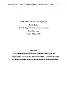
not m-coumaric acid, whose degradation occurred in a longer incubation after induction o f specific enzymes. However, when p-coumaric grown cells were in cubated on ferulic acid we observed accumulation of vanillic acid which re mained in the medium for several hours after all the ferulic acid was completely consumed (Figure 1, panels A and A ’). This compound is an intermediary meta bolite in the ferulic degradative pathway and it accumulated transiently when cells were grown on ferulic acid (it disappears between 2 and 3 h; Figure 1 A’). By measuring the kinetic of vanillic acid depletion we could confirm that this molecule is degraded by p-coumaric grown cells, when these cells were incu bated on medium containing 0.1% vanillic acid, only at a longer incubation time (24 h) after induction of specific enzymes. B F I3 cells grown on ferulic acid were on the contrary able to degrade promptly both p-coumaric (Figure 1, panel B) and 4-hydroxybenzoic acid. In fact, 4-hydroxybenzoic acid is produced transiently as expected for a metabolic intermediate (Figure 1, panel B ’). Cell-free extracts experiments The activity of vanillate O-demethylase, which catalyses the conversion of vanillate to protocatechuate, was not detected in crude extract of BF13 cells grown on p-coumaric acid as well as in extract of glucose grown cells. This result ex plains the vanillate accumulation observed during incubation of p-coum aric grown cells on ferulic acid (Figure 1, panel A ’). However, since a complete de pletion of vanillic acid occurred after prolonged incubation times when cells were exposed to vanillic acid, such enzyme is induced in BFI 3 cells by vanillic acid. In growth experiments we observed that BF13 cells duplicate slowly in me dium containing ferulic acid as a carbon source (Table 1), in spite of their abi92
ferulic acid depletion
p-coumaric acid depletion
vanillic acid formation
4-hydroxybenzoic acid formation
FIG. I - Kinetics of disappearance of the substrates (panels A and B) and transient ac cumulation of the benzoic intermediary metabolites (panels A’ and B’) by cell suspensions from ferulic (O) and p-coumaric ( • ) grown cultures. Wa shed cells were incubated at 30°C on saline phosphate buffer (see Materials and Methods) containing 0.1% ferulic or p-coumaric acid.
lity to consume this compound rapidly in whole cell experiments. The duplica tion time o f BF13 cells on vanillic acid was (3.5 h) comparable to that on ferulic acid (3.5 h; Table 1), thus suggesting that vanillate demethylation might be a rate limiting step for both vanillate and ferulate metabolism. A lthough the degradative pathw ays o f ferulic and p-coum aric acid have some steps in com mon, the rates o f substrate depletion are slightly different using cells pre-grown on ferulic rather than p-coumaric acid (figure 1, panels A and B). The com plete conversion into the corresponding benzoic acid derivative was always faster when cells were grown on ferulic acid, but in both cases pcoumaric acid was depleted more rapidly than ferulic acid. Similar results were obtained in experiments carried out with cell-free extracts, where slightly higher 93
TABLE 3 - The influence o f selected cofactors on the conversion offerulic acid to va nillic acid and o f p-coumaric acid in 4-hydroxybenzoic acid by cell-free extracts o f BFI3. reaction mixture
yield (mg/ml) of vanillic acid*
4-hydroxybenzoic acid**
ferulic-grown cells: 0.15
Extract + CoA/ATP + NAD+
0.1
Extract + CoA/ATP
0.082
nd
Extract + NAD+
0
nd
Extract only
0
nd
p-coumaric-grown cells: 0.06
0.11
Extract + CoA/ATP
nd
0.085
Extract + NAD+
nd
0
Extract only
nd
0
Extract + CoA/ATP + NAD+
Incubations were carried out as described in Materials and Methods for 2 h in 2 ml vo lumes containing: *2 mg/ml of ferulic acid, **2 mg/ml of p-coumaric acid, nd = not de termined.
am ounts of vanillic acid and 4-hydroxybenzoic acid were obtained using ex tracts prepared from ferulic-grown cells. Both reactions required the same co factors: CoA, ATP and NAD+ (Table 3). Such results could be explained postulating that an higher induction rate is obtained using ferulic acid rather than p-coumaric acid as inducer. The slight differences in the conversion rates o f the substrate, which were observed both in experiments carried out with whole cells (Fig. 1, panels A and B) and in experi ments with cell-free extracts (Table 3) should not be, instead, due to the sub strate uptake. The cofactor requirements are similar to those reported by Huang et al. (16) for the bioconversion o f ferulic acid in vanillic acid by cell extracts of Rhodotorula rubra. These authors suggested that this reaction is mechanistically sim ilar to the (3-oxidation of fatty acids and involves the remotion of an acetate moiety from ferulic acid in order to give vanillic acid. W e think that the same mechanism might be proposed here for the degrada tion o f ferulic acid and p-coumaric acid by P. fluorescens BF13. Further genetic work will help us to confirm this mechanism and analyse more precisely the regulation of the enzymes which are involved in the degrada tion o f these compounds.
The authors thank M rs Paola Tamantini fo r her skilled technical assistance.
94
REFERENCES (1) Crawford D.L., Crawford R.L.: Microbial degradation o f lignin. Enzyme Microb. Techno]., 2, 11-22 (1980). (2) Crawford R.L.: Lignin Biodegradation and Transformation. John Wiley & Sons, Inc., New York (1981). (3) Kirk T.K.: Degradation and conversion o f ligninocelluloses, in “Microbial Degra dation of Organic Compounds” (Gibson D.T. ed), pp. 399-347, Marcel Dekker, Inc., New York (1984). (4) Subba Rao P.V., Nambudiri A.M.D., Bhat J.V.: Microbial degradation o f phenylpropanoid compounds. J. Sci. Ind. Res., 30, 663-679 (1971). (5) Cain R.B.: The uptake and catabolism o f lignin-related aromatic compounds and their regulation in microorganisms, in “Lignin Biodegradation: Microbiology, Chemistry and Potential Applications” (Kirk T.K., Higuchi T„ Chang H.M. eds.), Vol 1, pp. 21-30, CRC Press, New York (1980). (6) Blakey E.R., Simpson F.J.: The microbial metabolism o f cinnamic acid. Can. J. Microbiol., 10, 175-185 (1964). (7) Coulson C.B., Evans W.C.: Microbiological degradation o f trans-cinnamic acid by soil pseudomonads. Chem. Ind., 1959, 543-544 (1959) (8) Sutherland J.B., Crawford D.L., Pometto 111 A.L.: Metabolism o f cinnamic, pcoumaric, andferulic acids by Streptomyces setonii. Can. J. Microbiol., 29, 12531257 (1983). (9) Whiting G.C., Carr J.G.: Metabolism o f cinnamic and hydroxycinnamic acids by Lactobacillus pastorianus var. quinicus. Nature (London), 184, 1427-1428 (1959). (10) Kawakami H.: Degradation o f lignin-related aromatics and lignins by several pseudomonads, in “Lignin Biodegradation: Microbiology, Chemistry and Potential Applications” (Kirk T.K., Higuchi T., Chang H.M. eds.), Vol. 2, pp. 103-125, CRC Press, New York (1980). (11) Andreoni V., Gall i E., Galliani G.: Metabolism o f ferulic acid by a facultatively anaerobic strain o f Pseudomonas cepacia. Syst. Appl. Microbiol., 5, 299-304 (1984). (12) Grbic-Galic D.: Fermentative and oxidative transformation o f ferulate by a facul tatively anaerobic bacterium isolated from sewage sludge. Appl. Environ. Micro biol., 50, 1052-1057 (1985). (13) Huang Z., Dostal L., Rosazza J.P.N.: Microbial transformations offerulic acid by Saccharomyces cerevisiae and Pseudomonas fluorescens. Appl. Environ. Micro biol., 59, 2244-2250(1993). (14) Toms A„ Wood J.M.:. The degradation o f trans-ferulic acid by Pseudomonas acidovorans. Biochemistry, 9, 337-343 (1970). (15) Huang Z., Dostal L., Rosazza J.P.N.: Purification and characterization o f a ferulic acid decarboxylase from Pseudomonas fluorescens. J. Bacteriol., 176, 5912-5918 (1994). (16) Huang Z., Dostal L., Rosazza J.P.N.: Mechanisms o f ferulic acid conversions to vanillic acid and guaiacol by Rhodotorula rubra. J. Biol. Chem., 268, 2395423958 (1993). (17) Bergey’s Manual of Systematic Bacteriology. Holt JG (Ed in chief). Williams & Wilkins, Baltimore (1984). (18) Sambrook J., Fritsch E.F., Maniatis T.: Molecular cloning: a laboratory manual, 2nd ed. Cold Spring Harbor Laboratory Press. Cold Spring Harbor, New York (1989). 95
(19) Miller J.H.: Experiments in molecular genetics. Cold Spring Harbor Laboratory, Cold Spring Harbor, New York (1972) (20) Buswell J.A.. Ribbons D.W.: Vanillate O-demethylase from Pseudomonas species, in “Methods in enzymology” (Wood W.A., Kellogg S.T, eds.), Vol 161, pp. 294301, Academic Press, New York. (1988).
96