Fifty pairs of hardboard boxes served as start and goal boxes. These boxes fitted into the end of each arm of the maze, forming a total arm length of 26 cm.
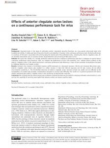
Behavioral Neuroscience 1989, Vol. 103, No. 5, 962-974
Copyright 1989 by the American Psychological Association, lnc. 0735-7044/89/$00.75
Effects of Amygdaloid and Amygdaloid-Hippocampal Lesions on Object Recognition and Spatial Working Memory in Rats J. P. Aggleton and H. S. Blindt
J. N. P. Rawlins
University of Durham Durham, England
University of Oxford Oxford, England
Neurotoxic lesions of the amygdala did not affect the postoperative acquisition of a nonspatial test of object recognition (delayed nonmatching to sample) even when retention delays were increased from 0 s to 20 or 60 s, or when test stimuli were deliberately repeated within a session. Although these amygdaloid lesions did not alter forced-choicespatial alternation, they slightly increased neophobic responses to novel foods and environments. In contrast, combined amygdalohippocampal (A + H) lesions impaired performance on the object recognition task when the retention intervals were increased beyond 0 s and when test stimuli were repeated within a session. The A + H rats were also severely impaired on the spatial alternation task, and they showed reduced neophobia. Comparisons with a previous study show that damage to the amygdala or hippocampus does not affect object recognition, whereas A + H damage produces clear deficits.
In recent years, Mishkin and his coworkers have shown that in monkeys, removal of the amygdala and the hippocampus produces severe impairment on a one-trial test of object recognition, delayed nonmatching to sample (DNMS; Mishkin, 1978; Murray & Mishkin, 1984). Similar results have been found when both of the major efferent pathways from these two structures, the ventral amygdalofugal pathway and the fornix, are damaged (Bachevalier, Parkinson, & Mishkin, 1985). These results contrast dramatically with the mild effects that are observed when just one of these limbic structures or pathways is removed (Mishkin, 1978; Murray & Mishkin, 1984). On the basis of these and related findings, investigators have proposed that the most severe syndromes of temporal lobe amnesia are the consequence of combined damage to these two limbic structures (Mishkin, 1978; Mishkin, Spiegler, Saunders, & Malamut, 1982). In this study, our goal was to determine whether in rats, like in monkeys, combined amygdaloid-hippocampal (A + H) damage is necessary to disrupt object recognition. In a previous study, we showed that by rewarding rats for selecting the unfamiliar goal box in a Y maze, it is possible to teach a one-trial DNMS task that appears analogous to the tasks given to monkeys (Aggleton, 1985). In addition, we have reported that removal of the hippocampus does not affect acquisition of this same recognition task (Aggleton, Hunt, & Rawlins, 1986). That study also showed that hippocampectomy had no effect when the recognition task was made harder by the imposition of retention delays of either 20 or 60 s between sample presentation and test, or when stimuli were
repeated within a session, thereby taxing recency judgements. One possible explanation for the lack of effects after hippocampectomy is that for some classes of memory task, recognition, for example, the hippocampus is just one of several structures that are involved and that, as a consequence, damage to multiple structures is required to produce full impairment. In addition to testing object recognition, we also examined performance on a spatial forced-alternation task. This spatial test of working memory (Honig, 1978; Olton, Becket, & Handelmann, 1979), which can also be described as a spatial DNMS task, was chosen because it is highly sensitive to hippocampal system damage (Rawlins & Olton, 1982) and hence would help confirm the effects of the hippocampal surgeries in the A + H group. The rats were also tested on their responsiveness to several novel foods. These tests were performed because there is evidence that amygdaloid damage can disrupt neophobic responses (Aggleton, Petrides, & Iversen, 1981; Nachman & Ashe, 1974; Rolls & Rolls, 1973), and such tests might help measure the effects of the amygdaloid surgeries. Unlike in comparable studies with monkeys, the amygdaloid lesions in this study were made with the neurotoxin ibotenic acid. This method helps minimize damage to fibers of passage, a feature of particular importance given recent evidence that some of the "well-established" effects ofamygdaloid lesions in the rat vanish when fibers of passage are spared (Dunn & Everitt, 1988).
Experiment 1: DNMS This work was supported by Grant G 8400477 from the Medical Research Council. We thank J. Emery, P. Hunt, M. Rollins, and S. Whitely for their assistance. Correspondence concerning this article should be addressed to J. P. Aggleton,Department of Psychology,Universityof Durham, South Road, Durham DH1 3LE, England.
The task was modeled on DNMS tests of object recognition that have proved sensitive to limbic damage in monkeys (Mishkin, 1978; Squire & Zola-Morgan, 1983). The rats were tested in a ¥ maze in which the start box and the two goal boxes were removable. In each trial the start box matched one of the goal boxes and differed from the other. The rats 962
LIMBIC LESIONS AND MEMORY
963
were r e w a r d e d for selecting the goal box that differed f r o m the start box (nonmatching). T h i s task taxed working m e m ory in that the start and goal boxes were changed after every trial (Figure 1), and although the task m i g h t be regarded as a simultaneous oddity problem, previous observations have s h o w n that rats a l m o s t n e v e r a t t e m p t to m a k e a direct c o m parison o f all three boxes before m a k i n g a choice. In this and all subsequent experiments, there were four e x p e r i m e n t a l groups: A, a m y g d a l o i d lesions alone; A + H, a m y g d a l o i d lesions c o m b i n e d with surgical aspiration o f the h i p p o c a m p u s , a surgery that i n v o l v e d r e m o v i n g a small section o f dorsal cortex and corpus callosum to allow visualization o f the structure; ACORT, a m y g d a l o i d lesions c o m b i n e d with visualization, but not r e m o v a l , o f the h i p p o c a m p u s ; and sham, a group o f s h a m a m y g d a l o i d lesions. A group with j u s t h i p p o c a m p a l lesions was not included because the effects o f this surgery are addressed in a p r e v i o u s study using identical procedures and apparatus (Aggleton et al., 1986).
Method Subjects. The subjectswere 60 naive, male rats of the pigmented DA strain (Bantin and Kingman, Hull, England). They were caged individually and were fed approximately 15 g/day of laboratory diet (Labsure ERM) so that they did not drop below 85% of normal body weight. The rats were approximately 3 months old and weighed 170255 g. Apparatus. Each arm of the aluminum ¥ maze was 13 cm wide and 20 cm high. Fifty pairs of hardboard boxes served as start and goal boxes. These boxes fitted into the end of each arm of the maze, forming a total arm length of 26 cm. The appearance of the boxes in each pair was made as similar as possible, but each pair was distinct from every other pair. This distinction was achieved by painting the walls and floors of the boxes in different colors and patterns and lining the floors with various materials, such as sandpaper, wooden strips, metal, Perspex, and cloth. In addition, each pair contained an identical object, such as a plastic cup, metal bracket, or wooden block, although no two pairs contained the same object. The floors of the boxes, which extended toward the center of the maze, began 8 cm from a Y-shaped aluminum guillotine door at the center of the ¥ maze (Figure 1). Food pellets (45 rag, Campden Instruments) could be dispensed under the back of each box. The ¥ maze was illuminated by a fluorescent ceiling light 215 cm above the apparatus. Further details of the apparatus have been published (Aggleton, 1985). Surgery. Each rat was anesthetized by injection of 3 ml/kg ip chloral hydrate-pentobarbital sodium mixture (containing 42 mg/ ml chloral hydrate and 9.7 mg/ml Nembutal). The animal was then placed in a stereotaxic head holder, and the scalp was retracted to expose the skull. A dental drill was used to make an opening exposing the dura mater above the amygdaloid and hippocampal regions. For the animals receiving amygdaloid lesions, two injections of ibotenic acid (1 mg/100 tA, Sigma), dissolved in phosphate buffer (pH = 7.2), were made through a 1-~1 Hamilton syringe in each hemisphere. In the first series of animals (sham = 6, A = 7, ACORT = 5, A + H = 5), each injection contained 0.35 ~1. In two subsequent series of animals, we attempted to reduce the likelihood of extraamygdaloid damage by reducing each injection to 0.3 ul (sham = 2, A = 2, ACORT 2, A + H = 2) and 0.22 tzl (sham = 4, A = 10, ACORT= 7, A + H = 9) ofibotenic acid. Each injection took 5 min, and the needle was left in position for another 5 min before being retracted. The injection coordinates relative to Ear bar 0 with the incisor bar set at 5.0 were: AP = +5.1, HT = +1.6, LAT = _+3.6; =
Figure 1. Representation of the delayed nonmatching-to-sample procedure. (Arrows indicate direction of correct response.)
and AP = +4.1, HT = +1.6, LAT = _+3.7. An identical procedure was used for the sham-operated controls, except that the needle was only lowered to a height of 4.0 and withdrawn immediately. After amygdaloid surgery, the A + H and ACORTrats were transferred to a specially designed head holder (Rawlins & Bennett, 1980). The dura mater was sectioned, and the minimum neocortex and corpus callosum were removed to allow visualization of the alveus layer of the dorsal hippocampus. For the A + H animals, the hippocampus was then aspirated under visual control with a Wild M650 operating microscope. Both groups had the wound packed with Sterispon gelatin foam soaked in physiological saline. In all four groups on completion of the surgery, sulfanilamide powder was applied, and the skin was sutured. Histology. At the end of the study, every rat was perfused intracardially with 5% formol-saline. The brains were subsequently blocked, embedded in wax (Paraplas0, and cut in 10-#m coronal sections. Every 10th section was mounted and stained with a Nissl stain (cresyl violet). Every adjacent section was also stained, but with a fiber stain (alcian fast blue). The use of different amounts ofibotenic acid (0.35, 0.3, or 0.22 ~l) in each injection site resulted in differing degrees of amygdaloid damage. Consequently, we estimated the extent of every amygdaloid lesion in the A, ACORT,and A + H groups by plotting the extent of cell loss onto 5 standard coronal sections (AP = 6.2, 5.6, 5.2, 4.6, 4.2) taken from the atlas of Pellegrino and Cushman (1967). A planimeter was then used to measure the extent of total damage and the amount of damage restricted to the amygdala. The sum of these areas from the 5 sections provided an estimate of the total extent of all ibotenate damage and the total extent of amygdaloid damage. The extent and range ofamygdaloid damage in the A, ACORT,and A + H groups were highly comparable (Figures 2 and 3). In all three groups, the larger amygdaloid lesions (0.35 or 0.3 ~1 ibotenic acid) damaged nearly all of the major nuclei, and estimates of overall damage indicated that between 30% and 75% of the structure had been damaged (means: A, 43%; ACORT,49%; A + H, 60%). There was, however, a tendency for some lesions to be asymmetrical so that there was sparing in the more lateral nuclei in one hemisphere and in the more medial nuclei in the other hemisphere. The smaller
964
J. AGGLETON, H. BLINDT, AND J. RAWLINS
Figure 2. Representation ofamygdalohippocampal lesions(A + H), amygdaloid lesionswith exposed hippocampus (ACORT), and amygdaloid lesions (A). (Top left:Reconstruction of smallest [solid]and largest [stippled] hippocampal lesion. Top right and bottom: Extent of amygdaloid damage in A + H, ACORT, and A groups. Shown are the median cases of animals that received "large" [stippled]or "small" [cross hatched] injections of ibotenic acid. All lesions are depicted on coronal sections, and the numbers refer to the corresponding plate from A Stereotaxic Atlas of the Rat Brain by L. J. Pellegrino and A. J. Cushman, 1967. New York: Appleton-Century-Crofts. Copyright 1967 by the publisher. Adapted by permission.)
LIMBIC LESIONS A N D MEMORY amygdaloid lesions (0.2 ~,1) were centered bilaterally in the rostral lateral and basolateral nuclei so that there was consistent sparing of the more medial and more caudal nuclei (Figure 2). These lesions were considerably more restricted, typically damaging between 10% and 30% of the structure (means: A, 23%; ACORT, 15%; A + H, 16%). The alcian fast blue stain confirmed that the neurotoxin had spared fibers of passage. Extra-amygdaloid damage was confined to three sites. The pyriform cortex received variable unilateral damage in 8 A, 4 A + H, and 6 ACORTrats; in some animals this damage was slight, although in a few it involved most of the tissue below the rhinal sulcus. That part of the putamen immediately above the amygdala revealed slight unilateral or bilateral damage in 6 A, 4 A + H, and 2 ACORTcases, whereas the part of the olfactory tubercle adjacent to the rostral pole of the amygdala was slightly damaged in approximately half of all the amygdaloid lesions. Throughout this study the term hippocampus refers to the dentate gyrus, the CA fields, and the subiculum, but not the entorhinal cortices. The hippocampal surgeries produced extensive bilateral damage to the hippocampus, and in all cases, most of the structure was removed (Figures 2 and 3). In addition, the fimbria and fornix were either completely sectioned or displayed dense gliosis. Only the most caudal and the most ventral portions of the hippocampus were spared, and in some cases, even these regions were damaged. Therefore, only the subiculum showed consistent sparing (Figures 2 and 3). In all but one case, there were small infarcts in the thalamus. These infarcts, which were usually bilateral, were concentrated typically in the lateral dorsal nucleus and, to a lesser extent, in the anterior nuclei. Two A + H animals that failed to master the DNMS task were excluded from all subsequent analyses because they had suffered excessive cortical damage after either an infection or an infarct. Surgical damage to the cortex appeared comparable in the hippocampal-lesioned and ACORT animals, and in all of the ACORT animals, the corpus callosum was completely sectioned. In 1 ACORT animal there was significant bilateral damage to the hippocampus, and this animal, which failed to master the DNMS task, was removed from all subsequent analyses. O f the remaining 12 ACORTcases, there was evidence of minor damage to the alveus, the most superficial portion of the hippocampus, in 3 cases. Nearly all of the ACORTcases had small infarcts in the thalamus; these were similar but typically more restricted than those seen in the A + H animals. Behavior. After a period of pretraining, which involved handling the rats daily and training them to run in the ¥ maze for food pellets, the experiment began. To start each daily test session, the rat was placed in an arm with a featureless start box. The central door was then raised, and the animal was allowed to choose between two arms that contained a matching pair of distinctive goal boxes (A1 ÷ and A2 +, Figure la). The rat was deemed to have made a choice when all four paws entered an arm, whereupon the guillotine door was lowered. On this first run, the animal was rewarded with three food pellets in whichever box it entered. The animal was confined to this box (AI) for 20 s, while the other two test boxes were replaced. The central door was then raised, revealing a familiar box (A2-) in one arm and a novel box (BI ÷) in the other (Figure lb). The animal was rewarded with three food pellets if it entered the novel box B 1. After 20 s of confinement in Box B1, the second trial began (Figure lc). The central door was raised, and the animal chose between the familiar appearance of Box B2- and Novel Box C 1+. This sequence was repeated with new pairs of boxes for a total of 10 trials, and selection of the novel box was always rewarded (Trial 3, C2- versus D 1+ . . . Trial 10, J2- versus K1 +). A balanced schedule determined whether the correct response was to the fight or left. The sequence of test boxes was varied after every 50 trials so that any particular box appeared, on average, in every fifth session.
965
Figure 3. Top: Photomicrograph of hippocampal lesions in the group receiving lesions in the amygdala and hippocampus. Bottom: Photomicrograph of coronal section (Nissl stain), showing shrunken, acellular appearance ofamygdaloid region after injection ofibotenic acid. (OT = optic tract; P = pyriform cortex; T = needle tract. See Plate A.) If an animal made an incorrect choice, correction trials were run with the same set of goal boxes until the animal selected the novel box. During these correction trials, the goal boxes were rearranged so that entering the positive box required the same body turn as in the test trial. These correction trials were necessary because they ensured that the animal entered the goal box that was to become the next start box. When a rat reached the criterion score of 40 or more correct responses in 5 consecutive sessions (80%), the second phase of the experiment began. If a rat failed to reach this criterion score within 500 trials (50 sessions), it was excluded from the rest of the experiment. Rats that reached criterion performed 150 more trials, in which retention intervals of 0 (training condition), 20, and 60 s were imposed. For the two longer retention intervals, the animals were once again confined in the start box for 20 s, but the box was then removed and the animal was tipped into the arm of the Y maze. The start box was then immediately replaced by a blank, featureless box with no floor. The animal was not handled during this procedure. After another 20 or 60 s of confinement in the arm with this blank box, the central door was raised, revealing a novel goal box and one that resembled the original start box. As before, the animal was
966
J. AGGLETON, H. BLINDT, AND J. RAWLINS
U') 500
**
**
::x:
"~
0i
~
70
Z O 400 11,4
/
~, 3oo
L.;
U
o
I-- 20(}
60
•
o I~ J"
100
~.
Z
0
SHAM
A
ACORT A + H
,u •
A
5
• HIPPOCAMPUS i. . "~. . 0
CHANCEl
20
DELAY
60
S
Figure 4. Nonmatching-to-sample results. (Left: Trials to 80% criterion level [excluding criterion trials]. Animals failing to learn within 500 trials are marked by asterisks. Right: Performance with increasing retention delays between stimulus presentation and recognition test. Vertical bars show standard errors. The mean performance of 9 rats with hippocampal lesions from an earlier study [A; Aggleton, Hunt, & Rawlins, 1986] are shown for comparison. A = group receiving amygdaloid lesions; ACOgT = group receiving amygdaloid lesions with surgery to visualize hippocampus; A + H = group receiving amygdalohippocampal lesions.)
rewarded for choosing the novel box, and an incorrect choice was followed by correction trials. This procedure meant that the animal had to retain the memory of the start box for at least 20 or 60 s. The animals underwent 5 days of testing at each of the three conditions in a counterbalanced order. In the final test condition, the rats underwent 12 trials a day for l0 sessions. Half of these sessions were the same as those during the acquisition phase, with session-unique stimuli being used on every trial. However, on alternate sessions, the rats were tested with a limited set of just four pairs of goal boxes, a different set of four pairs being used in each of the 5 sessions. During these sessions, each pair was used as the correct "unfamiliar" stimulus three times. On the first occasion (Trials 1-4), each goal box was indeed unfamiliar; on Trials 5-8, each box had been entered once already; and on Trials 9-12, each box had been entered twice. Thus, a sequence of correct goal boxes over the 12 trials might be as follows; A, B, C, D, A, C, D, B, D, A, C, B. Consequently, on Trials 5-12, the animal was required to make a relative recency rather than a recognition judgement. A standard series of five different goal-box sequences was given to each animal. In all other respects the testing conditions, including correction trials, were precisely the same as in the original acquisition procedure and in the intervening comparison sessions.
Results After histological analysis, 3 animals with lesions that involved extensive cortical damage were excluded from the study (1 ACORT, 2 A + H), leaving 12 sham, 19 A, 12 ACORT, and 14 A + H animals. Figure 4 shows the number o f trials preceding the 80% criterion learning score. The apparent lack o f difference be-
tween the acquisition scores o f the four groups was confirmed with the Kruskal-Wallis test (H = 0.60), nonparametric statistics being used in response to the animals that had reached the arbitrary limit o f 500 trials. Additional analyses using the Spearman test showed no evidence o f a significant positive correlation between the extent o f amygdaloid damage and the number o f acquisition trials within any o f the three experimental groups. A similar lack o f correlation was found between the total extent of temporal lobe damage (amygdala and adjacent conical and subcortical regions) and the number o f acquisition trials. Although more o f the animals in the A + H group failed to reach the criterion score (Figure 4), the proportion was not significant, and normal rats sometimes failed to master the D N M S task. In another analysis, we compared the number o f correct responses made over the first 20 trials (2 days) o f the D N M S task. This comparison followed from the discovery that normal rats will spontaneously nonmatch (Aggleton, 1985). We found that the mean scores o f the four groups were above the chance score o f 10 (sham = 12.2, A -- I 1.8, ACORT = 11.7, A + H = I 1.7), and there were no differences among the four groups ( F < 1). In contrast, a clear group difference emerged when animals that had reached the 80% learning criterion were tested with 50 trials at each o f three retention delays; that is, 0 (training condition), 20, and 60 s (Figure 4). A n analysis of variance (ANOVA) confirmed that increasing the retention interval impaired performance, F(2, 90) = 38.14, p < .001, although this effect was only pronounced for the 60-s delays. There
967
LIMBIC LESIONS AND MEMORY
,0[
1-1 Normal
31
~--)
u Z
=
7
_
30
O
25
I-LJ
20 .
.
.
.
oA 6
"".
,/ ,,'~
~'
//
Z
4
,
A
/
~ ACORT a A+H
O
15
SHAM
• SHAM
ACORT A+H
e
i
1
2
,
|
l
3
4
5
"J"
10÷
NUMBER INTERVENING TEST BOXES Figure 5. Recency test results. (Left: Mean performance over 40 trials in which the "novel" stimuli were either session unique or repeated within a session. Right: Performance plotted against number of intervening goal boxes between repetition of particular goal box. Vertical bars show standard errors. Abbreviations for groups are defined in Figure 4 caption.) was, however, a significant lesion effect, reflecting the poor performance o f the A + H animals, F(3, 45) = 6.28, p < .01. Although there were no significant interactions between the various groups, F(6, 90) = 2.13, .1 > p > .05, the A + H group did not differ from the other three groups at 0 s, whereas a difference was present at 20 s (minimum t = 2.45, p < .05) and 60 s (minimum t = 2.72, p < .05). Further confirmation of a lesion effect was found when the total errors made over the 150 test trials, including those on correction trials, were compared, F(3, 45) = 6.19, p < .01, this group difference reflecting the larger number of total errors made by the A + H animals. All o f the animals that had been tested over the three delays were then tested on their ability to perform the DNMS task at 0-s delays, when the goal boxes were repeated within a session. Figure 5 (left panel) shows the mean performance of the four groups over 40 trials (the last 8 trials from five sessions) in which each correct unfamiliar stimulus had already been used within that session. These scores are compared with 40 equivalent trials from the intervening normal sessions in which every goal box was used only once within a session. An ANOVA comparing performance under these two conditions confirmed that the animals made considerably more errors when stimuli were repeated within a session, F(I, 45) -- 85.06, p < .001. In addition, there was evidence o f a lesion effect, reflecting the poor performance of the A + H group when test stimuli were repeated, F(3, 45) = 4.29, p < .025. There was, however, no interaction between lesion and the two test conditions, F(3, 45) = 1.48. A more detailed way o f presenting the same data is also shown in Figure 5 (right panel), in which recognition performance is plotted against the number o f different test boxes intervening between the reoccurrence o f the same goal box. The test sequences were arranged so that every rat underwent a total o f 8 trials at each o f the different numbers of intervening boxes (1-5). The results for 10 or more intervening
stimuli came from the corresponding 40 trials on the 5 intervening days, that is, the last 8 in each session, the score being divided by 5 to make it comparable with those with repeated boxes. An ANOVAcomparing performance with one to five intervening boxes confirmed that the task became easier as the number of intervening stimuli increased, F(4, 180) = 16.70, p < .001. The same analysis showed that there was a lesion effect, F(3, 45) = 4.19, p < .025, and a significant interaction F (12, 180) = 2.10, p < .025. The lesion effect and the interaction clearly reflect the relatively poor performance of the A + H group when tested with three, four, and five intervening boxes (Figure 5). This effect was exemplified by the poor performance of the A + H group with five intervening boxes when compared with the other three groups, miminum t(18) = 3.84, p < .01. In contrast, there were no group differences, F(3, 45) = 1.68, on the alternate control days (10 or more intervening boxes). No evidence was found that either the total extent o f amygdaloid or temporal lobe damage correlated with the overall performance of the A + H group with the repeated goal boxes.
Discussion Rats with amygdaloid lesions (A, ACORT) or amygdaloidhippocampal lesions (A + H) were able to learn a nonspatial test o f object recognition, DNMS. However, the A + H animals were impaired when the retention interval was increased to 20 or 60 s. This deficit probably cannot solely be attributed to hippocampal damage, because a previous study in which exactly the same methods were used gave no evidence that hippocampectomy alone affected performance (Aggleton et al., 1986). For comparison, the mean scores from that study are included in Figure 4. Indeed, because the performance scores o f the sham-operated groups from the two
968
J. AGGLETON, H. BLINDT, AND J. RAWLINS
studies did not differ on the three retention delays, F ( I , 12) = 2.01, comparisons were made between the A + H group from this study and the hippocampal lesion group from the previous study. These comparisons confirm that the A + H animals performed significantly worse than those with just hippocampal lesions, F(1, 17) --- 12.69, p < .01, although there was no Delay x Lesion interaction, F(2, 34) = 1.20. Repetition o f the test boxes within a session, which increased proactive interference and taxed recency rather than recognition, had no differential effect on the A or ACORX animals (Figure 5). In contrast, the A + H group performed close to chance, even when the number of intervening stimuli increased, thereby making the recency task easier. The chance performance o f all groups under the hardest conditions, in other words, when the unfamiliar stimulus had been used recently, helps rule out the use o f unintentional cues that may have allowed the animals to "cheat." The slightly lower than chance scores with one intervening item appear to reflect the animals' reluctance to enter a goal box that not only had the same appearance as that used in the previous trial but was in the same arm o f the maze (Aggleton et al., 1986). The lack o f difference between the groups at 0 s during the delay conditions (Figure 4) and on the alternate control days in the recency test (Figure 5) shows that A + H animals that mastered the task were able to perform normally when the m e m o r y demands were minimal. This result implies that the subsequent impairments were not a consequence o f motivational or sensory deficits and hence probably reflected m e m o r y deficits that did not affect the learning o f the general rule but disrupted remembrance o f particular stimuli. At first, finding a unitary explanation to account for the performance of the A + H animals is difficult, especially given the nature o f the lesions. The most straightforward explanation is that the A + H lesions resulted in more rapid decay o f m e m o r y traces for the test stimuli than the other lesions. There are, however, two major problems with this interpretation. First, the interaction between groups and delays failed to reach significance (. l > p > .05), and the forgetting curves for the four groups are nearly parallel between 20 and 60 s. Second, and more important, the abnormal performance of the A + H animals on the recency task cannot easily be accounted for in terms of faster trace decay alone (Figure 5). Indeed, one might expect rapid forgetting to lessen the confusion between recently used test stimuli and so aid performance by producing a release from proactive interference. The finding that the A + H animals performed close to chance on the recency test when there were three, four, or five intervening stimuli (Figure 5) implies that the animals could r e m e m b e r both stimuli but could not distinguish them on the basis o f temporal order. Support for this notion comes from other evidence that hippocampal lesions disrupt tasks involving temporal discriminations (Moore, 1979; Olton, Meck, & Church, 1987; Rawlins, 1985; Solomon, 1980), although there is much disagreement over the interpretation o f such effects (Rawlins, 1985). Furthermore, we have shown that hippocampectomy alone does not disrupt an almost identical test o f recency (Aggleton et at., 1986), suggesting that the additional amygdaloid damage made an important contribution.
The above explanation is a specific case o f the more general proposal that A + H lesions increase sensitivity to proactive interference and thereby impair memory. Because each box was reused every 5 days and there were inevitable similarities between different boxes (e.g., the same surface on the floors), there is considerable potential for interference effects. These results are, of course, exaggerated in the recency test, in which each stimulus was presented three times within each test session. Unfortunately, we could not determine whether the A + H rats were differentially impaired the second or third time that an item was repeated within a session (Trials 5 12), because this analysis was contaminated by floor effects. Nevertheless, altered sensitivity to proactive interference, primarily as a consequence of altered temporal tagging of events, appears to offer the best explanation for the pattern o f results (Moore, 1979; Solomon, 1980). Note, however, that there was no clear positive correlation within the A + H group between performances under the delay (20-s and 60-s) and recency conditions. In Experiment 2, we examined whether the A + H lesions were suflicient to disrupt a m e m o r y task known to be sensitive to hippocampal damage, hence testing the functional effectiveness of the hippocampal lesions. The animals were therefore trained on a rewarded alternation task run as a test o f working memory. In this task the animal was forced to enter one arm o f the 3" maze and then, on the next run, to enter the other arm to receive a food reward. Previous studies have confirmed that this task is highly sensitive to hippocampal or fornix ablations (Aggleton et al., 1986; Rawlins & Olton, 1982). E x p e r i m e n t 2: S p a t i a l N o n m a t c h i n g t o S a m p l e Method Subjects and apparatus. The subjects were the same as those used in the previous experiment, and they included the rats that failed to reach the learning criterion on the nonmatching task. The floors of the T maze were 10 cm wide and made of aluminum. The stem of the maze was 80 cm long, with a guillotine door 33 cm from the beginning. The crosspiece was 136 cm long, and at each end there was food well 4 cm in diameter and 0.75 cm deep. The walls of the maze were 17 cm high and made of clear Perspex. The maze was supported on two stands that were 93 cm high. Testing was carried out in a large room (different from that used in Experiment 1) in which tables, sinks, and empty cage racks provided salient room cues. Behavior. Testing began between 9 clays and 8 weeks after completion of Experiment 1 and between 2 and 8 clays after completion of the first of the two tests of food neophobia in Experiment 3. Each animal required only 1 or 2 clays of pretraining to run reliably down the stem of the T maze to find food pellets in both of the food wells. At the start of each trial, which consisted of two stages, three food pellets were placed in each food well, and a wooden block was placed in one arm close to the choice point. The rat was then placed in the start box, and the guillotine door was raised. On this information run, the animal was forced by the wooden block to enter the open arm and allowed to eat the food there. No retracing was permitted. The animal was then picked up and placed back in the start box, and the wooden block was removed. The guillotine door was then raised, and the rat was free to enter either arm. On this choice run, the rat was deemed to have chosen when it placed a back foot on either choice arm, whereupon the wooden block was placed behind
969
LIMBIC LESIONS AND MEMORY the rat to stop retracing. If the rat alternated, that is, entered the arm it had not visited on the information run, it was allowed to eat the food and return to its cage. If the other arm was chosen, that is, the same arm as on the information run, the rat was confined to the arm for approximately 10 s and then returned to its holding cage. In this manner, each animal was rewarded for selecting the arm it had not entered on the information run. The rats were tested in groups of 4, with each rat having 1 trial in turn. As a consequence, the intertrial interval ranged 3-5 min. The animals underwent 6 trials each day, each consisting of two runs through the maze. Every rat received 3 forced-right and 3 forcedleft trials every day, although consecutive rats were run on different schedules. The animals were tested for a total of 12 days (72 trials).
17 16
15I- ACORT af I
e~
i-Go
14
I-U
12
I,M
11
..v
All animals, including those that did not achieve the acquisition criterion score on the object recognition task were tested for 12 sessions on a spatial nonmatching-to-sample task. Figure 6 shows the mean performances of the four groups over four successive blocks of 3 test sessions (l 8 trials). The A + H group showed a striking impairment, which was confirmed by an ANOVA,F(3, 53) -- 97.71, p < .001. There was, in addition, a block effect, showing that the animals were improving over the four blocks of trials, F(3, 159) = 7.19, p < .001, although there was no Lesion x Block interaction (F < l). Whereas the mean percentage correct over all 12 days for three of the groups was 92.3 (sham), 90.9% (A), and 87.6% (ACORT), the A + H animals averaged only 55.2% correct. The same overall pattern of results was obtained when only the animals that mastered the object recognition task were considered.
13
I~ lO
O
Results
SHAM
Z .