BEHAVIOURAL BRAIN RESEARCH Behavioural Brain Research 81 (1996) 115 121
ELSEVIER
Research report
Effects of parietal cortex lesions on spatial problem solving in the rat C. Thinus-Blanc ", E. Save "'*, B. Poucet a, N. Foreman b a Centre de Recherche en Neurosciences Cognitives, Centre National de la Recherche Scientifique, 31 chemin Joseph-Aiguier, 13402 Marseille, Cedex 20, France b Department of Psychology, University of Leicester, Leicester, UK
Received 29 December 1995; revised 25 March 1996; accepted 25 March 1996
Abstract
The Maier 3-table task was used to examine spatial representations in rats with lesions of the parietal cortex. Some animals had anteriorly placed lesions, some posteriorly placed in cortical areas, sometimes regarded as 'parietal' in earlier studies. After 5 days of familiarization, animals were given 18 days of testing on the standard Maier task. Both parietal groups were initially impaired, but reached the same level of performance as controls by the end of the test period. Learning occurred both within and between sessions for the anterior group, but only between sessions for the posterior group. There was no major functional differentiation apparent on this task between the two 'parietal' areas. Rate of exploration increased in both parietal groups across test sessions as task performance improved. It is argued that the change in exploratory activity across sessions in parietal groups may reflect the adoption of a compensatory strategy which improved performance, but that improvement could also have been due to neural changes, as structures, such as the frontal cortex or hippocampus, assume some functions normally mediated by the parietal area. Keywords: Parietal cortex; Spatial learning; Rat
1. Introduction
For over 20 years, the hippocampus has been considered by m a n y to be the only brain structure involved in the high-level processing of spatial information in the rat [21,22]. However, increasing evidence suggests that other structures play complementary roles to that of the hippocampus. Interest in the study of these structures reflects a wider interest in the neural substrates of spatial processing that arises from cooperation and interplay between various brain structures, but with a key role being occupied by the hippocampus. The associative parietal cortex (APC) has recently been proposed as one likely crucial component for spatial processing functions. Behavioral studies have shown that lesions of the APC in rodents induce deficits in spatial learning tasks like place navigation and 8-arm maze tasks [5,14-17], as well as in behaviors that occur in response to a spatial change after exploration [1,27], in effortful processing
* Corresponding author. Tel.: (33) 9116-4358; Fax: (33) 9177-4969; E-mail:
[email protected] 0166-4328/96/$15.00 © 1996 Elsevier Science B.V. All rights reserved PII S0166-4328 (96) 00050-2
[4], and in visuospatial attention [ 10]. More recently, bilaterally lesioned rats were found to be impaired in a path-integration-based navigation task, suggesting that the APC is, at least, involved in the processing of kinesthetic information [28]. This is consistent with the results of a unit recording study in the freely moving rat showing that some APC cells fired selectively when specific movements and views of the environment were combined [20]. Head-direction cells, i.e., cells firing when the rat's head is oriented towards a specific direction, whatever its location in the environment, have also been recorded in this area. Their firing was found to be sensitive to vestibular and visual inputs [2,3]. Overall, these results show that the APC as a center for multimodal sensory integration might be involved at various levels of spatial information processing. Surprisingly, the neuroanatomical boundaries of the APC remain unclear. In the rat, Kolb and Walkey [-15], using retrograde labeling, delineated an area which could be structurally analogous to the posterior parietal cortex of primates on the basis of its connectivity with other structures, such as the visual, somatosensory and frontal
116
C. Thinus-Blanc et al./Behavioural Brain Research 81 (1996) 115-121
cortices. Lesions in this area resulted in retrograde degeneration in the dorsolateral and lateroposterior nuclei of the thalamus. It was concluded that this region is likely to be homologous with Krieg's area 7 [18]. However, there is still some disagreement about the localization of the APC, since different authors have defined it in a more anterior region than that defined by Kolb and Walkey [4,5,13,14]. In the present study, we were concerned with the distinction between these two regions (referred to as APC-POST and APC-ANT, for the sake of clarity). As a matter of fact, we consider this issue particularly important in relation to the past conclusions that have been drawn from the behavioral effects of APC lesions. A previous study has suggested that each of these regions has different functions [28] and is involved, to a different extent, in the construction and use of allocentric spatial representations. Rats were freely allowed to explore an open field containing 5 objects. After a habituation phase, the spatial arrangement of objects was modified by moving two of them and the animals' exploratory reactions to this change were examined. The APC-ANT-lesioned rats re-explored all the objects whereas the APC-POST-lesioned rats failed to renew exploration. In contrast, control rats re-explored only the displaced objects. It appears then, that the APC plays an important role in spatial information processing, especially when the animals have to spontaneously collect sensory information and form a spatial representation of the environment. The opportunity to freely explore the environment has been shown to be crucial for spatial problem solving in the familiar 3-table task. This task, originally described by Maier [19], involves two training phases followed by a test trial. During the first training phase, the animal is allowed to explore the apparatus which consists of 3 elevated platforms connected to each other by 3 pathways, converging on a central choice point. During the second phase, the rat is fed on one of the tables. During the test trial, it is placed on one of the remaining tables and is required to return to the table where it was just fed. Each day the food table and the start table are varied in a pseudorandomized manner so that the animal cannot rely on a specific orientation to reach the food. A correct response is determined by the integration of the feeding experience and the representation of the 'problem space' built up during the first phase [8,9]. It has been demonstrated that problem solving is affected by lesions of the septohippocampal formation and frontal cortex 1-25]. Therefore, it was of interest to determine whether parietal damage would also result in a 3-table problem-solving deficit. As mentioned above, the present study was further aimed at comparing the effects of lesions of the APC-ANT and APC-POST on, firstly, the time course of exploratory activity, and secondly on spatial problem-solving ability.
2. Materials and methods 2.1. Subjects
Twenty-four naive male Long-Evans hooded rats, purchased from a commercial laboratory animal supplier (Elevage Janvier, St Berthevin, France) served as subjects. Upon arrival, they were housed individually in transparent cages (40x26 x 15 cm) in a colony room with natural lighting. They were approximately 90 days of age at the time of surgery. Once the experiment began, rats were placed on a 23-h food deprivation schedule, with water ad libitum. Their weight was reduced to an average of 90% of pre-experimental body weight (mean weight, 350 g). 2.2. Apparatus
The apparatus consisted of 3 tables (40x20cm) interconnected by a Y-shaped runway system. Each segment was 1 m long. Obstruction screens at the front of each table prevented the animals from seeing the contents of a table when they were either on a different table or at the intersection of the runways. 2.3. Surgery and histology
Prior to the experiment, rats were randomly assigned to groups with bilateral lesions of the anterior region of the APC (using DiMattia and Kesner's coordinates [4,5]; APC-ANT group, n=7), of the posterior region of the APC (using Kolb and Walkey's coordinates [ 15]; APC-POST group, n = 7) and to a sham-operated group (CNT group, n= 10). After an injection of 0.3 ml of atropine sulfate, rats were deeply anesthetized with an i.p. injection of sodium pentobarbital (55 mg/kg) and placed in a Kopf stereotaxic instrument. After a midline incision of the scalp was made, the skin and muscles were retracted. Lesions of the posterior parietal cortex were performed by thermocoagulation. Two windows were opened in the skull at the following stereotaxic coordinates according to bregma: for the APC-ANT group, AP=from +0.5 to - 3 . 5 m m , L = f r o m +1.5 to +5.5mm; and for the APC-POST group, AP=from - 2 to - 6 m m , L = f r o m +1.5 to +5.5 mm. The underlying cortex was destroyed by using heating resistance applied directly to the dura. Particular care was made not to damage the dura. CNT animals were shamoperated (i.e., they were anesthetized, had their skin cut, and were then sutured). 2.4. Histological procedure
After behavioral testing, rats with brain damage were given a lethal dose of sodium pentobarbital and perfused transcardially with 10% formalin. Each brain was photo-
C. Thinus-Blanc et al. / Behavioural Brain Research 81 (1996) 115-121
graphed from above to evaluate the external extent of the lesions. The brains were then sectioned on a freezing microtome. Every fifth 40-pm section was stained with Cresyl fast violet. The slides were examined microscopically to determine the dorsoventral extent of the lesion.
117
~' 11
i~ 3
2.5. Behavioral procedure During the 10-day postoperative period, animals received daily 10-min handling sessions. During the last 5 days of this period, the animals were placed on a straight runway suspended between two chairs in the experimental room and allowed to run to the end of the runway for food. The runway was the same length and width as those used in the experiment. The rats were given 10 such trials on each of the 5 days. Testing on the 3-table task took place over the following 18 days. Each day, groups of 3 animals were placed on the unbaited apparatus and allowed to explore the tables and runway configuration for 15 min. Following the exploratory phase, subjects were individually given a 1-min feeding experience on what was to be the baited table for that animal that day. After the 1-min feeding phase, the rat was placed on one of the two unbaited tables, and its task was to return to the place where it had just been fed. Each rat was given 3 test trials per day. Each day, the start and baited tables changed for each animal in the normal and parietal groups. The animals were allowed 3 min to leave the start table and reach the goal table. An error was scored whenever an animal either failed to leave the start or went to the unbaited table. In the latter instance, the rat was allowed to self-correct. Detailed records of the runway segments traversed by each animal were taken, and a detailed analysis performed on the exploratory behavior of each of the 3 groups. The 15-min daily exploratory period was subdivided into 5 successive 3-min periods. Particular attention was paid to the number of tables entered and to the number of entrances recorded up to the point when all 3 tables had been visited (the latter being referred to as the 'index of exhaustive exploration').
/ /
I
I 6
4 //s--~'v
7
\,
8
POST1
"
2
5
3
6
i
\
4
7
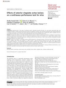
Fig. 1. Cortical surface diagrams showing the extent of the anterior (APC-ANT) and posterior (APC-POST) parietal lesions for all brains.
brains. Coronal reconstructions of typical anterior and posterior parietal lesions at 3 levels corresponding to + 0.5, - 1.8 and - 3.6 mm (anterior lesion) and - 1.8, - 4 . 3 and - 5 . 6 m m (posterior lesion) from bregma according to the Paxinos and Watson atlas [23] are shown in Fig. 2. The neocortex was completely destroyed within the intended coordinates. Most importantly, there was no damage to the underlying hippocampus.
3. Results
3.2. Behavioral findings
3.1. Anatomical findings
3.2.1. Test trial performance Fig. 3 shows the mean number of correct choices on the first test trial for the 3 groups of animals over the course of the 18-day test period. The data have been blocked into 6-day periods. Particular attention was paid to the first test trial of each daily session because it corresponds to the immediate understanding of the task. Trials 2 and 3 may incorporate learning (cf. Table 1). CNT animals performed at or above the 75% level (first daily choice) over the course of the experiment.
Fig. 1 shows the dorsal aspect (drawn from photographs) of all APC-lesioned brains. Coronal reconstructions of typical anterior and posterior parietal lesions at 3 levels corresponding to +0.5, - 1 . 8 and - 3 . 6 m m (anterior lesion) and - 1.8, - 4 . 3 and - 5 . 6 mm (posterior lesion) from bregma according to the Paxinos and Watson atlas [-23] are shown in Fig. 2 shows the dorsal aspect (drawn from photographs) of all APC-lesioned
118
C. Thinus-Blanc et al./Behavioural Brain Research 81 (1996) 115-121
+O~jj. .1.8~
B Fig. 2. Coronal sections of a typical anterior (A) and posterior (B) parietal lesion corresponding to the levels +0.5, - 1 . 8 and - 3 . 6 m m , and - 1 . 8 , - 4 . 3 and - 5 . 6 mm from the bregma point, respectively (redrawn after Paxinos and Watson [23]). APC-ANT -" APC-POST -----t:3--- CNT
6-
4-
~6
2-
~o
z 1-6
7-12 Blocks of trials
13-18
Fig. 3. Mean number of correct first choices (entering the baited tables) in the first test trial over 6-day blocks for CNT, A P C - A N T , and APCP O S T groups (standard errors are shown as vertical bars).
In contrast, both APC groups were impaired during the first 6-trial block, although they displayed a clear improvement in first-choice performance level across the three 6-trial blocks, starting from about 45% correct to 73% by the last block. A repeated measures VAR 3 analysis of variance [26] with surgical treatment as the between-groups measure and blocks of days and trials as the repeated within-groups measures confirmed these impressions. Each of the 3 factors was found to have a significant effect (Group, Fz,22=8.03, P