Autonomic Neuroscience: Basic and Clinical 197 (2016) 46–55
Contents lists available at ScienceDirect
Autonomic Neuroscience: Basic and Clinical journal homepage: www.elsevier.com/locate/autneu
Effects on hemodynamic variables and echocardiographic parameters after a stellate ganglion block in 15 healthy volunteers Katia Puente de la Vega Costa a, Miquel A. Gómez Perez b,c, Cristina Roqueta c,1, Lorenz Fischer a,⁎ a b c
Department of Neural Therapy, IKOM, University of Bern, Inselspital, PH 4, 3010 Bern, Switzerland Cardiology Department, Hospital del Mar, Passeig Marítim 25-29, 08003 Barcelona, Spain Medicine Department, Faculty of Medicine, Universitat Autonoma de Barcelona, Edifici W - UD de Medicina de la Vall d'Hebron, Passeig Vall d'Hebron 119, 08035 Barcelona, Spain
a r t i c l e
i n f o
Article history: Received 3 January 2016 Received in revised form 6 March 2016 Accepted 11 April 2016 Keywords: Stellate ganglion block Local anesthesia Neural therapy Procaine Hemodynamic parameters Echocardiographic parameters
a b s t r a c t Background: The sympathetic nervous system has an important role in generating pain. Various pathomechanisms are involved that respond well to the application of local anesthetics (LA), for example to the stellate ganglion block (SGB). Objectives: We wanted to know more about the effects of SGB on cardiovascular parameters. Methods: We included 15 healthy volunteers; another 15 healthy volunteers as a control group (sham injection of LA). In order to produce a more precise SGB, we employed only a small volume of LA (3 mL), a LA with a lower permeability (procaine 1%), and a modified injection technique. Systolic and diastolic blood pressure (SBP, DBP), heart rate (HR), and echocardiographic parameters were recorded before and after SGB. We also investigated whether there are side differences (left and right SBG). Results: At baseline all parameters were within the normal range. After performing right and left SGB DBP significantly increased (on the right side from 68.73 ± 8.61 to 73.53 ± 11.10, p = 0.015; on the left side from 70.66 ± 13.01 to 77.93 ± 10.40, p = 0.003). In the control group no increase in DBP was observed. No side-specific differences were found, except a significant reduction in the maximum velocity of myocardial contraction during the systole with left-sided SGB. Conclusions: Even with our methods we could not prevent the simultaneous occurrence of a partial parasympatholytic effect. For this reason, the SGB has only minor hemodynamic effects, which is desirable as it enhances the safety of the SGB. © 2016 The Authors. Published by Elsevier B.V. This is an open access article under the CC BY-NC-ND license (http://creativecommons.org/licenses/by-nc-nd/4.0/).
1. Introduction 1.1. Background The autonomic nervous system plays an important role in the regulation of blood pressure, heart rate, myocardial contractility and coronary perfusion. The balance of activity of its sympathetic and parasympathetic branches has a key part in this (Alston et al., 2011; Drew and Sinoway, 2012; Esler, 2010; Jänig, 2006; Low, 1993; Paton
Abbreviations: DBP, diastolic blood pressure; EF, ejection fraction; HR, heart rate; IVRT, isovolumetric relaxation time; LA, local anesthetics; LVEF, left ventricular ejection fraction; SBP, systolic blood pressure; SD, standard deviation; SG, stellate ganglion; SGB, stellate ganglion block; WDR, wide dynamic range. ⁎ Corresponding author at: Department of Neural Therapy, IKOM, University of Bern, Inselspital, PH 4, CH-3010 Bern, Switzerland. E-mail addresses:
[email protected] (K. Puente de la Vega Costa),
[email protected] (M.A. Gómez Perez),
[email protected] (C. Roqueta), lorenz.fi
[email protected] (L. Fischer). 1 Present address: Geriatric Medicine Department, Centro Fórum, Hospital del Mar, Hospital de la Esperança, Sant Josep de la Muntanya 12, 08024 Barcelona, Spain.
and Spyer, 2013; Schwartz and De Ferrari, 2011; Sunagawa et al., 1998; Yanowitz et al., 1966). An imbalance in the autonomic cardiac fibers can, for example, lead to cardiac arrhythmias (Lopez-Sendon et al., 2004; Nademanee et al., 2000; Ogawa et al., 2007; Schwartz, 1984; Schwartz, 1998; Schwartz and Zaza, 1986; Shen et al., 2011) or hypertension (Fernandez et al., 2012; Grassi, 2010; Grassi and Seravalle, 2012). An imbalance of the sympathetic and parasympathetic branches of the autonomic nervous system, however, can also cause and maintain pain and inflammation (Baron, 2006; Baron and Jänig, 1998; Levick et al., 2010; Ricker, 1924; Stanton-Hicks et al., 1995; Straub et al., 2006; Tracey, 2002; Zhang et al., 2009). Here, peripheral and central memory and sensitization processes take place that implicate the sympathetic branch (Jänig and Baron, 2011). Various pathomechanisms are involved that can be influenced by local anesthesia, including, for example, the following: • Long-term potentiation in the sympathetic ganglia (Alkadhi et al., 2005) • Sympathetic sprouting (Almarestani et al., 2008; Chartier et al., 2014; Chung and Chung, 2002; Docimo et al., 2008; Garcia-Larrea and Magnin,
http://dx.doi.org/10.1016/j.autneu.2016.04.002 1566-0702/© 2016 The Authors. Published by Elsevier B.V. This is an open access article under the CC BY-NC-ND license (http://creativecommons.org/licenses/by-nc-nd/4.0/).
K. Puente de la Vega Costa et al. / Autonomic Neuroscience: Basic and Clinical 197 (2016) 46–55
•
•
• •
• •
2008; Martinez-Lavin and Solano, 2009; McLachlan et al., 1993; McLachlan and Hu, 2014; Price and Mudge, 1983; Ramer et al., 1999: Yen et al., 2006) Sympathetic-afferent coupling (Baron and Jänig, 1998; Baron and Raja, 2002; Devor and Jänig, 1981; Devor et al., 1994; Jänig and Koltzenburg, 1991; Jänig and Koltzenburg, 1992; Jänig and McLachlan, 1994; Jänig et al., 1996; Schattschneider et al., 2003) Positive neuronal feedback loops, with the sympathetic nervous system being implicated on a spinal and supraspinal level (Eggli and Fischer, 2011; Fischer, 1998; Fischer, 2003; Fischer, 2013; Jänig, 2011) Sensitization of wide dynamic range (WDR) neurons (Zieglgänsberger, 2010) Release of pro-inflammatory neuropeptides from sympathetic fibers (Baron and Jänig, 1998; Cassuto et al., 2006; Goadsby and Edvinsson, 1993; Herbert and Holzer, 2002; Jänig, 2006; Miao et al., 1996a; Miao et al., 1996b; Strittmatter et al., 1996; van de Beek et al., 2001) Vasomotor-related inflammation (Ricker, 1924; Jänig, 2006) Disruption of the interaction between the sympathetic nervous system and the immune system (Elenkov et al., 2000; Hori et al., 1995; Jänig, 2006; Madden and Felten, 1995; Marvar and Harrison, 2012; Pongratz and Straub, 2010; Pongratz et al., 2012; Straub et al., 2006; Watkins et al., 2007; Yokoyama et al., 2000).
Local anesthetics are gaining ground in the treatment of these disorders of the autonomic nervous system. For example, studies have demonstrated that the stellate ganglion block using LA (Gadhinglajkar et al., 2013; Garneau et al., 2011; Huang et al., 2013; Mata Francisco et al., 2013; Nademanee et al., 2000; Patel et al., 2011; Vaseghi et al., 2012) or stellectomy (Schwartz, 1984) have a beneficial effect on cardiac arrhythmias. Also, LAs can, on the one hand, by exerting an immediate pharmacological effect favorably influence pain and inflammation (Cassuto et al., 2006; Hollmann and Durieux, 2000; Kirillova et al., 2011; Koppert et al., 1998; Pietruck et al., 2003; Ricker, 1924; Spiess, 1906); and, on the other hand, by being delivered to the stellate ganglion favorably influence pain (Kohjitani et al., 2002; Liu et al., 2013; Masuda and Okamoto, 2005; Noma et al., 2013; Peterson et al., 2009; Pfister and Fischer, 2009; Price et al., 1998; Rosenquist and Vrooman, 2013; Salvaggio et al., 2008; Shanthanna, 2013; Wang et al., 2005; Yoo et al., 2012) and inflammation (Liu et al., 2013; Masuda and Okamoto, 2005; Noma et al., 2013; Pfister and Fischer, 2009; Wang et al., 2005; Yoo et al., 2012). The same beneficial effect on pain and inflammation has also been reported for sympathectomy (Leriche, 1958; Noble et al., 2006). The often longstanding analgesic action of local anesthetics cannot only be explained by their pharmacological effect, but also by various other mechanisms such as, for example, by indirectly reducing longterm potentiation (Kansha et al., 1999; Tan et al., 1999), by decreasing sympathetic sprouting (Chung and Chung, 2001; Takatori et al., 2006; Xie et al., 2007; Zhang et al., 2004), by decreasing the sympathetically mediated activity of WDR neurons (Roberts and Foglesong, 1988), and by temporarily disrupting the positive neuronal feedback loops (“reset”) and subsequently re-organizing the systems involved (Fischer, 1998; Jänig, 2011; McQuay and Moore, 1999; Pfister and Fischer, 2009; Rosenquist and Vrooman, 2013). This is why injections of local anesthetic and the stellate ganglion block are gaining importance, especially in the context of diagnostic and therapeutic local anesthesia (neural therapy) and interventional pain therapy. Against this background, it is essential to the safety of our patients to gain more insights into the effects of the SGB on cardiovascular parameters. A review of previous work in this field revealed heterogeneous and in part inconsistent results. One possible explanation for these greatly varying results, which we will use as our hypothesis, is that different but always relatively large volumes of LA have been delivered in these studies so that together with the stellate ganglion a varying
47
number of parasympathetic nerves in the vicinity have also been anesthetized. This effect is enhanced by both the diffusion properties of the amide-based LAs (such as lidocaine), which have been primarily administered in previous studies, and by injection techniques where the needle tip comes to rest close to parasympathetic structures. 1.2. Objectives The first aim of this study was a) to elucidate whether we can generate more conclusive results on the changes in cardiovascular parameters by more accurately targeting the stellate ganglion and by avoiding as many adjacent neuronal structures as possible, and b) to learn whether these changes are the specific result of the SGB or whether they are unspecific. Our study efforts therefore focused on performing the stellate ganglion block by using 1. the smallest possible volume of local anesthetic; 2. a less permeable local anesthetic; 3. an injection technique employing the largest possible distance between the needle and the parasympathetic structures (as closely as possible to the SG), and 4. a control group (sham injection), recruited subsequently to compare only important (significant) changes (studies on SGB so far, as we found, were conducted without such a control group). The second objective was to find out whether a difference in cardiovascular parameters can be observed between right and left SGB in the same study participants. The literature search revealed that so far no studies have been published where the same study participants received a bilateral stellate ganglion block and were then monitored by echocardiography: Cinca et al. (1985) and Egawa et al. (2001) performed a bilateral SGB, but conducted electrophysiological instead of echocardiographic examinations (Cinca et al., 1985; Egawa et al., 2001). All other previous studies in the field investigated the effects of injections to the right and left side of the stellate ganglion in different individuals. 2. Methods 2.1. Study participants The study included 15 healthy volunteers (American Society of Anesthesiologists' Physical Status Classification [ASA PS I]; 12 women, 3 men; mean age 46.0 ± 13.49 years), whose written consent had been obtained. None of the participants took medication that is known for producing effects on the cardiovascular system. A control group (another 15 healthy volunteers, ASA PS I; 8 women, 7 men; mean age 54.7 ± 14.93 years) received a subcutaneous sham injection into the vicinity of the stellate ganglion (single-blind). Written informed consent was obtained from all participants. The ethical and scientific commission of IFMANT (International Federation of Medical Associations of Neural Therapy) approved of our study design and measurements. Our study is in full compliance with the statements of the Helsinki Declaration. 2.2. Material We used procaine 1%, an amino ester-type local anesthetic (which has a lower (membrane) permeability than amide-type LAs such as lidocaine). Procaine has a short duration of action (20 to 30 min), and it is metabolized by the unspecific pseudocholinesterase in nearly every tissue. Its therapeutically active metabolites include para-amino benzoic acid (PABA) and diethylaminoethanol (DEAE). Among other things, they produce vasodilation, and they exhibit sealing effects on the capillary walls as well as membrane stabilizing effects. We used a 20 mm × 0.4 mm needle.
48
K. Puente de la Vega Costa et al. / Autonomic Neuroscience: Basic and Clinical 197 (2016) 46–55
2.3. Stellate ganglion block A special technique was used for the purpose mentioned above (Fischer, 2014). We modified the approach described by Leriche and Fontaine (1934) and Leriche (1958) and Dosch (1986). The operator uses his or her middle finger to palpate the sternocleidomastoid muscle at the junction between its middle and distal third and shift the muscle medioventrally, thus shifting also the neurovascular bundle of the neck lying underneath (common carotid artery, internal jugular vein, vagus nerve) into the same direction, i.e., away from the injection zone. This enables the operator to palpate the anterior tubercle of the transverse process of the sixth cervical vertebra (carotid or Chassaignac's tubercle). Next, the patient's cervical spine is slightly extended and rotated 45 degrees to the contralateral side of the block, with the palpating finger being left on the Chassaignac's tubercle. The point of needle puncture lies 1 mm below Chassaignac's tubercle. The needle is directed 45 degrees caudally, 45 degrees medially and 45 degrees dorsally. The needle is inserted to a depth of no N2 cm. After a negative aspirate for blood and cerebrospinal fluid, 3 mL of procaine 1% were injected. Proper placement of the injection to the stellate ganglion was verified with the presence of ipsilateral Horner's syndrome and the simultaneous rise in the skin temperature of the ipsilateral upper extremity. All SGBs were performed by the same physician (KP). 2.4. Sham injection In the participants of the control group, who received a sham injection, the same palpation procedure and the same point of needle puncture was used. 3 mL of procaine 1% were then subcutaneously injected dorsal to the sternocleidomastoid muscle. Prior to treatment, the participants of the control group did not know whether they had been assigned to receive a stellate or a subcutaneous injection (single-blind). 2.5. Hemodynamic and echocardiographic measurements and calculations In addition to systolic blood pressure (SBP; mm Hg), diastolic blood pressure (DBP; mm Hg), and heart rate (HR; beats per min), echocardiographic parameters were recorded using the General Electric Vivid 7 (with a 3.5 MHz transducer probe). The left ventricular ejection fraction (LVEF) was measured to assess systolic function: it is calculated from the difference between the left ventricular end-diastolic volume (LVEDV) and the left ventricular endsystolic volume (LVESV) and usually expressed as a percentage:
were recorded approx. 4 min after the injection in the presence of Horner's syndrome. In the control group (see Section 2.4) we injected to the right side in 7, and to the left side in 8 study participants. Their diastolic blood pressure (see Discussion) was measured a few minutes before and 4 min after the injection. 2.7. Statistical analysis The data are presented as means (±standard deviation, SD). Statistical significance was defined in terms of p values b 0.05. Hemodynamic and echocardiographic values were analyzed using the non-parametric Wilcoxon test to evaluate the difference between paired samples, and the U of Mann-Whitney test statistic was employed to compare the mean values of the left and the right side. Statistical analysis of the DBP values in the control group was conducted using the Wilcoxon-Mann-Whitney test (p values b 0.05) and the paired t-test (p values b 0.05). 3. Results Baseline (prior to SGB) levels of heart rate and blood pressure (SBP and DBP) were within normal ranges in all study participants. Following both right and left SGB a significant increase in DBP was observed (right side injection: from 68.73 ± 8.61 mm Hg to 73.53 ± 11.10 mm Hg, p = 0.015 [Table 1]; left side injection: from 70.66 ± 13.01 mm Hg to 77.93 ± 10.40 mm Hg, p = 0.003 [Table 2]). No significant changes occurred in SBP and HR after both right- and left-sided SGB (Tables 1 and 2). Compared to baseline, the right SGB did not produce a significant change in the parameters of left-ventricular contractile strength (ejection fraction and myocardial systolic velocity, Sa). With left SGB there was also no change in EF, whereas the Sa wave velocity significantly decreased from 15.46 ± 4.03 cm/s to 13.40 ± 3.46 cm/s (p = 0.017). Concerning the parameters of diastolic function, no statistically significant differences were found between right and left SGB. Also, no side differences were observed after right and left SGB in the same individuals (Table 3). Comparing the difference in means of the hemodynamic parameters (HF, SBP, DBP) we found no difference in either EF or the parameters of diastolic function. However, there was a difference in left ventricular contraction (Sa wave) between the two sides Table 1 Hemodynamic and echocardiographic parameters before and after right-side SGB (n = 15).
LVEF ¼ LVEDV–LVESV=LVEDV: A tissue Doppler was used to derive the peak systolic myocardial velocity (Sa), as well as the peak early diastolic myocardial relaxation velocity (Ea) and the peak myocardial velocity associated with atrial contraction (Aa). Furthermore, the peak velocities of the early filling wave (wave E; caused by ventricular relaxation) and the left atrial filling wave (wave A; caused by left atrial contraction) were recorded as a measure of mitral inflow. In addition, we recorded another parameter of left ventricular diastolic function, i.e., the isovolumetric relaxation time (IVRT). All hemodynamic measurements were performed by the same cardiologist (MG) with many years of experience in this field. In the control group, the comparison was restricted to the pre- and post-injection DBP values which were increased in the treatment group. 2.6. Study protocol On day 1, all study participants received an injection to the right SG; on day 2, the same procedure was repeated on the left side. At baseline, i.e., a few minutes prior to each injection, both hemodynamic and echocardiographic measurements were conducted. The same parameters
Baseline valuesa (n = 15) Hemodynamic parameters Arterial SBP (mm Hg) 118.20 ± 16.91 Arterial DBP (mm Hg) 68.73 ± 8.61 HR (bpm) 71.93 ± 6.50
Values after right-side SGBa (n = 15)
p
119.13 ± 18.53 73.53 ± 11.10 70.46 ± 8.53
0.825 0.015 0.319
Systolic function LVEF (%) Doppler Sa (cm/s)
68.06 ± 5.31 15.00 ± 4.08
67.06 ± 4.71 15.53 ± 3.79
0.529 0.550
Diastolic function E wave (m/s) A wave (m/s) Doppler Ea (cm/s) Doppler Aa (cm/s) E/A ratio IVRT (ms)
0.79 ± 0.25 0.59 ± 0.32 24.80 ± 7.63 16.86 ± 5.84 0.03 ± 0.01 85.73 ± 17.90
0.75 ± 0.26 0.59 ± 0.34 22.26 ± 5.02 19.46 ± 5.37 0.03 ± 0.02 88.40 ± 17.03
0.378 0.925 0.176 0.082 0.477 0.932
DBP: diastolic blood pressure; Doppler Aa: peak myocardial velocity during atrial contraction; Doppler Ea: peak early diastolic velocity during myocardial relaxation; Doppler Sa: peak systolic velocity during myocardial contraction; HR: heart rate; IVRT: isovolumetric relaxation time; SBP: systolic blood pressure; SGB: stellate ganglion block; A wave: peak blood flow velocity during late diastolic filling (due to atrial contraction); E wave: peak blood flow velocity during early diastolic filling; cm: centimeters; s: second; m: meters; ms: milliseconds. a Mean ± SD.
K. Puente de la Vega Costa et al. / Autonomic Neuroscience: Basic and Clinical 197 (2016) 46–55 Table 2 Hemodynamic variables and echocardiographic findings prior to and after left-side SGB (n = 15). Baseline valuesa (n = 15)
Values after left-side SGBa (n = 15)
p
119.06 ± 17.87 70.66 ± 13.01 71.60 ± 7.22
122.37 ± 18.99 77.93 ± 10.40 70.40 ± 6.25
0.292 0.003 0.800
Systolic function LVEF (%) Doppler Sa (cm/s)
64.86 ± 6.10 15.46 ± 4.03
65.73 ± 5.84 13.40 ± 3.46
0.479 0.017
Diastolic function E wave (m/s) A wave (m/s) Doppler Ea (cm/s) Doppler Aa (cm/s) E/A ratio IVRT (ms)
0.75 ± 0.27 0.59 ± 0.30 22.53 ± 8.18 15.73 ± 4.92 0.03 ± 0.02 85.73 ± 17.90
0.74 ± 0.28 0.62 ± 0.37 20.73 ± 7.15 15.60 ± 5.36 0.04 ± 0.02 88.73 ± 17.03
0.700 0.154 0.251 0.384 0.394 0.462
Hemodynamic variables Arterial SBP (mm Hg) Arterial DBP (mm Hg) HR (bpm)
DBP: diastolic blood pressure; Doppler Aa: peak myocardial velocity during atrial contraction; Doppler Ea: peak early diastolic velocity during myocardial relaxation; Doppler Sa: peak systolic velocity during myocardial contraction; HR: heart rate; IVRT: isovolumetric relaxation time; SBP: systolic blood pressure; SGB: stellate ganglion block; A wave: peak blood flow velocity during late diastolic filling (due to atrial contraction); E wave: peak blood flow velocity during early diastolic filling; cm: centimeters; s: second; m: meters; ms: milliseconds. a Mean ± SD.
(difference in means for left SGB: 2.06 ± 2.84 cm/s and difference in means for right SGB: 0.53 ± 2.61 cm/s; p = 0.021). Apart from a mild, transient (lasting a few minutes) dizziness, no other side effects or complications occurred. In the control group, the participants' diastolic blood pressure did not increase before or after the injection (p = 0.230 and p = 0.283, respectively). 4. Discussion Previous studies investigating the stellate ganglion block (SGB) have produced variable results regarding: • Blood pressure Blood pressure elevations following left and right SGB were reported Table 3 Differences in post-SGB changes of hemodynamic and echocardiographic parameters between left and right nerve block (n = 15). Change lefta (n = 15) Hemodynamic variables Arterial SBP (mm Hg) Arterial DBP (mm Hg) HR (bpm)
3.66 ± 10.86 7.26 ± 7.71 −1.2 ± 7.90
Change righta (n = 15)
p
0.93 ± 9.30 4.80 ± 6.95 −1.92 ± 6.18
0.394 0.329 0.456
Systolic function LVEF (%) Doppler Sa (cm/s)
0.86 ± 3.72 −2.06 ± 2.84
−1.00 ± 6.41 0.53 ± 2.61
0.426 0.021
Diastolic function E wave (m/s) A wave (m/s) Doppler Ea (cm/s) Doppler Aa (cm/s) E/A ratio IVRT (ms)
0.001 ± 0.08 0.03 ± 0.09 −1.80 ± 4.66 −0.13 ± 4.29 0.001 ± 0.007 2.66 ± 14.28
−0.03 ± 0.12 0.003 ± 0.07 −2.53 ± 6.82 2.33 ± 5.19 0.0006 ± 0.01 1.60 ± 15.80
0.289 0.503 0.560 0.203 0.309 0.519
DBP: diastolic blood pressure; Doppler Aa: peak myocardial velocity during atrial contraction; Doppler Ea: peak early diastolic velocity during myocardial relaxation; Doppler Sa: peak systolic velocity during myocardial contraction; HR: heart rate; IVRT: isovolumetric relaxation time; SBP: systolic blood pressure; SGB: stellate ganglion block; A wave: peak blood flow velocity during late diastolic filling (due to atrial contraction); E wave: peak blood flow velocity during early diastolic filling; cm: centimeters; s: second; m: meters; ms: milliseconds. a Mean ± SD.
49
by Kweon et al. (2006; for DBP and SBP) as well as Park et al. (2010; only mean blood pressure readings were documented). No changes in either DBP or SBP were found by Goh et al. (1990) and Lobato et al. (2000). • Heart rate With right-side SGB, the heart rate was either decreased (Egawa et al., 2001; Goh et al., 1990; Kashima et al., 1981; Park et al., 2010; Rogers et al., 1978), unchanged (Fujiki et al., 1999; Kim et al., 2010; Lobato et al., 2000), or increased (Kweon et al., 2006). With left-side SGB the heart rate was either decreased (Egawa et al., 2001), unchanged (Fujiki et al., 1999; Garneau et al., 2011; Goh et al., 1990; Kashima et al., 1981; Kim et al., 2010; Lobato et al., 2000; Rogers et al., 1978; Ogawa et al., 2007), or increased (Kweon et al., 2006; Park et al., 2010). • Ventricular function (echocardiography) No change in ejection fraction (EF) was observed with right-side SGB (Gardner et al., 1993; Garneau et al., 2011; Lobato et al., 2000). With left-side SGB, there have been reports of increased left ventricular end-diastolic and end-systolic volumes (Lobato et al., 2000), reduced contractility (Milne et al., 1982), a reduction in afterload, an increased stroke volume as well as a prolonged isovolumetric relaxation period (Schlack and Dinter, 2000).
Heterogeneous results have also been published for a variety of other parameters that were not recorded in our study, including heart rate variability after right or left SGB (Fujiki et al., 1999; Kim et al., 2010; Kweon et al., 2006; Taneyama and Goto, 2009). Different findings have also been revealed regarding the electrocardiographically documented QT interval (prolongation, shortening or no change) following right- and/or left-side SGB (Cinca et al., 1985; Egawa et al., 2001; Fujii et al., 2004; Gardner et al., 1993; Garneau et al., 2011; Kashima et al., 1981; Milne et al., 1982; Rogers et al., 1973; Saxena et al., 2004; Solti et al., 1978; Wong and Wang, 1999; Yanowitz et al., 1966). From our point of view, the markedly different findings obtained in the various previous studies are related to the fact that these studies exclusively used highly permeable LAs of different, but in all cases relatively large volumes (5–15 mL), leading to the blockade of not only sympathetic but parasympathetic nerve fibers as well. Moreover, different injection techniques were used in these studies. As a result, the SGB injections produced a variable spread of the LA, and thus included a variable number of sympathetic fibers of the sympathetic trunk and, at the same time, a variable number of parasympathetic nerve fibers as well. Despite the otherwise heterogeneous results of previous studies, there is some trend indicating that the right stellate ganglion fibers are responsible for heart rate control (sinuatrial node), while the left stellate ganglion fibers supply the ventricles (coronary vessels, myocardium; Fujiki et al., 1999; Rogers et al., 1973; Schwartz, 1984; Song et al., 2009; Vaseghi et al., 2012). This is consistent with the fact that we found a significant decrease in Sa velocity after left SGB (Table 2), which is also compatible with the increase in LVEF observed after stimulation of the left SG (Wong and Wang, 1999). Otherwise, there were no side differences following right and left SGB. The majority of studies did not find the same significant increase in DBP after right and left SGB that we measured in our study, except Kweon et al. Interestingly, the significant increase of DBP in their study can be traced back to using relatively small volumes of LA (compared to the volumes injected in most of the other studies; Kweon et al., 2006). This discrepancy cannot be easily explained since we do not know exactly which sympathetic and parasympathetic nerve fibers (Fig. 1) have been blocked by the LA. And given the anatomic variability and the complex network of the thin afferent and efferent sympathetic cardiac nerves and the parasympathetic superior and inferior cervical cardiac branches (Fig. 2), it cannot be readily elicited by contrastenhanced imaging techniques either, even though the spread of the
50
K. Puente de la Vega Costa et al. / Autonomic Neuroscience: Basic and Clinical 197 (2016) 46–55
local anesthetic following SGB has been visualized by ultrasound (Kapral et al., 1995), magnetic resonance imaging (MRI; Hogan et al., 1992), computerized tomography (CT; Feigl et al., 2007), and latex injections (Honma et al., 2000). Furthermore, the regulation of DBP is a complex process. Among other factors, the DBP is related to the peripheral vascular resistance. Depending on their concentration and receptor (tissue-specific), norepinephrine and epinephrine can either constrict or dilate the resistance vessels. The same applies to NO, O2, CO2, lactate, K+, endothelin, serotonin etc. (Burnstock, 2012; Lombard and Cowley, 2012). Some of these substances are perfusion-dependent, and the perfusion is also altered after SGB. Thus, a feedback mechanism might be involved. Since an obvious explanation was lacking we wondered whether the mental stress that study participants suffered during the injection might have a role in increasing the DBP. Another theoretical possibility would be a pharmacological effect of procaine (almost all other studies used amid-type LAa). Thus, a control group (sham injection) was added to exclude these two possibilities. We informed the participants in the control group that we would either perform a stellate ganglion block or just a subcutaneous injection
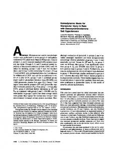
close to the stellate ganglion. This sham injection included the same volume of procaine, the sensation of pinprick in the same anatomical region and the palpation of the stellate ganglion (which we performed in identical fashion in the two groups). Both true and sham injections evoke the same kind of anticipation (that is, the study participants would have to reckon with an injection). Since the diastolic blood pressure readings before and after the injection were unchanged in these study participants, we may safely assume that the increase in DBP in the SGB treatment group is the specific result of the stellate ganglion block in our study. In our opinion, the complex anatomy of the stellate ganglion and its connections, which is concentrated in a very small area, inhibits an exclusive nerve block of just the sympathetic structures (despite the small volumes of slowly diffusing LA and the special injection technique that we used in our experiment), and hence the interpretation of the findings. The sympathetic and the parasympathetic nerves merge at the cardiac plexus (Fig. 1; Clara, 1959; Janes et al., 1986; Jänig, 2006; Kawashima, 2005; Samandari, 1994; Sinelnikov, 1981). The parasympathetic fibers mainly originate from the recurrent nerve and the cervical vagus nerve. However, the cardiac nerves (i.e., the superior, middle and
Fig. 1. Diagram showing the autonomic input to the cardiac plexus. For clarity of presentation, the spinal cord is displayed on the side rather than in the middle. The thoracic cardiac nerves travel to the cardiac plexus, independently of the cervical ganglia, and are not affected by stellate ganglion block.
K. Puente de la Vega Costa et al. / Autonomic Neuroscience: Basic and Clinical 197 (2016) 46–55
inferior cervical cardiac nerves and the superior, medial and inferior cervical cardiac branches [Fig. 2]) contain both sympathetic and parasympathetic fibers, even in their more proximal portions (Clara, 1959;
51
Kawashima, 2005). Moreover, there are large individual differences in the composition and number of cardiac fibers; the reason is that the sympathetic cardiac nerves soon after their origin can carry
52
K. Puente de la Vega Costa et al. / Autonomic Neuroscience: Basic and Clinical 197 (2016) 46–55
parasympathetic fibers from the vagus nerve and, vice versa, the parasympathetic cervical cardiac branches can carry sympathetic fibers (Fig. 2; Clara, 1959). In addition, there are larger, defined interconnections between the sympathetic and the parasympathetic branches, such as, for example, the jugular nerve connecting the superior cervical ganglia and the vagus nerve (Fig. 2). Even if we very specifically targeted the stellate ganglion, the nerve block would interrupt the fibers traveling to the superior cervical ganglia and thus the jugular nerve activity. Other connections from the sympathetic to the parasympathetic branch include the communicating branches to the vagus nerve and to the recurrent laryngeal nerve, both of which arise from the stellate ganglion (Fig. 2; Clara, 1959; Sinelnikov, 1981). Thus, the injection of a local anesthetic into sympathetic nerve tissue will at the same time automatically produce a partial block of parasympathetic components. The sympathetic regulation of the heart is not the sole responsibility of the cervical ganglia (i.e., inferior [stellate ganglion], middle cervical ganglion and superior cervical ganglion): Janes et al. described sympathetic mediastinal ganglia that are also contributing to cardiac regulation. Sympathetic fibers arising from the ganglia T2 to T5 of the paravertebral chain are directly destined to the heart and join the cardiac plexus (thoracic cardiac nerves), without making a “detour” via the cervical ganglia (Fig. 1; Clara, 1959; Janes et al., 1986; Kawashima, 2005; Samandari, 1994; Sinelnikov, 1981; Song et al., 2009). And even after a cervical ganglion block, these will remain active. Hence, the sympathetic fibers extending down to the heart will not be completely blocked by applying a local anesthetic to the cervical ganglia. In our opinion, the same considerations also apply to stellate ganglionectomy (Leriche, 1958; Xie et al., 2011; Yoshimoto et al., 2008). For all these reasons, the balance between the sympathetic and parasympathetic divisions of the autonomic nervous system is hardly disrupted by stellate ganglion injections. Otherwise, we would expect the changes in cardiovascular parameters, which occur as a result of the theoretically isolated elimination of sympathetic cardiac fibers, to be much more pronounced. 5. Limitations Sympathetic and parasympathetic activity within the cardiovascular system also goes along with changes of heart rate variability. By addressing the heart rate variability (using spectral analysis), we could have possibly made additional statements about sympathetic and parasympathetic activity after SGB. Another limitation is the fact that we performed the right and left SGB in the same sequence in all subjects (on day 1, all study participants received an injection to the right SG; on day 2, the same procedure was repeated on the left side). As a result, a potential leftover effect, or subjects' anticipation of effect, may have influenced the results. 6. Conclusions The heterogeneous results concerning the hemodynamic parameters following SGB that were observed in several studies can be attributed to: 1. Different injection techniques, 2. Different injection volumes, 3. Different diffusion properties of the various LAs administered;
4. Anatomical variability and different baseline values for the sympathetic and parasympathetic tones (which gain more significance with single-case reports or small numbers of study participants). But even the precise infiltration of the stellate ganglion (special injection technique) with a small volume of procaine (a local anesthetic that is known for its low permeability) that we used in our study cannot avoid a partial parasympathicolytic effect. Also, there are sympathetic fibers from the thoracic segments lying inferiorly to the stellate ganglion that travel to the heart directly and that are unaffected by small-volume injections of LA. For these reasons, the hemodynamic effects are negligible. This does not mean though that the therapeutic effects following from SGB are diminished since the simultaneous anesthesia of parasympathetic fibers will not impair the beneficial antiarrhythmic and pain inhibiting effects (it may even have a synergistic effect). Although some changes in hemodynamic and echocardiographic parameters following SGB meet the level of statistical significance, they do not have any clinical significance. In conclusion, the SGB is safe to the cardiovascular system, and there are hardly any contraindications to the application of small volumes (3–5 mL) of procaine 1%. From the researcher's point of view we will have to accept though that isolated sympatholysis is not possible with stellate ganglion block. Acknowledgements We are grateful to Dr. Med. Sabina Ludin for technical assistance in producing the manuscript and critical input, to Raphael Fischer for technical assistance, to Karin Beifuss for translating the text, to Hans Holzherr for producing the illustrations and to Prof. Dr. Med. Peter Eggli, anatomist and Dean of the Medical Faculty, University of Bern, for his critical review of the anatomic descriptions. References Alkadhi, K., Alzoubi, K., Aleisa, A., 2005. Plasticity of synaptic transmission in autonomic ganglia. Prog. Neurobiol. 75, 83–108. Almarestani, L., Longo, G., Ribeiro-da-Silva, A., 2008. Autonomic fiber sprouting in the skin in chronic inflammation. Mol. Pain 4, 56. http://dx.doi.org/10.1186/1744-8069-4-56. Alston, E.N., Parrish, D.C., Hasan, W., Tharp, K., Pahlmeyer, L., Habecker, B.A., 2011. Cardiac ischemia-reperfusion regulates sympathetic neuropeptide expression through gp130-dependent and independent mechanisms. Neuropeptides 45, 33–42. http:// dx.doi.org/10.1016/j.npep.2010.10.002. Baron, R., 2006. Complex regional pain syndromes. In: McMahon, S.B., Koltzenburg, M. (Eds.), Wall & Melzack's Textbook of Pain, fifth ed. Elsevier Churchill Livingstone, Edinburgh, pp. 1011–1027. Baron, R., Jänig, W., 1998. Pain syndromes with causal participation of the sympathetic nervous system. Anästhesist 4–23. Baron, R., Raja, S.N., 2002. Role of adrenergic transmitters and receptors in nerve and tissue injury related pain. In: Malmberg, A.B., Chaplan, S.R. (Eds.), Mechanisms and Mediators of Neuropathic Pain. Birkhäuser, Basel, p. 174. Burnstock, G., 2012. Cotransmission. In: Robertson, D., Biaggioni, I., Burnstock, G., Low, P.A., Paton, J.F.R. (Eds.), Primer on the Autonomic Nervous System, third ed. Academic Press, London, Waltham, San Diego, pp. 27–33. Cassuto, D., Sinclair, R., Bonderovic, M., 2006. Anti-inflammatory properties of local anesthetics and their present and potential clinical implications. Acta Anaesthesiol. Scand. 50, 265–282. Chartier, S.R., Thompson, M.L., Longo, G., Fealk, M.N., Majuta, L.A., Mantyh, P.W., 2014. Exuberant sprouting of sensory and sympathetic nerve fibers in non-healed bone fractures and the generation and maintenance of chronic skeletal pain. Pain 155, 2323–2336. http://dx.doi.org/10.1016/j.pain.2014.08.026. Chung, K., Chung, J.M., 2001. Sympathetic sprouting in the dorsal root ganglion after spinal nerve ligation: evidence of regenerative collateral sprouting. Brain Res. 895, 204–212.
Fig. 2. A greatly simplified schematic representation of the autonomic efferent and afferent innervation of the heart, adapted from various sources (Clara, 1959; Gilman, 2007; Janes et al., 1986; Jänig, 2006; Kawashima, 2005; Lanz and Wachsmith, 2004; Mathias and Bannister, 2013; Netter, 1994; Robertson et al., 2012; Rohen, 2001; Samandari, 1994; Schiebler and Korf, 2007; Sinelnikov, 1981; Thomas and Gerdisch, 1990; Trepel, 2012; Waldeyer and Mayet, 1993; Williams and Bannister, 1995; Wilson Pauwels et al., 1997). Note the close proximity of sympathetic and parasympathetic fibers. Soon after their origin, the sympathetic cardiac nerve fibers can carry parasympathetic fibers, and vice versa, the parasympathetic cervical cardiac branches can carry sympathetic fibers (Clara, 1959; Kawashima, 2005). Other connections between the sympathetic and the parasympathetic divisions include the rami communicantes (for example, the jugular nerve, and branches arising from the stellate ganglion and connecting it with the vagus nerve and the recurrent laryngeal nerve; Clara, 1959; Sinelnikov, 1981). Note: The number and course of the cardiac nerves and rami can vary across individuals, and this is reflected by the partly inconsistent terminology in the literature. For example, some of the references do not mention the ramus cardiacus medius (the middle cervical cardiac ramus). The diagram is intended to illustrate how parasympathetic fibers can be (inadvertently) affected by SGB along with the SGB's sympathetic targets.
K. Puente de la Vega Costa et al. / Autonomic Neuroscience: Basic and Clinical 197 (2016) 46–55 Chung, J.M., Chung, K., 2002. Importance of hyperexcitability of DRG neurons in neuropathic pain. Pain Pract. 2, 87–97. Cinca, J., Evangelista, A., Montoyo, J., Barutell, C., Figueras, J., Valle, V., Rius, J., Soler-Soler, J., 1985. Electrophysiologic effects of unilateral right and left stellate ganglion block on the human heart. Am. Heart J. 109, 46–54. Clara, M., 1959. Das Nervensystem des Menschen. 3. Aufl. Barth, Leipzig. Devor, M., Jänig, W., 1981. Activation of myelinated afferents ending in a neuroma by stimulation of the sympathetic supply in the rat. Neurosci. Lett. 24, 43–47. Devor, M., Jänig, W., Michaelis, M., 1994. Modulation of activity in dorsal root ganglion neurons by sympathetic activation in nerve-injured rats. J. Neurophysiol. 71, 38–47. Docimo Jr., S., Piccolo, C., Van Arsdale, D., Elkowitz, D.E., 2008. Pathology-dependent histological changes of the left stellate ganglia: a cadaveric study. Clin. Med. Pathol. 1, 105–113. Dosch, P., 1986. Lehrbuch der Neuraltherapie nach Huneke. 12. Aufl. Haug, Stuttgart. Drew, R.C., Sinoway, L.I., 2012. Autonomic control of the heart. In: Robertson, D., Biaggioni, I., Burnstock, G., Low, P.A., Paton, J.F.R. (Eds.), Primer on the autonomic nervous system, 3rd ed. Academic Press, London, pp. 177–180. Egawa, H., Okuda, Y., Kitajima, T., Minami, J., 2001. Assessment of QT interval and QT dispersion following stellate ganglion block using computerized measurements. Reg. Anesth. Pain Med. 26, 539–544. Eggli, P., Fischer, L., 2011. Vegetatives Nervensystem. In: Fischer, L., Peuker, E. (Eds.), Lehrbuch Integrative Schmerztherapie. Haug, Stuttgart, pp. 17–26. Elenkov, I.J., Wilder, R.L., Chrousos, G.P., Vizi, E.S., 2000. The sympathetic nerve – an integrative interface between two supersystems: the brain and the immune system. Pharmacol. Rev. 52, 595–638. Esler, M., 2010. The 2009 Carl Ludwig Lecture: pathophysiology of the human sympathetic nervous system in cardiovascular diseases: the transition from mechanisms to medical management. J. Appl. Physiol. 108, 227–237. http://dx.doi.org/10.1152/ japplphysiol.00832.2009. Feigl, G.C., Rosmarin, W., Stelzl, A., Weninger, B., Likar, R., 2007. Comparison of different injectate volumes for stellate ganglion block: an anatomic and radiologic study. Reg. Anesth. Pain Med. 32, 203–208. Fernandez, S., Srikakarlapudi, S., Izzo, J.L., 2012. Blood pressure variability. In: Robertson, D., Biaggioni, I., Burnstock, G., Low, P.A., Paton, J.F.R. (Eds.), Primer on the Autonomic Nervous System, third ed. Academic Press, London, Waltham, San Diego, pp. 355–357. Fischer, L., 1998. Neuraltherapie nach Huneke. Grundlagen, Technik, praktische Anwendung. 1. Aufl. Hippokrates, Stuttgart. Fischer, L., 2003. Pathophysiology of pain and neural therapy. Praxis 92, 2051–2059. Fischer, L., 2013. Neuraltherapie. In: Baron, R., Koppert, W., Strumpf, M., WillweberStrumpf, A. (Eds.), Praktische Schmerztherapie, 3. Aufl. Springer, Heidelberg, pp. 191–199. Fischer, L., 2014. Neuraltherapie. Neurophysiologie, Injektionstechnik und Therapievorschläge. 4. Aufl. Haug, Stuttgart. Fujii, K., Yamaguchi, S., Egawa, H., Hamaguchi, S., Kitajima, T., Minami, J., 2004. Effects of head-up tilt after stellate ganglion block on QT interval and QT dispersion. Reg. Anesth. Pain Med. 29, 317–322. Fujiki, A., Masuda, A., Inoue, H., 1999. Effects of unilateral stellate ganglion block on the spectral characteristics of heart rate variability. Jpn. Circ. J. 63, 854–858. Gadhinglajkar, S., Sreedhar, R., Unnikrishnan, M., Namboodiri, N., 2013. Electrical storm: role of stellate ganglion blockade and anesthetic implications of left cardiac sympathetic denervation. Indian J. Anaesth. 57, 397–400. http://dx.doi.org/10.4103/00195049.118568. Garcia-Larrea, L., Magnin, M., 2008. Pathophysiology of neuropathic pain: review of experimental models and proposed mechanisms. Presse Med. 37, 315–340. http://dx. doi.org/10.1016/j.lpm.2007.07.025. Gardner, M.J., Kimber, S., Johnstone, D.E., Shukla, R.C., Horacek, B.M., Forbes, C., Armour, J.A., 1993. The effects of unilateral stellate ganglion blockade on human cardiac function during rest and exercise. J. Cardiovasc. Electrophysiol. 4, 2–8. Garneau, S.Y., Deschamps, A., Couture, P., Levesque, S., Babin, D., Lambert, J., Tardif, J.C., Perrault, L.P., Denault, A.Y., 2011. Preliminary experience in the use of preoperative echo-guided left stellate ganglion block in patients undergoing cardiac surgery. J. Cardiothorac. Vasc. Anesth. 25, 78–84. http://dx.doi.org/10.1053/j.jvca.2010.03.007. Gilman, S., 2007. Neurobiology of disease. Boston, Elsevier Academic Press, Amsterdam. Goadsby, P.J., Edvinsson, L., 1993. The trigeminovascular system and migraine: studies characterizing cerebrovascular and neuropeptide changes seen in humans and cats. Ann. Neurol. 33, 48–56. Goh, J.S., Min, B.W., Kim, H.D., 1990. Blood pressure, pulse rate and temperature changes of the ipsilateral upper extremity after unilateral stellate ganglion block. Korean J. Pain 3, 27–33. Grassi, G., 2010. Sympathetic neural activity in hypertension and related diseases. Am. J. Hypertens. 23, 1052–1060. http://dx.doi.org/10.1038/ajh.2010.154. Grassi, G., Seravalle, G., Low, P.A., Paton, J.F.R., 2012. Sympatho-vagal imbalance in hypertension. In: Robertson, D., Biaggioni, I., Burnstock, G. (Eds.), Primer on the Autonomic Nervous System, third ed. Academic Press, London, Waltham, San Diego, pp. 345–348. Herbert, M.K., Holzer, P., 2002. Die neurogene Entzündung. I. Grundlegende Mechanismen, Physiologie und Pharmakologie. II. Pathophysiologie und klinische Implikationen. Anasthesiol. Intensivmed. Notfallmed. Schmerzther. 2002 (37), 314–325 (386-394). Hogan, Q.H., Erickson, S.J., Haddox, J.D., Abram, S.E., 1992. The spread of solutions during stellate ganglion block. Reg. Anesth. 17, 78–83. Hollmann, M.W., Durieux, M.E., 2000. Local anesthetics and the inflammatory response: a new therapeutic indication? Anesthesiology 93, 858–875. Honma, M., Murakami, G., Sato, T.J., Namiki, A., 2000. Spread of injectate during C6 stellate ganglion block and fascial arrangement in the prevertebral region: an experimental study using donated cadavers. Reg. Anesth. Pain Med. 25, 573–583.
53
Hori, T., Katafuchi, T., Take, S., Shimizu, N., Niijima, A., 1995. The autonomic nervous system as a communication channel between the brain and the immune system. Neuroimmunomodulation 2, 203–215. Huang, H.D., Tamarisa, R., Mathur, N., Alam, M., Makkar, A., Birnbaum, Y., AfsharKharaghan, H., 2013. Stellate ganglion block: a therapeutic alternative for patients with medically refractory inappropriate sinus tachycardia? J. Electrocardiol. 46, 693–696. http://dx.doi.org/10.1016/j.jelectrocard.2012.12.010. Janes, R.D., Brandys, J.C., Hopkins, D.A., Johnstone, D.E., Murphy, D.A., Armour, J.A., 1986. Anatomy of human extrinsic cardiac nerves and ganglia. Am. J. Cardiol. 57, 299–309. Jänig, W., 2006. The Integrative Action of the Autonomic Nervous System. Cambridge University Press, Cambridge. Jänig, W., 2011. Rolle von motorischen Rückkopplungsmechanismen in der Erzeugung von Schmerzen. In: Fischer, L., Peuker, E. (Eds.), Lehrbuch Integrative Schmerztherapie. Haug, Stuttgart, pp. 81–89. Jänig, W., Baron, R., 2011. Regionale und generalisierte Schmerzsyndrome. In: Fischer, L., Peuker, E. (Eds.), Lehrbuch Integrative Schmerztherapie. Haug, Stuttgart, pp. 474–491. Jänig, W., Koltzenburg, M., 1991. Plasticity of sympathetic reflex organization following cross-union of inappropriate nerves in the adult cat. J. Physiol. 436, 309–323. Jänig, W., Koltzenburg, M., 1992. Possible ways of sympathetic afferent interaction. In: Jänig, W., Schmidt, R.F. (Eds.), Reflex Sympathetic Dystrophy. Pathophysiological Mechanisms and Clinical Implications. Weinheim, New York, VCH Verlagsgemeinschaft, pp. 213–243. Jänig, W., McLachlan, E.M., 1994. The role of modifications in noradrenergic peripheral pathways after nerve lesions in the generation of pain. In: Fields, H.L., Liebeskind, J.C. (Eds.), Pharmacological Approaches to the Treatment of Pain: New Concepts and Critical IssuesProgress in Pain Research and Management vol. 1. IASP Press, Seattle. Jänig, W., Levine, J.D., Michaelis, M., 1996. Interactions of sympathetic and primary afferent neurons following nerve injury and tissue trauma. Prog. Brain Res. 113, 161–184. Kansha, M., Nagata, T., Irita, K., Takahashi, S., 1999. Dibucaine and tetracaine inhibit the activation of mitogen-activated protein kinase mediated by L-type calcium channels in PC12 cells. Anesthesiology 91, 1798–1806. Kapral, S., Krafft, P., Gosch, M., Fleischmann, D., Weinstabl, C., 1995. Ultrasound imaging for stellate ganglion block: direct visualization of puncture site and local anesthetic spread. A pilot study. Reg. Anesth. 20, 323–328. Kashima, T., Tanaka, H., Minagoe, S., Toda, H., 1981. Electrocardiographic changes induced by the stellate ganglion block in normal subjects. J. Electrocardiol. 14, 169–174. Kawashima, T., 2005. The autonomic nervous system of the human heart with special reference to its origin, course, and peripheral distribution. Anat. Embryol. 209, 425–438. Kim, J.J., Chung, R.K., Lee, H.S., Han, J.I., 2010. The changes of heart rate variability after unilateral stellate ganglion block. Korean J. Anesthesiol. 58, 56–60. http://dx.doi. org/10.4097/kjae.2010.58.1.56. Kirillova, I., Teliban, A., Gorodetskaya, N., Grossmann, L., Bartsch, F., Rausch, V.H., Struck, M., Tode, J., Baron, R., Jänig, W., 2011. Effect of local and intravenous lidocaine on ongoing activity in injured afferent nerve fibers. Pain 152, 1562–1571. http://dx.doi.org/ 10.1016/j.pain.2011.02.046. Kohjitani, A., Miyawaki, T., Kasuya, K., Shimada, M., 2002. Sympathetic activity-mediated neuropathic facial pain following simple tooth extraction: a case report. Cranio 20, 135–138. Koppert, W., Zeck, S., Sittl, R., Likar, R., Knoll, R., Schmelz, M., 1998. Low-dose lidocaine suppresses experimentally induced hyperalgesia in humans. Anesthesiology 89, 1345–1353. Kweon, T.K., Han, C.M., Kim, S.Y., Lee, Y.-W., 2006. The changes of blood pressure, heart rate and heart rate variability after stellate ganglion block. Korean J. Pain 19, 202–206. Lanz, T., Wachsmith, W., 2004. Praktische Anatomie. Springer, Berlin, Göttingen, Heidelberg. Leriche, R., 1958. Die Chirurgie des Schmerzes. Barth, Leipzig. Leriche, R., Fontaine, R., 1934. L'anesthésie du ganglion étoile: sa technique, ses indications, ses résultats. Presse méd 42, 849–850. Levick, S.P., Murray, D.B., Janicki, J.S., Brower, G.L., 2010. Sympathetic nervous system modulation of inflammation and remodeling in the hypertensive heart. Hypertension 55, 270–276. http://dx.doi.org/10.1161/HYPERTENSIONAHA.109.142042. Liu, M.H., Tian, J., Su, Y.P., Wang, T., Xiang, Q., Wen, L., 2013. Cervical sympathetic block regulates early systemic inflammatory response in severe trauma patients. Med. Sci. Monit. 19, 194–201. http://dx.doi.org/10.12659/MSM.883833. Lobato, E.B., Kern, K.B., Paige, G.B., Brown, M., Sulek, C.A., 2000. Differential effects of right versus left stellate ganglion block on left ventricular function in humans: an echocardiographic analysis. J. Clin. Anesth. 12, 315–318. Lombard, J.H., Cowley Jr., A.W., 2012. Neural control of blood vessels. In: Robertson, D., Biaggioni, I., Burnstock, G., Low, P.A., Paton, J.F.R. (Eds.), Primer on the Autonomic Nervous System, third ed. Academic Press, London, Waltham, San Diego, pp. 187–191. Lopez-Sendon, J., Swedberg, K., McMurray, J., Tamargo, J., Maggioni, A.P., Dargie, H., Tendera, M., Waagstein, F., Kjekshus, J., Lechat, P., Torp-Pedersen, C., 2004. Expert consensus document on beta-adrenergic receptor blockers. Eur. Heart J. 25, 1341–1362. Low, P.A., 1993. Composite autonomic scoring scale for laboratory quantification of generalized autonomic failure. Mayo Clin. Proc. 68, 748–752. Madden, K.S., Felten, D.L., 1995. Experimental basis for neural-immune interactions. Physiol. Rev. 75, 77–106. Martinez-Lavin, M., Solano, C., 2009. Dorsal root ganglia, sodium channels, and fibromyalgia sympathetic pain. Med. Hypotheses 72, 64–66. http://dx.doi.org/10.1016/j. mehy.2008.07.055. Marvar, P.J., Harrison, D.G., 2012. Inflammation, immunity and the autonomic nervous system. In: Robertson, D., Biaggioni, I., Burnstock, G., Low, P.A., Paton, J.F.R. (Eds.), Primer on the Autonomic Nervous System, third ed. Academic Press, London, Waltham, San Diego, pp. 325–329.
54
K. Puente de la Vega Costa et al. / Autonomic Neuroscience: Basic and Clinical 197 (2016) 46–55
Masuda, Y., Okamoto, K., 2005. Management and treatment of headache in the pain clinic. Nihon Rinsho 63, 1802–1807. Mata Francisco, N.C., Gómez, P.E., Ruiz, L.N., Alvarez López, J.C., Jorge-Monjas, P., 2013. Treatment of a patient with refractory cardiac arrhythmias using stellate ganglion block. Access by the classical and ultrasound-guided approach. Rev. Esp. Anestesiol. Reanim. 13, 00228–00234. http://dx.doi.org/10.1016/j.redar.2013.08.007 (pii: S0034-9356). Mathias, C.J., Bannister, R. (Eds.), 2013. Autonomic Failure: A Textbook of Clinical Disorders of the Autonomic Nervous System, fifth ed. Oxford University Press, Oxford, New York. McLachlan, E.M., Hu, P., 2014. Inflammation in dorsal root ganglia after peripheral nerve injury: effects of the sympathetic innervation. Auton. Neurosci. 182, 108–117. http:// dx.doi.org/10.1016/j.autneu.2013.12.009. McLachlan, E.M., Jänig, W., Devor, M., Michaelis, M., 1993. Peripheral nerve injury triggers noradrenergic sprouting within dorsal root ganglia. Nature 363, 543–546. McQuay, H.J., Moore, R.A., 1999. Local anesthetics and epidurals. In: Wall, P.D., Melzack, R. (Eds.), Textbook of Pain, fourth ed. Churchill Livingstone, New York. Miao, F.J., Green, P.G., Coderre, T.J., Jänig, W., Levine, J.D., 1996a. Sympathetic-dependence in bradykinin-induced synovial plasma extravasation is dose-related. Neurosci. Lett. 205, 165–168. Miao, F.J., Jänig, W., Levine, J., 1996b. Role of sympathetic postganglionic neurons in synovial plasma extravasation induced by bradykinin. J. Neurophysiol. 75, 715–724. Milne, J.R., Ward, D.E., Spurrell, R.A., Camm, A.J., 1982. The long QT syndrome; effects of drugs and left stellate ganglion block. Am. Heart J. 104, 194–198. Nademanee, K., Taylor, R., Bailey, W.E., Rieders, D.E., Kosar, E.M., 2000. Treating electrical storm: sympathetic blockade versus advanced cardiac life support-guided therapy. Circulation 2, 742–747. Netter, F.H., 1994. The CIBA collection of medical illustrations. Vol. I: Nervous System. Part I: Anatomy and Physiology. W.B. Saunders Company. Noble, M.D., Romac, J., Wang, Y., Hsu, J., Humphrey, J.E., Liddle, R.A., 2006. Local disruption of the celiac ganglion inhibits substance P release and ameliorates caerulein-induced pancreatitis in rats. Am. J. Physiol. Gastrointest. Liver Physiol. 291, G128–G134. Noma, N., Kamo, H., Nakaya, Y., Dezawa, K., Young, A., Khan, J., Imamura, Y., 2013. Stellate ganglion block as an early intervention in sympathetically maintained headache and orofacial pain caused by temporal arteritis. Pain Med. 14, 392–397. http://dx.doi.org/ 10.1111/pme.12040. Ogawa, M., Zhou, S., Tan, A.Y., Song, J., Gholmieh, G., Fishbein, M.C., Luo, H., Siegel, R.J., Karagueuzian, H.S., Chen, L.S., Lin, S.F., Chen, P.S., 2007. Left stellate ganglion and vagal nerve activity and cardiac arrhythmias in ambulatory dogs with pacinginduced congestive heart failure. J. Am. Coll. Cardiol. 50, 335–343. Park, H.M., Kim, T.W., Choi, H.G., Yoon, K.B., Yoon, D.M., 2010. The change in regional cerebral oxygen saturation after stellate ganglion block. Korean J. Pain 23, 142–146. http://dx.doi.org/10.3344/kjp.2010.23.2.142. Patel, R.A., Priore, D.L., Szeto, W.Y., Slevin, K.A., 2011. Left stellate ganglion blockade for the management of drug-resistant electrical storm. Pain Med. 12, 1196–1198. http://dx.doi.org/10.1111/j.1526-4637.2011.01167.x. Paton, J.F.R., Spyer, K.M., 2013. Central nervous control of the cardiovascular system. In: Mathias, C.J., Bannister, R. (Eds.), Autonomic Failure: A Textbook of Clinical Disorders of the Autonomic Nervous System, fifth ed. Oxford University Press, Oxford, New York. Peterson, R.C., Patel, L., Cubert, K., Gulati, A., 2009. Serial stellate ganglion blocks for intractable postherpetic itching in a pediatric patient: a case report. Pain Physician 12, 629–632. Pfister, M., Fischer, L., 2009. The treatment of the complex regional pain syndrome (CRPS 1 and CRPS 2) of the upper limb with repeated local anaesthesia to the stellate ganglion. Praxis 98, 247–257. Pietruck, C., Grond, S., Xie, G.X., Palmer, P.P., 2003. Local anesthetics differentially inhibit sympathetic neuron-mediated and C fiber-mediated synovial neurogenic plasma extravasation. Anesth. Analg. 96, 1397–1402. Pongratz, G., Straub, R.H., 2010. The B cells arthritis and the sympathetic nervous system. Brain Behav. Immun. 24, 186–192. http://dx.doi.org/10.1136/ard.2011.153056. Pongratz, G., Melzer, M., Straub, R.H., 2012. The sympathetic nervous system stimulates anti-inflammatory B cell in collagen-type II-induced arthritis. Ann. Rheum. Dis. 71, 432–439. http://dx.doi.org/10.1136/ard.2011.153056. Price, J., Mudge, A.W., 1983. A subpopulation of rat dorsal root ganglion neurones is catecholaminergic. Nature 301, 241–243. Price, D.D., Long, S., Wilsey, B., Rafii, A., 1998. Analysis of peak magnitude and duration of analgesia produced by local anesthetics injected into sympathetic ganglia of complex regional pain syndrome patients. Clin. J. Pain 14, 216–226. Ramer, M.S., Thompson, S.W., McMahon, S.B., 1999. Causes and consequences of sympathetic basket formation in dorsal root ganglia. Pain (Suppl. 6), S111–S120. Ricker, G., 1924. Pathologie als Naturwissenschaft – Relationspathologie. Springer, Berlin. Roberts, W.J., Foglesong, M.E., 1988. Spinal recordings suggest that wide-dynamic-range neurons mediate sympathetically maintained pain. Pain 34, 289–304. Robertson, D., Biaggioni, I., Burnstock, G., Low, P.A., Paton, J.F.R. (Eds.), 2012. Primer on the Autonomic Nervous System, third ed. Academic Press, London, Waltham, San Diego. Rogers, M.C., Abildskov, J.A., Preston, J.B., 1973. Cardiac effects of stimulation and block of the stellate ganglion. Anesthesiology 39, 525–533. Rogers, M.C., Battit, G., McPeek, B., Todd, D., 1978. Lateralization of sympathetic control of the human sinus node: ECG changes of stellate ganglion block. Anesthesiology 48, 139–141. Rohen, J.W., 2001. Funktionelle Neuroanatomie. 6. Aufl. Schattauer, Stuttgart. Rosenquist, R.W., Vrooman, B.M., 2013. Chronic pain management. In: Butterworth, J.F., Mackey, D.C., Wasnick, J.D. (Eds.), Morgan and Mikhail's Clinical Anesthesiology, fifth ed. McGraw-Hill Medical, New York (Chapter 47).
Salvaggio, I., Adducci, E., Dell'Aquila, L., Rinaldi, S., Marini, M., Zappia, L., Mascaro, A., 2008. Facial pain: a possible therapy with stellate ganglion block. Pain Med. 9, 958–962. http://dx.doi.org/10.1111/j.1526-4637.2008.00515.x. Samandari, F., 1994. Funktionelle Anatomie der Hirnnerven und des vegetativen Nervensystems für Mediziner und Zahnmediziner. De Gruyter, Berlin, New York. Saxena, A.K., Saxena, N., Aggarwal, B., Sethi, A.K., 2004. An unusual complication of sinus arrest following right-sided stellate ganglion block: a case report. Pain Pract. 4, 245–248. Schattschneider, J., Wasner, G., Binder, A., Siebrecht, D., Baron, R., 2003. Das Symptom sympathisch unterhaltenen Schmerzes. Schmerz 17, 317–324. Schiebler, T.H., Korf, H.-W., 2007. Anatomie. 10. Aufl. Steinkopff, Heidelberg. Schlack, W., Dinter, W., 2000. Haemodynamic effects of a left stellate ganglion block in ASA I patients. An echocardiographic study. Eur. J. Anaesthesiol. 17, 79–84. Schwartz, P.J., 1984. The rationale and the role of left stellectomy for the prevention of malignant arrhythmias. Ann. N. Y. Acad. Sci. 427, 199–221. Schwartz, P.J., 1998. The autonomic nervous system and sudden death. Eur. Heart 19 (Suppl. F), F72–F80. Schwartz, P.J., De Ferrari, G.M., 2011. Sympathetic-parasympathetic interaction in health and disease: abnormalities and relevance in heart failure. Heart Fail. Rev. 16, 101–107. http://dx.doi.org/10.1007/s10741-010-9179-1. Schwartz, P.J., Zaza, A., 1986. The rational basis and the clinical value of selective cardiac sympathetic denervation in the prevention of malignant arrhythmias. Eur. Heart 7 (Suppl. A), A107–A118. Shanthanna, H., 2013. Utility of stellate ganglion block in atypical facial pain: a case report and consideration of its possible mechanisms. Case Rep. Med. 2013, 293826. http:// dx.doi.org/10.1155/2013/293826. Shen, M.J., Shinohara, T., Park, H.W., Frick, K., Ice, D.S., Choi, E.K., et al., 2011. Continuous low-level vagus nerve stimulation reduces stellate ganglion nerve activity and paroxysmal atrial tachyarrhythmias in ambulatory canines. Circulation 123, 2204–2212. http://dx.doi.org/10.1161/CIRCULATIONAHA.111.018028. Sinelnikov, R.D., 1981. Atlas of Human Anatomy vol. 3 (Moscow). Solti, F., Balogh, A., Czakó, E., 1978. Effect of the stellate ganglion blockade upon the electrical systole (Q-T interval) of the heart. Cor Vasa 20, 264–270. Song, J.G., Hwang, G.S., Lee, E.H., Leem, J.G., Lee, C., Park, P.H., Shin, J.W., 2009. Effects of bilateral stellate ganglion block on autonomic cardiovascular regulation. Circ. J. 73, 1909–1913. Spiess, G., 1906. Die Bedeutung der Anästhesie in der Entzündungstheorie. MMW Munch Med Wochenschr 53, 345–351. Stanton-Hicks, M., Jänig, W., Hassenbusch, S., Haddox, J.D., Boas, R., Wilson, P., 1995. Reflex sympathetic dystrophy: changing concepts and taxonomy. Pain 63, 127–133. Straub, R.H., Wiest, R., Strauch, U.G., Harle, P., Scholmerich, J., 2006. The role of the sympathetic nervous system in intestinal inflammation. Gut 55, 1640–1649. Strittmatter, M., Grauer, M.T., Fischer, C., Hamann, G., Hoffmann, K.H., Blaes, F., Schimrigk, K., 1996. Autonomic nervous system and neuroendocrine changes in patients with idiopathic trigeminal neuralgia. Cephalalgia 16, 476–480. Sunagawa, K., Kawada, T., Nakahara, T., 1998. Dynamic nonlinear vago-sympathetic interaction in regulating heart rate. Heart Vessel. 13, 157–174. Tan, Z., Dohi, S., Ohguchi, K., Nakashima, S., Nozawa, Y., 1999. Local anaesthetics inhibit muscarinic receptor-mediated activation of extracellular sign regulated kinases in rat feochromocytoma PC12 cells. Anaesthesiology 9, 1014–1024. Taneyama, C., Goto, H., 2009. Fractal cardiovascular dynamics and baroreflex sensitivity after stellate ganglion block. Anesth. Analg. 109, 1335–1340. http://dx.doi.org/10. 1213/ane.0b013e3181b018d8. Takatori, M., Kuroda, Y., Hirose, M., 2006. Local anesthetics suppress nerve growth factormediated neurite outgrowth by inhibition of tyrosine kinase activity of TrkA. Anesth. Analg. 102, 462–467. Thomas Jr., J.X., Gerdisch, M.W., 1990. Topical organization of the cardiac sympathetic nervous system. Basic Res. Cardiol. 85 (Suppl. 1), 3–8. Tracey, K.J., 2002. The inflammatory reflex. Nature 420, 853–859. Trepel, M., 2012. Neuroanatomie. 5. Aufl. Urban & Fischer/Elsevier, München. van de Beek, W.J., Remarque, E.J., Westendorp, R.G., van Hilten, J.J., 2001. Innate cytokine profile in patients with Complex Regional Pain Syndrome is normal. Pain 91, 259–261. Vaseghi, M., Zhou, W., Shi, J., Ajijola, O.A., Hadaya, J., Shivkumar, K., Mahajan, A., 2012. Sympathetic innervation of the anterior left ventricular wall by the right and left stellate ganglia. Heart Rhythm. 9, 1303–1309. http://dx.doi.org/10.1016/j.hrthm.2012.03.052. Waldeyer, A., Mayet, A., 1993. Anatomie des Menschen. De Gruyter, Berlin, New York. Wang, Q.X., Wang, X.Y., Fu, N.A., Liu, J.Y., Yao, S.L., 2005. Stellate ganglion block inhibits formalin-induced nociceptive responses: mechanism of action. Eur. J. Anaesthesiol. 22, 913–918. Watkins, L.R., Hutchinson, M.R., Milligan, E.D., Maier, S.F., 2007. “Listening” and “talking” to neurons: implications of immune activation for pain control and increasing the efficacy of opioids. Brain Res. Rev. 56, 148–169. Williams, P.L., Bannister, L.H. (Eds.), 1995. Gray's Anatomy: The Anatomical Basis of Medicine and Surgery, 38th ed. Churchill Livingstone, New York. Wilson Pauwels, L., Stewart, P.A., Akesson, E.J., 1997. Autonomic Nerves. B. C. Decker, Inc., Hamilton, London. Wong, C.W., Wang, C.H., 1999. Left stellate stimulation increases left ventricular ejection fraction in patients with essential palmar hyperhidrosis. J. Auton. Nerv. Syst. 78, 64–67. Xie, W., Strong, J.A., Li, H., Zhang, J.M., 2007. Sympathetic sprouting near sensory neurons after nerve injury occurs preferentially on spontaneously active cells and is reduced by early nerve block. J. Neurophysiol. 97, 492–502. Xie, X., Visweswaran, R., Guzman, P.A., Smith, R.M., Osborn, J.W., Tolkacheva, E.G., 2011. The effect of cardiac sympathetic denervation through bilateral stellate ganglionectomy on electrical properties of the heart. Am. J. Physiol. Heart Circ. Physiol. 301, H192–H199. http://dx.doi.org/10.1152/ajpheart.01149.2010.
K. Puente de la Vega Costa et al. / Autonomic Neuroscience: Basic and Clinical 197 (2016) 46–55 Yanowitz, F., Preston, J.B., Abildskov, J.A., 1966. Functional distribution of right and left stellate innervation to the ventricles. Production of neurogenic electrocardiographic changes by unilateral alteration of sympathetic tone. Circ. Res. 18, 416–428. Yen, L.D., Bennett, G.J., Ribeiro-da-Silva, A., 2006. Sympathetic sprouting and changes in nociceptive sensory innervation in the glabrous skin of the rat hind paw following partial peripheral nerve injury. J. Comp. Neurol. 495, 679–690. Yokoyama, M., Nakatsuka, H., Itano, Y., Hirakawa, M., 2000. Stellate ganglion block modifies the distribution of lymphocyte subsets and natural-killer cell activity. Anesthesiology 92, 109–115. Yoo, S.D., Jung, S.S., Kim, H.S., Yun, D.H., Kim, D.H., Chon, J., Hong, D.W., 2012. Efficacy of ultrasonography guided stellate ganglion blockade in the stroke patients with complex regional pain syndrome. Ann. Rehabil. Med. 36, 633–639. http://dx.doi.org/10. 5535/arm.2012.36.5.633. Yoshimoto, M., Wehrwein, E.A., Novotny, M., Swain, G.M., Kreulen, D.L., Osborn, J.W., 2008. Effect of stellate ganglionectomy on basal cardiovascular function and
55
responses to beta1-adrenoceptor blockade in the rat. Am. J. Physiol. Heart Circ. Physiol. 295, H2447–H2454. http://dx.doi.org/10.1152/ajpheart.00958.2008. Zhang, J.M., Li, H., Munir, M.A., 2004. Decreasing sympathetic sprouting in pathologic sensory ganglia: a new mechanism for treating neuropathic pain using lidocaine. Pain 109, 143–149. Zhang, Y., Popovic, Z.B., Bibevski, S., Fakhry, I., Sica, D.A., Van Wagoner, D.R., Mazgalev, T.N., 2009. Chronic vagus nerve stimulation improves autonomic control and attenuates systemic inflammation and heart failure progression in a canine high-rate pacing model. Circ. Heart Fail. 2, 692–699. http://dx.doi.org/10.1161/CIRCHEARTFAILURE. 109.873968. Zieglgänsberger W. (2010) Neuronale Plastizität, Schmerzgedächtnis und chronischer Schmerz. In: Weinschenk S. Hrsg. Handbuch Neuraltherapie. München: Elsevier Urban & Fischer.