Drug treatment was well tolerated, and selection occurred in myeloid, lymphoid ...... Seattle Cancer Care Alliance for analysis of serum samples for the busulfan ...
Research article
Efficient and stable MGMT-mediated selection of long-term repopulating stem cells in nonhuman primates Brian C. Beard,1 Grant D. Trobridge,1,2 Christina Ironside,1 Jeannine S. McCune,1,3 Jennifer E. Adair,1 and Hans-Peter Kiem1,2 2Department
1Clinical Research Division, Fred Hutchinson Cancer Research Center, Seattle, Washington, USA. of Medicine, Division of Hematology, and 3Department of Pharmacy, University of Washington, Seattle Washington, USA.
HSC transplantation using genetically modified autologous cells is a promising therapeutic strategy for various genetic diseases, cancer, and HIV. However, for many of these conditions, the current efficiency of gene transfer to HSCs is not sufficient for clinical use. The ability to increase the percentage of gene-modified cells following transplantation is critical to overcoming this obstacle. In vivo selection with mutant methylguanine methyltransferase (MGMTP140K) has been proposed to overcome low gene transfer efficiency to HSCs. Previous studies have shown efficient in vivo selection in mice and dogs but only transient selection in primates. Here, we report efficient and stable MGMTP140K-mediated multilineage selection in both macaque and baboon nonhuman primate models. Treatment consisting of both O6-benzylguanine (O6BG) and N,N′-bis(2-chloroethyl)-N-nitroso-urea (BCNU) stably increased the percentage of transgene-expressing cells from a range of initial levels of engrafted genetically modified cells, with the longest follow-up after drug treatment occurring over 2.2 years. Drug treatment was well tolerated, and selection occurred in myeloid, lymphoid, and erythroid cells as well as platelets. Retrovirus integration site analysis before and after drug treatments confirmed the presence of multiple clones. These nonhuman primate studies closely model a clinical setting and should have broad applications for HSC gene therapy targeting human diseases of malignant, genetic, and infectious nature, including HIV. Introduction HSC transplantation using allogeneic cells has been a successful treatment modality for a variety of genetic diseases, because the graft stably produces phenotypically normal cells for the lifetime of the individual (1). However, many patients do not have suitable donors for allogeneic transplantation. In addition, allogeneic transplantation is often associated with significant morbidity and mortality from graft-versus-host disease and infectious complications due to immunosuppressive treatment (2). Thus, the infusion of genetically corrected autologous stem cells would circumvent these limitations of allogeneic transplantation (3). Furthermore, new treatment modalities using autologous HSCs harboring a potent anti-HIV transgene could be applied alone or in parallel with highly active antiretroviral treatment for HIV patients with advanced disease to limit disease progression. For all of these genemodified autologous HSC strategies to be successful, high levels of genetically modified cells are required long term to provide a prolonged clinical benefit. For treatment of hematopoietic diseases that affect a variety of lineages, such as erythrocytes (hemoglobinopathies and thalassemia) and platelets (PLTs) (Wiskott-Aldrich syndrome), genetic modification at the HSC level is desirable to provide lifelong production of “corrected” hematopoietic lineages. A similar strategy of genetic modification of HSCs is favored to provide lifelong “protected” macrophages and lymphocytes in HIV-infected patients. Successful HSC gene therapy has been unequivocally demonstrated for patients with X-linked SCID (SCID-X1) (4), Conflict of interest: The authors have declared that no conflict of interest exists. Citation for this article: J Clin Invest. 2010;120(7):2345–2354. doi:10.1172/JCI40767.
adenosine deaminase deficient SCID (5), X-linked chronic granulomatous disease (X-CGD) (6), and adrenoleukodystrophy (ALD) (7). Numerous advances in transduction protocols, including transduction on fibronectin fragment CH-296 (8), the use of multiple cytokines (9, 10), and the use of different vector pseudotypes (11–13), have contributed to gene transfer levels of 20%–30% in clinically relevant nonhuman primates (14). Even with the gene marking levels (defined as the percentage or fraction of genetically modified cells) achieved clinically ranging from approximately 9%–15% in peripheral blood with reduced-intensity busulfan (6) and high-intensity cyclophosphamide and busulfan (7) and levels in the nonhuman primate ranging from 20%–30% in peripheral blood with total body irradiation (TBI) (14), for numerous clinical applications gene marking levels will still likely be below therapeutic thresholds (for review see ref. 15). Without an intrinsic growth advantage provided by gene-modified cells, such as that observed for SCID-X1, efficient and stable posttransplant in vivo selection will likely be required to increase the percentage of these cells into a therapeutic range for diseases such as thalassemia and other hemoglobinopathies. This approach has several therapeutic applications, such as using anti-HIV transgenes to block infection or limit propagation of HIV or using drug resistance transgenes to limit myelosuppression from chemotherapy during cancer therapy to allow dose intensification. The system that currently meets the criteria for all of these applications is mutant methylguanine methyltransferase (MGMTP140K) (16–20), because of stable multilineage in vivo selection that has been previously described in mice (21) and dogs (22, 23). MGMTP140K-mediated selection does not require chronic administration of pharmacologic agents but instead can be achieved with limited treatments
The Journal of Clinical Investigation http://www.jci.org Volume 120 Number 7 July 2010
2345
research article using the wild-type MGMT inhibitor O6-benzylguanine (O6BG) in combination with either N,N′-bis(2-chloroethyl)-N-nitrosourea (BCNU) or temozolomide (TMZ). It is important to extend MGMTP140K-mediated in vivo selection studies to nonhuman primates and test durable sustained selection, because of the ability to directly translate transduction procedures (i.e., antibodies for HSC enrichment, growth factor cocktail, and vector pseudotype), similar dosing of drugs used for conditioning, and in vivo selection to clinical applications. In addition, the nonhuman primate model is a powerful model for diseases such as HIV, which can be modeled using SHIV (24). With regard to safety, studies in the nonhuman primate are critical to assess the long-term implications of cytotoxic drug treatment (BCNU and TMZ), for which the method of action is DNA damage, before proceeding to clinical applications. This long-term data will be especially important for application to nonmalignant disease, for which drug treatments with O6BG and BCNU or TMZ would be new treatment modalities. Given these considerations, successful studies in the nonhuman primate should readily translate to clinical applications. Here, we tested MGMTP140K-mediated in vivo selection and chemoprotection using gammaretrovirus- and HIV-derived self-inactivating (SIN) lentivirus-based vectors in baboons (Papio cynocephalus) and pigtailed macaques (Macaca nemestrina). We also extended these studies, using bicistronic and tricistronic vectors that allow for in vivo selection of gene-modified cells that also express antiHIV transgenes, and also included reduced-intensity conditioning, using fractionated busulfan in the macaque. To accompany these efficacy studies and given the adverse events associated with gammaretrovirus-based vectors in clinical trials for X-linked immunodeficiency (SCID-X1) (25) and clonal expansion in X-CGD (6), we also carried out retrovirus integration site (RIS) mapping, before and after drug treatment, to monitor any early indication of clonal restriction that may lead to leukemic transformation. The combination of these studies, along with long-term analysis following drug treatment, is critical to demonstrate the feasibility, safety and efficacy of MGMTP140K-mediated in vivo selection in an important preclinical model. Our data supports the use and continued development of this strategy for chemoprotection during human cancer therapy or to mediate in vivo selection for genetic or infectious diseases affecting the hematopoietic system. Results Efficient MGMTP140K-mediated in vivo selection and chemoprotection in baboons and macaques following transplantation with cells genetically modified with gammaretrovirus vectors. HSC gene therapy for a variety of severe hematopoietic genetic, infectious, and malignant diseases will likely require an efficient and robust in vivo selection strategy to attain therapeutic levels of gene marking (defined as the presence of genetically modified cells). Here, we tested the MGMTP140Kmediated in vivo selection in the well-established and clinically relevant baboon and macaque nonhuman primate models. We first evaluated gammaretrovirus vectors expressing MGMTP140K alone or with a fluorescent reporter EGFP and, in some cases, included cells genetically modified with enhanced yellow fluorescent protein (YFP) only as a control arm (26). Drug treatment was initiated after stable hematopoietic recovery and relatively stable gene marking, at least 90 days after transplantation (Supplemental Table 1; supplemental material available online with this article; doi:10.1172/JCI40767DS1). Initially, baboons M01044 and M00228 were given 3 cycles and 1 cycle, respectively, 2346
of TMZ, with a single dose of O6BG before TMZ (Supplemental Table 2). This resulted in transient in vivo selection (Figure 1, A and B, and Supplemental Figure 1B, dashed arrows) with no pronounced myelosuppression (Figure 1C). Both monkeys went on to receive TMZ dose escalation, adding a second dose of O6BG (O6BG-2X) 7–8 hours after TMZ administration to maximize inhibition of wild-type MGMT, further sensitizing unmodified cells (27) (Supplemental Table 2). This drug regimen strategy also resulted in transient in vivo selection (Figure 1, A and B, and Supplemental Figure 1B), with no pronounced myelosuppression (Figure 1C), even with TMZ doses of up to 1,100 mg/m2 (M01044) and 1,400 mg/m2 (M00228). As an alternative drug regimen for in vivo selection, we next tested the combination of O6BG-2X and BCNU, which we had previously shown to be very effective in the dog model, to facilitate MGMTP140K-mediated in vivo selection in primates (23, 28). A drug treatment consisting of O6BG-2X and BCNU resulted in substantial in vivo selection of MGMTP140K gene-modified cells (Figure 1, A and B, and Supplemental Figure 1, see selection) and nearly a complete elimination of the YFP-only (unprotected) gene-modified cells (Figure 1, A and B, Supplemental Figure 1, see elimination, and Supplemental Figure 2). Gene marking was monitored in baboons M01044 and M00228 by flow cytometry of the GFP- and YFP-positive cells (representative gene marking plot and flow cytometry in Supplemental Figure 3 of another baboon, M01277). To verify in vivo selection of the MGMTP140K-GFP cells and elimination of the YFP-only cells using a DNA-based assay, we tracked cells using GFP-specific and YFPspecific real-time PCR (RT-PCR) primers and probes. As depicted in Figure 1D, following O6BG-2X and BCNU treatment, the trend of MGMTP140K-GFP in vivo selection and YFP-only elimination determined using RT-PCR followed the same trends determined using flow cytometry for the same samples in Figure 1, A and B. We then wanted to extend the MGMTP140K-mediated in vivo selection studies to the pigtail macaque (M. nemestrina), as this is a pertinent preclinical model for anti-HIV strategies (29). After stable hematopoietic engraftment, we observed gene marking, with a provirus copy number of approximately 0.04, determined using RT-PCR, in total wbc of animal M02426. We then dose-escalated O6BG-2X and BCNU treatments (Supplemental Table 2). Following 4 cycles of O6BG-2X and BCNU treatments, gene marking stabilized at a provirus copy number of approximately 0.55 (Supplemental Figure 4A). The increase in gene marking was verified with retrovirus-specific PCR of individual CFUs following the last cycle of drug treatment in which 62% of individual CFUs contained provirus (data not shown). Following the final dose of O6BG-2X and BCNU, there was no pronounced neutropenia, while a control macaque with no MGMTP140K gene marking showed pronounced neutropenia at a lower dose of BCNU (Supplemental Figure 4B). Gene-modified cells maintain multilineage hematopoietic repopulation potential following MGMTP140K-mediated in vivo selection. Many important hematopoietic and infectious diseases require stable multilineage in vivo selection, because more than one hematopoietic lineage is affected. To achieve stable multilineage selection, the strategy must select HSCs/early progenitors. To establish multilineage selection, we evaluated the level of gene marking in multiple hematopoietic lineages (CD13+ granulocytes, CD13+ monocytes, CD20+ lymphocytes, and CD34+ BM cells) before and after MGMTP140K- mediated in vivo selection. Granulocyte and lymphocyte selection was clearly demonstrated in overall hematopoietic populations, based on flow cytometric analysis of MGMTP140K-GFP+ and YFP+
The Journal of Clinical Investigation http://www.jci.org Volume 120 Number 7 July 2010
research article
Figure 1 Efficient MGMTP140K-mediated in vivo selection and chemoprotection in the baboon (M00228). (A) Gene marking in MGMTP140K-GFP granulocytes (closed circles), MGMTP140K-GFP lymphocytes (open circles), YFP granulocytes (closed squares), and YFP lymphocytes (open squares), before and after in vivo selection with either O6BG and TMZ or BCNU. (B) A truncated representation of the time following in vivo selection with O6BG and TMZ or BCNU. Granulocytes are the only subset represented for clarity of selection (MGMTP140K-GFP [MGMT-GFP]) and elimination (YFP). (C) Graphs are the corresponding neutrophil and PLT counts of the same baboon during the drug treatment cycles. (D) Provirus copy number data of MGMTP140K-GFP peripheral blood cells (closed circles) and YFP peripheral blood cells (open circles) of the flow cytometry data represented in A and B. (A–D) Dashed arrows represent in vivo selection with either O6BG and TMZ, and solid arrows represent in vivo selection with BCNU.
forward- and side-scatter cell gates (Figure 1, A and B, and Supplemental Figures 1, 2, and 5). Further analysis of more distinct hematopoietic subsets showed the same trend as the overall population, with selection of all the MGMTP140K-GFP+ subsets and loss of all unprotected YFP+ subsets (Figure 2). The increase in multiple hematopoietic lineages was very stable, as subset analyses were carried out more than 1 year (379 days) after the last drug treatment (O6BG-2X and BCNU). Efficient MGMTP140K-mediated in vivo selection and chemoprotection in macaques following transplantation with cells genetically modified with HIV-derived lentivirus vectors. Multiple retrovirus vectors have been used for stable gene transfer, including gammaretrovirus, HIVand SIV-derived lentivirus, and foamy virus vectors (30). A variety of factors contribute to the choice of one vector system over the others, but for clinical applications, the RIS profile may be the most important with regard to safety. Both in vitro (31, 32) and in vivo (33–35) RIS profile analyses suggest that lentivirus and foamy virus vectors have an improved safety profile, relative to gammaretrovirus vectors, based on integration in and around promoter regions, specifically those promoters driving protooncogene expression (33, 35). To this end, we initiated studies of MGMTP140K-mediated in vivo selection using HIV-derived lentivirus backbones in pigtailed macaques.
Hematopoietic recovery and long-term gene marking prior to MGMTP140K-mediated in vivo selection has been previously described for monkeys J02043, J02370, and T04228 (14). Monkey M05189, which received cells transduced with a tricistronic lentivirus vector containing an anti-HIV transgene (C46), MGMTP140K, and GFP, recovered complete blood counts, with normal engraftment kinetics (14) reaching an absolute neutrophil count (ANC) of more than 500 neutrophils per μl by day 17 (24). Gene marking stabilized in granulocytes and lymphocytes at 5.8% and 4.5%, respectively, by day 249 after transplantation and prior to initiating drug treatment on day 253 (Figure 3). Following each drug treatment, multilineage increases in gene marking were observed in monkeys with MGMTP140K and GFP (Figure 4) and a monkey with an anti-HIV transgene (C46) included in the expression cassette (Figure 3). The increase in gene marking was stable in monkey J02370 upon a follow-up of more than 14 months after the last drug treatment, which resulted in an increase in the gene marking level before drug treatment of 11.3% in granulocytes and 15.3% in lymphocytes to a gene marking level after drug treatment (2 cycles; Supplemental Table 2) of 76.9% in granulocytes and 49.0% in lymphocytes (Figure 4). Furthermore, stable increases in gene marking were also observed in rbc and PLTs, with a gene marking level before drug treatment of 5.6% and 6.7% (14), respectively, and a gene marking level after drug treatment of 15.2%
The Journal of Clinical Investigation http://www.jci.org Volume 120 Number 7 July 2010
2347
research article
Figure 2 Multilineage MGMTP140K-mediated in vivo selection. Bar graph representation of flow cytometry data from baboon M00228 gated on peripheral blood cells positive for MGMT-GFP or YFP and a monoclonal antibody for the indicated subset. White bars represent MGMT-GFP cells before drug treatment, and black bars represent MGMT-GFP cells after drug treatment (selection). Light gray bars represent YFP cells before drug treatment, and gray bars represent YFP cells after drug treatment (elimination).
and 64.0% (Figure 5), respectively. The EGFP signal is somewhat reduced in rbc, so the gene marking levels reported here are likely an underestimation. Finally, efficient MGMTP140K-mediated in vivo selection was achieved using an HIV-derived lentivirus backbone, with an internal promoter of either elongation factor 1 α (EF1α) (J02043; data not shown) or spleen-focus forming virus (SFFV) (Figures 3 and 4) expressing MGMTP140K, confirming previous in vitro selection data using similar HIV-derived lentivirus constructs and macaque CD34-selected cells (data not shown). Efficient MGMTP140K-mediated in vivo selection in macaques following reduced-intensity busulfan conditioning. In the studies described above, we demonstrated efficient and stable MGMTP140K-mediated in vivo selection and chemoprotection following TBI conditioning and transplantation. Since most of the current clinical gene therapy protocols for genetic diseases use a busulfan-based conditioning regimen, we tested targeted busulfan for pretransplant conditioning to provide sufficient myelosuppression and to facilitate engraftment of chemoprotected HSCs, while minimizing extrahematopoietic toxicity. Based on previous results testing busulfan conditioning in control monkeys (Supplemental Table 3), we proceeded with a busulfan dose of 4 mg/kg on 2 consecutive days. This dose was well tolerated in the control monkey and resulted in significant neutropenia, indicating efficacy. We hypothesized that this level of myelosuppression would be sufficient to allow for engraftment of gene-modified cells. We also hypothesized that the length of neutropenia observed would be shortened as a result of the genemodified cell infusion compared with the control animal that did not receive cells. Busulfan blood levels and clearance (Supplemental Methods and Supplemental Table 3) were similar to data previously reported in humans (36). Following conditioning with busulfan and infusion of gene-modified cells (approximately 1.7 × 10⁷ CD34-selected cells/kg), there was moderate cytopenia, with an ANC of less than 500 neutrophils per μl for 7 days, and thrombocytopenia, with a nadir of 18,000 PLTs per μl. Following stable hematopoietic recovery, we observed stable 2348
gene marking, determined using RT-PCR, of 0.04 provirus copies in peripheral blood mononuclear cells that, following a single cycle of O6BG-2X and BCNU treatment (Supplemental Table 2, see K05079), rose to 0.16 (Figure 6) in animal K05079. Currently, gene marking has been stable for more than 9 months following drug treatment. Using this reduced-intensity conditioning approach, a high dose of gene-modified cells per kilogram for infusion (37), and posttransplant in vivo selection, we were able to achieve engraftment and selection of MGMTP140K gene-modified cells. Polyclonal hematopoietic repopulation before and after in vivo selection. In the context of gene-modified in vivo selection following transplantation, either conferred intrinsically by a transgene (i.e., X-SCID) or conditionally during the presence of a biologic molecule or drug (i.e., MGMTP140K), it is important to assess the clonality of hematopoiesis, because of the potential to restrict the clonal repertoire as a result of in vivo selection. It is preferable to have multiple clones contributing to hematopoiesis following standard or genemodified HSC transplantation to maintain sufficient long-term repopulating cells and clonal repertoire. We have previously reported 235 unique lentivirus integration sites (RISs) from monkeys J02370 and J02043 prior to drug treatment (33). We have extended the analysis of monkeys J02370 and J02043 and included initial analysis of monkeys M05189, T04228, and K05079 prior to drug treatment and have identified 434 RISs that we believe to be unique (Table 1). Multiple RIS were identified in all monkeys prior to drug treatment; thus, we next wanted to determine whether multiple clones contributed to hematopoiesis following drug treatment, as we have done before in the dog model (38). In a limited RIS analysis of monkeys, J02370 (RIS analysis on day 883 after the initial transplant and 373 days after the first treatment with O6BG-2X and BCNU), J02043 (RIS analysis on day 1,129 after the initial transplant and 837 days after the second treatment with O6BG-2X and BCNU), and K05079 (RIS analysis on day 202 after the initial reduced-intensity transplant and 146 days after treatment with O6BG-2X and BCNU), we identified 78 sites that we believe to be unique. Of the 78 RISs identified in monkeys J02370, J02043, and K057079, 31 of the RISs were also identified in the initial RIS analysis prior to drug treatment (Table 1). It was clear, even in limited integration analysis following drug treatment, that multiple clones contributed to hematopoiesis in all monkeys analyzed. We wanted to extend this analysis, and considering the
Figure 3 Efficient MGMT P140K-mediated in vivo selection of cells harboring anti-HIV transgenes in the macaque (M05189). Gene marking in granulocytes (closed circles) and lymphocytes (open circles), before and after in vivo selection of MGMT P140K-GFP genetically modified cells in a macaque. Treatment with O6BG-2X and BCNU is denoted by solid arrows.
The Journal of Clinical Investigation http://www.jci.org Volume 120 Number 7 July 2010
research article
Figure 4 Efficient MGMTP140K-mediated in vivo selection in macaques (J02370 and T04228). Gene marking in granulocytes (closed circles) and lymphocytes (open circles), before and after in vivo selection of MGMTP140KGFP genetically modified cells in macaques. Treatment with O6BG-2X and BCNU is denoted by solid arrows.
recurring theme between leukemic transformations in the SCID-X1 clinical trials has been provirus integration near the transcription start site (TSS) of proto-oncogenes, we focused on RIS proximity to proto-oncogene TSSs for safety evaluation. To accomplish this, we carried out integration site analysis both before and after drug treatment (Table 1 and Supplemental Tables 4–6) and mapped the closest proto-oncogene TSS relative to unique RIS and the percentage of RISs within proto-oncogenes. We grouped unique RISs into 3 groups, before drug treatment, after drug treatment, and RISs found both before and after drug treatment (shared), and in all groups, the average distance to the closest proto-oncogene TSS was more than 1 Mb (Supplemental Tables 4–6). The percentages of RISs that mapped within proto-oncogenes from the same 3 groups were 5.2% (before drug treatment), 3.9% (after drug treatment), and 3.2% (shared). This suggests that, following MGMTP140K-mediated in vivo selection, there is not a profound enrichment for RISs in or near proto-oncogene TSSs. Discussion Here, we show stable and efficient selection of hematopoietic repopulating cells in a highly relevant nonhuman primate model. We demonstrate stable multilineage selection and chemoprotection in both the baboon (P. cynocephalus) and pigtailed macaque (M. nemestrina) after O6BG-2X and BCNU treatment, including selection in PLTs and rbc, with up to 27 months of follow-up after selection. We also demonstrate efficient selection with an HIVbased lentivirus vector, following a reduced-intensity conditioning regimen with single-agent busulfan. Before and after selection, hematopoiesis was polyclonal with both gamma and HIV-derived lentiviral vectors, as determined by linear amplification mediated– PCR (LAM-PCR) in all animals analyzed. Importantly, numerous clones were identified before and after selection, demonstrating that multiple MGMTP140K gene-modified clones can engraft in mon
keys long-term (14, 26, 33), survive multiple rounds of in vivo selection, and contribute to hematopoiesis following drug treatment. Most importantly, selection was well tolerated in all animals, with acceptable hematopoietic toxicity, transient liver transaminitis, and no pronounced extramedullary toxicity (23). Furthermore, normal hematopoiesis was confirmed before and after drug treatments by histopathology of bone marrow biopsies and bone marrow smears that demonstrated normal cellularity and no indication of malignant progression. These are important studies for the advancement of retrovirus-based gene therapy for clinical applications in which autologous gene-modified cells are appropriate, but in vivo selection strategies will be required to elicit a therapeutic benefit or to enhance chemoprotection during cancer therapy. For many single-gene hematopoietic diseases or infectious diseases, resolution of the disease will likely require high levels of gene marking (for review see ref. 15). For disorders affecting rbc (i.e., thalassemia and hemoglobinopathies) or PLTs (Wiskott-Aldrich syndrome), high engraftment levels are often required to help alleviate some of the indirect toxicities of the disease, such as iron overload and various organ pathology (39, 40). Also, high engraftment levels are likely required for applications using autologous cells genetically modified with anti-HIV transgenes to provide a significant portion of protected lymphocytes to limit HIV propagation. There are also technical issues that may reduce initial gene marking, resulting from relatively low viral titers and subsequently reduced gene transfer levels, including large complex expression cassettes (i.e., hemoglobinopathies), site-specific targeting vectors, and anti-HIV transgenes that target the HIV-derived lentivirus vector system, thereby directly reducing titers. Therefore, these scenarios will require a robust and stable in vivo selection strategy to increase gene marking to therapeutic thresholds and, ideally, a selection strategy that affects the level of HSCs, so the same system can be used in every situation instead of different systems for each application, depending on the hematopoietic lineage affected. Using both gammaretrovirus and HIV-derived lentivirus vectors, we demonstrate efficient selection of MGMTP140K gene-modified HSCs, based on long-term multilineage selection using the drug combination O6BG-2X and BCNU, in agreement with our previ-
Figure 5 Flow cytometric analysis of transgene-expressing rbc and PLTs following MGMT-mediated in vivo selection. Gating on rbc and PLTs using forward scatter (F-SC) and side scatter (S-SC) and EGFP-expressing rbc (top row) and PLTs (bottom row) plotted by side scatter is shown for a control macaque and macaque J02370.
The Journal of Clinical Investigation http://www.jci.org Volume 120 Number 7 July 2010
2349
research article
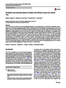
Figure 6 Efficient MGMTP140K-mediated in vivo selection in the macaque following reduced-intensity conditioning (K05079). Gene marking plotted is as provirus copy number from whole wbc, before and after in vivo selection of MGMTP140K-GFP genetically modified cells in a macaque. Treatment with O6BG-2X and BCNU is denoted by a solid arrow.
ous dog studies (23, 28). However, whereas in dogs, different drug doses are required, especially for BCNU, in monkeys, we used exactly what would be given and has been given to patients, suggesting our approach should readily translate to the clinic. A recent report by Larochelle et al. suggested that only transient selection could be achieved with MGMT P140K gene-modified cells in the rhesus macaque, with pronounced pulmonary and gastrointestinal (GI) toxicity (41), which contrasts with our studies, which resulted in substantial and stable increases in all hematopoietic lineages analyzed, with acceptable extramedullary toxicity. The major disparities between the studies described by Larochelle et al., our previous reports employing the dog model (22, 23), and the studies described herein include the gene marking reported prior to initiating drug treatment, which, reported by Larochelle et al., was below 3% in the PB of all but one monkey and less than 10% in the remaining monkey, and the drug treatment regimens used for in vivo selection (41). Low MGMT P140K gene marking levels (