Efficient isolation method for high‐ ... - Wiley Online Library
Recommend Documents
Zhan-Hui Zhang, Liang Yin, Yong-Mei Wang*. Department of Chemistry and the State Key Laboratory of Elemento-Organic Chemistry, Nankai University,.
Using this SS culture system, we calculated somite formation rate at early .... and scales of the pregnant snake each time another fer- tilized egg was isolated ...
Various intensity patterns were observed in lungs, kidney, liver, and thymus, whereas muscles ...... 36 Campbell RE, Tour O, Palmer AE et al. A monomeric red ...
echo [MPRAGE] and dedicated spine coil) and the new protocol; and 2) ..... server SD/CV for the SCA measurement derived from .... Rapid semi-automatic.
Feb 2, 2010 - Summary: When characterizing surfaces and searching for correlations to functional properties, such as friction, finding the right scale of ...
selective staining with fluorescent dyes as follows: ... of 1 µg/mL to stain and eliminate dead cells. ... sociation and vital staining procedures are summarized.
(see Basic Protocol 1 and Alternate Protocol) involves the isolation of DCs ... This technique for isolating dendritic cells has been used with only minor modifi-.
Sep 14, 2013 - The spring peeper (Pseudacris crucifer), a North American chorus frog from the ... ondary contact among mitochondrial lineages across the species range ..... did compromise locomotion causing tadpoles to swim in circles.
1980; Howard and. Bennet 1993; Abbott et al. 1994; Bock et al. 1994; Matte ... between Aeromonas and Vibrio (Austin et al. 1997). As biomolecular methods are ...
clinical outcome. A hybrid approach of combining box isolation with CFAE ablation is highly ..... also present a threat to patient quality of life and even longevity.
It is a land of high hopes and mystic ... when does 'apart, separate from others' begin and when does it end? To ..... In the Middle Ages, for instance, counts and ... heretic in 1539, his followers fled to Slovakia (where they were forced to .... Th
Mar 13, 2012 - E-mail: [email protected]. DOI: 10.1002/aenm.201100718. 1. Introduction. The two-phase morphology of bulk heterojunction (BHJ) pol-.
irradiation,[10] Co(III)-Salen complexes,[11] Lewis acids,[12] SiO2,[13] ..... 2 1992, 1603â1607; d) V. L. Arcus, C. D. Simpson, L. Main, J. Chem. Res. (S) 1992 .... 1960, 1406â1408; b) F. Robert, D. C. Nystroman, B. Rainera, J. Am. Chem. Soc.
desert locust, Schistocerca gregaria (Forskal) (Orthoptera: Acrididae), and fully identified. Its concentration is higher in crowd-reared (gregarious) animals than in.
Apr 15, 1992 - Stan- dard deviations in A and e for these isotherms are given in. Table 1 ...... Hfihnerfuss, H., W. Alpers, A. Cross, W. D. Garrett, W. C. Keller,.
Oct 6, 2010 - Thomas D. Pawlik, Denis Y. Kondakov, Michael E. Miller, Tommie L. Royster, and Dustin L. Comfort. Eastman Kodak Company, Rochester, ...
Sep 2, 2010 - promising type of solar energy-to-electricity conversion device. ... develop an iodine-free redox couple and also showed application in flexible ...
process, the rationale for using the item-mapping method is discussed. ... the standard-setting studies, rating data fro
Jan 17, 2018 - due to inappropriate embryo transfer methods, with post- transfer embryo expulsion occurring in 45%- 50% of cases, mainly due to the.
Fan Feng1,â and Christoph Pflaum1,ââ. 1 Department of Computer Science and Erlangen Graduate School in Advanced Optical Technologies (SAOT),.
Lessmüller OCT is an innovative technology for laser welding or brazing ..... KGaA, Weinheim. Process Monitoring. Laser Technik Journal 3/2016. 41. Authors.
Feb 18, 2013 - Technical Institute of Physics and Chemistry. Chinese Academy of Sciences. Beijing 100190, P.R. China. E-mail: [email protected].
Bacterial strains were tested by plate assay using. PVK and NBRIP media supplemented with 1.5%. Bacto-agar (Difco Laboratories, Detroit, MI,. USA).
such as indoprofen.[21] 2-Formylbenzoic acid could be convert- ed into N-substituted isoindolinones in good to excellent yields (80â98%) upon reaction with ...
Efficient isolation method for high‐ ... - Wiley Online Library
Aug 11, 2017 - Efficient isolation method for high- quality genomic DNA from ..... Ï2 = 47.62, df = 4, p < .001, for part Wald Ï2 = 37.28, df = 3, p < .001).
|
|
Received: 5 June 2017 Revised: 4 August 2017 Accepted: 11 August 2017 DOI: 10.1002/ece3.3398
ORIGINAL RESEARCH
Efficient isolation method for high-quality genomic DNA from cicada exuviae Hoa Quynh Nguyen1
| Ye Inn Kim1 | Amaël Borzée2
1 Department of Life Science, Ewha Womans University, Seoul, Korea 2 Department of Biological Science, Seoul National University, Seoul, Korea
| Yikweon Jang1
Abstract In recent years, animal ethics issues have led researchers to explore nondestructive methods to access materials for genetic studies. Cicada exuviae are among those
Correspondence Yikweon Jang, Department of Life Science, College of Natural Science, Ewha Womans University, Seoul, Korea. Email: [email protected]
materials because they are cast skins that individuals left after molt and are easily col-
Funding information National Research Foundation of Korea, Grant/Award Number: 2015R1A4A1041997
able commercial kits, extracting from four different exoskeleton parts. Furthermore,
lected. In this study, we aim to identify the most efficient extraction method to obtain high quantity and quality of DNA from cicada exuviae. We compared relative DNA yield and purity of six extraction protocols, including both manual protocols and availamplification and sequencing of genomic DNA were evaluated in terms of availability of sequencing sequence at the expected genomic size. Both the choice of protocol and exuvia part significantly affected DNA yield and purity. Only samples that were extracted using the PowerSoil DNA Isolation kit generated gel bands of expected size as well as successful sequencing results. The failed attempts to extract DNA using other protocols could be partially explained by a low DNA yield from cicada exuviae and partly by contamination with humic acids that exist in the soil where cicada nymphs reside before emergence, as shown by spectroscopic measurements. Genomic DNA extracted from cicada exuviae could provide valuable information for species identification, allowing the investigation of genetic diversity across consecutive broods, or spatiotemporal variation among various populations. Consequently, we hope to provide a simple method to acquire pure genomic DNA applicable for multiple research purposes. KEYWORDS
cicada exoskeleton, Crytptotympana atrata, exuviae, noninvasive DNA sampling
1 | INTRODUCTION
could have negative consequences on subsequent behavior and survival of sampled individuals. Extensive sampling is problematic for
Nondestructive sampling methods for DNA resources have recently
small colonies of social insects (Starks & Peters, 2002). Moreover,
attracted more attention from ethological, conservational, and popula-
lethal sampling potentially decreases population size and alters pop-
tion genetic studies. DNA extraction from specimens usually required
ulation structure (Starks & Peters, 2002), which is harmful for the
scarifying essential sections of the insects such as leg, thorax, or head
conservation of endangered species. Consequently, nondestructive
capsule. Such sampling methods could cause severe impacts on the
sampling methods are in need for various genetic analyses (Châline,
species at both individual and population levels. Invasive sampling
Ratnieks, Raine, Badcock, & Burke, 2004; Su et al., 2007).
Exuviae have been demonstrated to be reliable genetic sources
2012) and were stored at ambient temperature. DNA extraction work
for a variety of species, including popular taxa such as honey bees
on those exuviae was performed approximately 14 months after field
(Gregory & Rinderer, 2004), mosquitoes (Dhananjeyan et al., 2010),
collection.
and scarabs (Lefort, Boyer, Worner, & Armstrong, 2012), and endangered species such as dragonflies (Keller, Brodbeck, & Holderegger, 2009; Monroe, Lynch, Soluk, & Britten, 2010) and tarantulas (Petersen et al., 2007). Cicada exuviae are exoskeletons that remain after molt-
2.2 | DNA extraction procedure Six manual and kit protocols were employed to extract DNA from
ing of final instar nymphs. Such material can persist despite exposure
cicada exuviae. Manual protocols included (1) ethanol precipitation
to variable environmental conditions. One exuvia equals one success-
using sodium chloride (EtSC), (2) ethanol precipitation using ammo-
fully emerged adult individual. Exuviae can therefore serve as a useful
nol, via briefly vortexing the sample followed by carefully pipetting off
2012; Straub, Pepper, & Gerba, 1995).
the supernatant without dislodging the DNA pellet. Pellets were left
Our goals in this study are to evaluate protocols for DNA extraction from cicada exuviae regarding their quality and quantity of DNA yield and to suggest the best protocol for downstream applications. Six extraction protocols including available commercial kits and manual protocols were tested. We further identified those parts of the cicada exoskeleton from which high DNA yield was obtained.
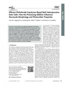
2 | METHODOLOGY 2.1 | Sample collection Exuviae of the black cicada (Cryptotympana atrata, Fig. 1) were collected in Gwangjin-gu, Seoul, Korea (37.533415°N, 127.070493°E), on 11 July 2015. The sampling location was an apartment complex where multiple cicada species coexisted, that is, C. atrata, Hyalessa fuscata, and Meimuna opalifera. After field collection, samples were identified for species based on morphological characters (Lee et al.,
F I G U R E 1 The black cicada (Cryptotympana atrata). This species is very common in urban areas in Korea. Photograph credit Yoonhyuk Bae
|
3
NGUYEN et al.
to dry in air for approximately 30 min and then resuspended in ultrapure water (Biosesang Inc., Gyeonggi-do, Republic of Korea). For EtAA samples, a precipitation step was carried on via addition of 200 μl of
2.4 | PCR amplification and purification Five hundred bp of the 16S region was amplified using two prim-
4 mol/L ammonium acetate followed by centrifugation at 18,000 xg
ers: LR-J-12887 (5′-CCGGTCTGAACTCAGATCACGT-3′) and LR-
for 20 min before transferring the top supernatant layer into a new
N-13398 (5′-CGCCTGTTTAACAAAAACAT-3′) (Simon et al., 1994)
1.5-ml tube. The samples were washed with ethanol as described for
by Takara Ex Taq (Takara Korea Biomedical Inc., Seoul, Republic
EtSC samples and resuspended in ultrapure water (Biosesang Inc.,
of Korea). For each PCR, 40 samples of each extraction proto-
Gyeonggi-do, Republic of Korea). Following the Ch5% protocol, the
col were run along with one negative control of ultrapure water
samples after incubation were further incubated at 100°C for 15 min
(Biosesang Inc., Gyeonggi-do, Republic of Korea) and one positive
and then centrifuged at 14,000 xg for 4 min, and the top layer super-
control of genomic DNA extracted from tissue of C. atrata. A total
natant was transferred to a new 1.5-ml tube.
of 25 μl amplified sample consisted of 2 μl template DNA and 23 μl of master mix (0.125 μl of Takara Ex Taq™ 5 U/μl, 2.5 μl of 10×
2.3 | Acquisition of UV–Vis spectra of DNA concentration
Buffer, 2 μl of dNTP Mix 2.5 mmol/L, 2 μl of MgCl2 25 mmol/L, 2 μl of each primer 10 mmol/L and ultrapure water (Biosesang Inc., Gyeonggi-do, Republic of Korea). PCR amplification initiated by
Extracted DNA samples were examined by gel electrophoresis in 1%
1-min initial denaturation at 94°C, followed by 30 cycles of 30 s
agarose gel (Biopure, Genomic Base, Seoul, Republic of Korea) visu-
denaturation at 94°C, 1-min annealing at 56°C, and 1-min elon-
alized on an UltraSlim LED Illuminator (MaestroGen Inc., Hsinchu,
gation at 72°C, finally completed by 2-min terminal elongation at
Taiwan) using MaestroSafe Nucleic Acid loading dye (MaestroGen
72°C. Three microliters of each PCR product were loaded on 1.5%
Inc., Hsinchu, Taiwan). Quantity and quality of DNA samples were
agarose gel and visualized using the same loading dye and LED il-
measured using a NanoDrop™ 2000/2000c spectrophotometer
luminator as described above. Samples with bands that appeared at
(Thermo Fisher Scientific Inc., Delaware, USA). In particular, the ratio
600-bp size, as in the positive control band (Fig. 2), were considered
of 260/280 indicated the presence of organic contaminants such as
as PCR success and were used for the gel purification procedure.
protein or phenol that strongly absorb at 280-nm wavelength; like-
We labeled 1 for successful amplification and 0 for amplification
wise, 260/230 indicated the appearance of other contaminants ab-
failure. Gel bands were excised using a sterile scalpel, and gel
sorbing at 230-nm wavelength such as EDTA or carbohydrates. A
purification was conducted using a QIAquick® Gel Extraction Kit
DNA sample was considered pure when both 260/280 and 260/230
(QIAGEN Group, Hilden, Germany). All samples were sequenced
ratios ranged between 1.8 and 2.0. For Ch5% samples, due to lack of
both in forward and in reverse directions by COSMO Genetech
baseline buffer, we used original Chelex 5% as baseline buffer, and the
Company (COSMO Genetech Co., Ltd., Seoul, South Korea), and
measurement of two ratios was employed only for purity comparison
sequencing success was labeled 1 as successful sequencing and 0
among protocols, but was not included in the statistical analysis.
for sequencing failure.
T A B L E 1 Cell lysis buffers and DNA extraction procedures of six protocols: ethanol precipitation using sodium chloride (EtSC), ethanol precipitation using ammonium acetate (EtAA), Chelex 5% (Ch5%), LaboPass™ Genomic DNA Purification Kit (LP), DNeasy® Blood and Tissue Kit (DN), and PowerSoil DNA Isolation Kit (PS). For each protocol, 40 samples from 10 cicada exuviae were employed
Protocol
Volume of cell lysis buffer
Volume of Proteinase K (20 mg/ml)
Incubation temperature (oC)
Ethanol precipitation using sodium chloride
600 μl of TNES buffer (Tris pH 7.5 10 mmol/L, NaCl 400 mmol/L, EDTA 100 mmol/L, and SDS 0.6%)
70 μl
50
Ethanol precipitation using ammonium acetate
600 μl of digestion buffer (NaCl 50 mmol/L, Tris pH 8.0 50 mmol/L, EDTA pH 8.0 20 mmol/L, and SDS 1%)
6 μl
55
Chelex 5%
360 μl of chelex 5% buffer
80 μl
57
LaboPass™ Genomic DNA Purification Kit
800 μl buffer TL
40 μl
56
DNeasy® Blood and Tissue Kit
360 μl buffer ATL
40 μl
56
PowerSoil DNA Isolation Kit
60 μl Solution C1
60 μl
65
|
NGUYEN et al.
4
2.5 | Statistical analysis
3.2 | DNA concentration
Generalized linear model (GLM) was performed to test for effects of
Results of GLM showed that both protocol and part contributed as
extraction protocols and exoskeleton parts on DNA quantity and pu-
significant factors affecting DNA concentration (for protocol Wald
rity ratios. Dependent variables included DNA concentration (ng/μl),