Reviews Electromagnetic Interference and Implanted Cardiac Devices: The Medical Environment (Part II)
Address for correspondence: Fred M. Kusumoto, MD Electrophysiology and Pacing Service Division of Cardiovascular Disease Mayo Clinic 4500 San Pablo Avenue Jacksonville, FL 32224
[email protected]
Juna Misiri, MD; Fred Kusumoto, MD; Nora Goldschlager, MD Department of Medicine (Misiri, Kusumoto), Division of Cardiovascular Disease, Electrophysiology and Pacing Service, Mayo Clinic, Jacksonville, Florida; Department of Medicine (Goldschlager), Cardiology Division, San Francisco General Hospital, and Department of Medicine, University of California, San Francisco, California
Electromagnetic interference produced by medical equipment can interact with implanted cardiac devices such as pacemakers and implantable cardioverter-defibrillators. The most commonly observed interaction is in the operating room with electrosurgery. The risk of interactions can often be mitigated by close communication between the cardiac-device specialist and the anesthesiology/surgical team to develop a patient-specific strategy that accounts for factors such as type of device, type of surgery, and whether the patient is pacemaker dependent. Although magnetic resonance imaging should generally not be used in patients with implanted cardiac devices, several published guidelines provide strategies and recommendations for managing risks if magnetic resonance imaging is required with no suitable diagnostic alternatives. Other common sources of electromagnetic interference in the medical environment are ionizing radiation and left ventricular assist devices.
Introduction There are multiple sources of electromagnetic interference (EMI) in the medical setting (Table 1), and recommendations for management in patients with cardiac implantable electronic devices (CIEDs) such as pacemakers and implantable cardioverter-defibrillators (ICDs) have been published in the Heart Rhythm Society/American Society of Anesthesiology Guidelines and in other reviews.1,2 The most common source of EMI in the medical environment is electrosurgery (Figure 1). It is also important for the clinician to understand possible interactions between CIEDs and commonly used medical sources of EMI such as magnetic resonance imaging (MRI), ventricular assist devices, therapeutic ionizing radiation, and cardioversion. Electrosurgery Electrosurgery uses plasma arcs generated from alternating current in the radiofrequency (RF) range (100–5000 kHz) to cut or coagulate tissue. Most commonly the current is delivered between a cauterizing instrument and a large ‘‘return’’ electrode placed on the skin (monopolar configuration), but
The authors have no funding, financial relationships, or conflicts of interest to disclose. Received: December 14, 2011 Accepted with revision: March 19, 2012
in some cases current is delivered in a bipolar configuration where electrical energy is delivered between the 2 electrodes at the tip of the surgical instrument.2 Because the EMI field is so small, bipolar electrosurgery does not interact with CIEDs and can be used without any special precautions. On the other hand, monopolar electrosurgery is the most common medical cause of EMI interaction with CIEDs. Interaction between CIEDs and electrosurgery can be effectively managed in most circumstances by taking a few simple steps. The most critical step for elective procedures is preoperative communication between the CIED management team and anesthesiologists. A de novo preoperative evaluation is generally not required. The likelihood and risk of CIED and electrosurgery interaction depend on patient/CIED factors and surgical factors. For example, interaction is less likely as the distance between the CIED and the surgical field increases. In fact, procedures below the umbilicus are unlikely to generate EMI sensed by the CIED if the CIED is placed in the usual position in the upper chest and the return pad for electrosurgery is placed on the lower body (thigh or gluteal region). In the largest published case series to date, EMI-CIED interaction was observed when the electrosurgery was performed within 8 cm of the CIED.3 In a more recent preliminary report of 171 patients with ICDs, EMI was identified in 9 of 22 surgical procedures performed above Clin. Cardiol. 35, 6, 321–328 (2012) Published online in Wiley Online Library (wileyonlinelibrary.com) DOI:10.1002/clc.21997 © 2012 Wiley Periodicals, Inc.
321
Table 1. Recommendations to Minimize Electromagnetic Interference in Medical Settings Electrosurgery 1. Maximize distance between site of monopolar electrosurgery and the CIED. Consider bipolar electrosurgery if required near the CIED. 2. Use the minimum power settings required for adequate electrosurgery. 3. For monopolar electrosurgery, place the return electrode at a site where the current path is kept as far as possible from the CIED. Often, the thigh on the leg contralateral to the CIED will be the best location. 4. For surgeries below the umbilicus, often no specific procedures are required for CIEDs. However, in some cases (patients with ICD or who are pacemaker dependent), reprogramming or magnet application may be considered. 5. Procedures above the umbilicus are more likely to be associated with EMI, and reprogramming or magnet application may be required, particularly if the patient has an ICD or is pacemaker dependent. 6. Using short bursts of electrosurgery may be required if inhibition is observed. 7. Continuously monitor the patient with plethysmography or arterial pressure. 8. After the surgery, address any preoperative programming changes that were made, and consider interrogation for any surgery with a higher likelihood of EMI. MRI (see Table 2) LVAD 1. Surgeons implanting the HeartMate II LVAD should be notified and be aware of possible loss of ICD telemetry in some types of ICDs. 2. Interrogate before and immediately after LVAD implantation. 3. If there is loss of ICD telemetry, metal shielding and/or implanting an ICD from a different manufacturer may be required. Radiation therapy 1. 2. 3. 4. 5. 6. 7. 8.
Avoid direct irradiation of the CIED. Consider relocation of the device if it is within the radiation field. Review with the manufacturer the susceptibility of the device to radiation effects. Establish the pacemaker dependency of the patient. Shield the pulse generator if possible. The absorbed dose to be received by the ICD should be estimated before treatment. Continuously monitor the patient’s ECG. Consider intermittent testing of the CIED during and after radiation therapy.
Cardioversion 1. Use an anterior-posterior patch position, with the patches positioned as far from the CIED as possible (>8 cm). 2. Evaluate CIED function after cardioversion. TENS 1. 2. 3. 4. 5. 6.
Assess the likelihood and patient risk of TENS for CIED interaction: location of TENS, pacemaker dependency, ICD vs pacemaker. Perform initial supervised testing of TENS use with monitoring to evaluate for interference. Set pacemaker sensing polarity to bipolar. Program OFF impedance-based sensors such as minute ventilation. Place the TENS electrodes close to each other and perpendicular to the device leads. Avoid treatment in the chest area; TENS can often be done safely in the lower extremities.
Radiofrequency ablation, lithotripsy, ECT 1. Generally, no specific programming is required. 2. It is reasonable to have a magnet available. 3. Cardiac monitoring is reasonable, particularly in those patients who are pacemaker dependent. Abbreviations: CIED, cardiovascular implantable electronic device; ECG, electrocardiogram; ECT, electroconvulsive therapy; EMI, electromagnetic interference; ICD, implantable cardioverter-defibrillator; LVAD, left ventricular assist device; MRI, magnetic resonance imaging; TENS, transcutaneous electrical nerve stimulation.
the umbilicus but in none of 53 procedures performed below the umbilicus.4 Often, a magnet is applied to ICDs during surgical procedures, because in most cases magnets will suspend tachycardia therapy. However, it is important to know that exceptions do exist and that in some cases the response to magnet application is a programmable option. Recently published guidelines suggest that for surgical procedures below the umbilicus, no intervention or magnet application are both reasonable options.1 For procedures above the umbilicus, where a higher likelihood of EMI
322
Clin. Cardiol. 35, 6, 321–328 (2012) J. Misiri et al: EMI interactions with ICDs: Part II Published online in Wiley Online Library (wileyonlinelibrary.com) DOI:10.1002/clc.21997 © 2012 Wiley Periodicals, Inc.
and CIED interaction exists, inactivation of ICDs by either programming or magnet application are both reasonable options. For pacing systems without ICD function, the best option will often depend on whether or not the patient is pacemaker dependent. In those patients who are not pacemaker dependent, often no programming changes are required. In patients who are pacemaker dependent, where inhibition of output may lead to asystole or profound bradycardia, asynchronous pacing by mode programming or magnet application may be necessary. Other strategies to
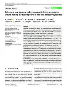
Figure 1. Atrial electrograms during electrosurgery used for contralateral shoulder surgery. During electrosurgery, atrial sensing of EMI (*) and intrinsic atrial activity (arrows) is observed. Abbreviations: AS, atrial sensing; EMI, electromagnetic interference.
minimize risk to the patient are to use bipolar electrosurgery if possible and, if monopolar electrosurgery is required, to position the indifferent electrode so that the current path between the surgical field and the indifferent electrode is as far from the CIED as possible, and to use only short bursts of electrosurgery with the lowest clinically effective energy. Intraoperatively, plethysmographic or arterial pressure monitoring is essential, because electrosurgery will often cause artifacts on electrocardiographic (ECG) monitoring equipment, rendering surface ECG leads uninterpretable. It is important to remember that after the surgery is completed, any preoperative programming changes that have been made must be addressed. In a retrospective single-center study, inadvertent inactivation of ICDs was found in 4 patients, 2 after surgery.5 In an evaluation of a database of ICD patients maintained by the US Food and Drug Administration (FDA) and device manufacturers, of 212 deaths documented between 1996 and 2003, 11 ICDs were deactivated, 3 after surgery.6
Magnetic Resonance Imaging Magnetic resonance imaging offers several advantages over other available imaging techniques but has been considered to be contraindicated in patients with CIEDs. It has been estimated that patients with pacemakers or ICDs have a 50%–75% likelihood of developing a clinical indication for MRI over the lifespan of the device, so manufacturers have been interested in developing ‘‘MRIcompatible/safe’’ implantable devices.7 All 3 components of the MRI—the static magnetic field, rapidly varying magnetic fields (100–200 Hz), and electromagnetic RF fields (60–70 MHz)—have the potential to affect the function of the CIED.7 – 24 The static magnetic field of the MRI will usually close the reed switch of the pacemaker, resulting in a magnet mode function that results in asynchronous pacing at a manufacturer-determined rate. Asynchronous pacing is usually tolerated well, with only rare cases of hemodynamic compromise or development of atrial or ventricular arrhythmias due to pacing stimuli being delivered in the vulnerable periods of atria and/or ventricles leading to repetitive beating.8 – 11 Theoretically, the static magnetic field could also cause sufficient torque in CIEDs within the device pocket, but no significant physical device movement has
been documented (although some patients have reported a vibrating sensation) in newer devices that use less ferromagnetic material than older ones.8 The alternating magnetic fields and the rapid RF pulses may induce inappropriate device function with rapid pacing corresponding to the frequency of pulsing due to effects of RF energy on the pacemaker output circuits.11 Electrode heating with tissue thermal injury at the electrodemyocardial tissue interface is another potential adverse effect.11,14 – 17 Heating is more pronounced with abandoned leads due to resonance from the similarity in the wavelength of the RF field of a 1.5-Tesla (T) MRI and the length of a standard lead.12 In addition, heating at the tip of an abandoned lead is higher than when the lead is connected to a pulse generator because the lead can be modeled as an ‘‘electrical open,’’ as the connector is usually insulated by a plastic cap.12 A substantial increase in capture threshold has been reported after MRI at 1.5 T in some patients with pacemakers, which was attributed to thermal injury in proximity to the pacemaker lead tip.8 – 10 If the results from 3 studies that used troponin release as a marker for myocardial injury associated with MRI are combined, an abnormal troponin elevation that correlated with increase in pacing capture threshold was observed in only 1 of 206 examinations.20 – 22 It is important to remember that patients with abandoned leads have generally been excluded from in vivo studies and that very little is known about the effects of MRI in these patients. To date, approximately 2000 patients with conventional CIEDs who have undergone MRI with no significant deleterious effects on the device are described in the literature.7 – 23 In the largest study to date, 555 MRI studies were performed in 438 patients with CIEDs (54% with pacemakers, 46% with ICDs).24 Patients with recently implanted leads (