www.nature.com/scientificreports
OPEN
Received: 7 September 2018 Accepted: 1 November 2018 Published: xx xx xxxx
Energy/Electron Transfer Switch for Controlling Optical Properties of Silicon Quantum Dots Mohammed Abdelhameed1, Shawkat Aly2, Jeremy T. Lant3, Xiaoran Zhang1 & Paul Charpentier1,2 The superior optical properties of Silicon Quantum Dots (SQDs) have made them of increasing interest for a variety of biological and opto-electronic applications. The surface functionalization of the SQDs with aromatic ligands plays a key role in controlling their optical properties due to the interaction of the ligands with the electronic wave function of SQDs. However, there is limited reports in literature describing the impact of spacer groups connecting the aromatic chromophore to SQDs on the optical properties of the SQDs. Herein, we report the synthesis of two SQDs assemblies (1.6 nm average diameter) functionalized with perylene-3,4,9,10-tetracarboxylic acid diimide (PDI) chromophore through N-propylurea and propylamine spacers. Depending on the nature of the spacer, the photophysical measurements provide clear evidence for efficient energy and/or electron transfer between the SQDs and PDI. Energy transfer was confirmed to be the operative process when propylurea spacer was used, in which the rate was estimated to be ~2 × 10⁹ s−1. On the other hand, the propylamine spacer was found to facilitate electron transfer process within the SQDs assembly. To illustrate functionality, the water soluble SQD-N-propylurea-PDI assembly was proven to be nontoxic and efficient for fluorescent imaging of embryonic kidney HEK293 cells and human bone cancerous U2OS cells. Silicon Quantum Dots (SQDs) have recently attracted tremendous attention due to their unique optical properties, including wide absorption spectra, excellent stability against photobleaching compared to conventional dyes, and size-dependent tuneable photoluminescence (PL)1. The SQDs have advantages over other quantum dots, including cadmium sulfide (CdS), due to their excellent biocompatibility and biodegradability, low toxicity, and the ease of their surface functionalization2. Thus, they are ideal candidates for a wide range of potential applications including bioimaging3, photodynamic therapy4, sensing5, photovoltaics6, and light-emitting diodes (LEDs)7. In general, the size-tuneable PL of SQDs is assigned to the quantum confinement effect where the PL is blue-shifted when the size of SQDs is more than ~3 nm. However, a deviation from this behaviour was observed for SQDs of size less than ~2 nm when the PL originates from surface relevant states8. The surface functionalization would then play a crucial role towards controlling the optical properties of SQDs9. It has been shown in previous studies that surface functionalization of SQDs with aromatic ligands helps tune their optical properties including their quantum yield and PL10,11. This strategy potentially can be utilized as a means to control the optical properties of SQDs. Moreover, the functionalization of SQDs with organic ligands increases their stability towards oxidation and prevents them from agglomeration and aggregation12. Only a few reports in literature investigated the influence of aromatic fluorophores covalently linked to SQDs and the role of the spacer connecting the fluorophore on the optical properties of SQDs. Interestingly, the utilization of aromatic fluorophores as a capping agent was found to play a key role to control the optical properties of SQDs10,13–16. Several methods have been reported in literature for preparing SQDs including laser pyrolysis17, etching of bulk silicon18, nonthermal plasma19, and preparation in supercritical fluids20. In this work, a facile solution-based reduction method has been adopted with minor modifications to prepare SQDs21. Triethoxysilane derivatives and 1
Department of Chemical and Biochemical Engineering, Western University, London, Ontario, N6A 5B9, Canada. Department of Mechanical and Materials Engineering, Western University, London, Ontario, N6A 5B9, Canada. 3 Department of Biochemistry, Western University, London, Ontario, N6A 5B9, Canada. Mohammed Abdelhameed and Shawkat Aly contributed equally. Correspondence and requests for materials should be addressed to P.C. (email:
[email protected]) 2
Scientific REPOrTS |
(2018) 8:17068 | DOI:10.1038/s41598-018-35201-0
1
www.nature.com/scientificreports/
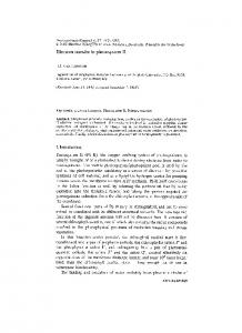
Figure 1. Synthesis route of SQDs (A) and their surface functionalization using perylene-3,4,9,10tetracarboxylic dianhydride (B). sodium citrate dihydrate were utilized as the silicon source and reducing agent, respectively while the synthesis was carried out in the glycerol green solvent at normal pressure and relatively high temperature (180 °C). Glycerol is a green solvent produced as a byproduct of biodiesel production, and with its high boiling point of 290 °C, and 3 available OH groups to help coordinate to the growing nanocrystals, is of interest for solvent engineering of SQDs. Here, we report the synthesis of SQDs functionalized with perylene-3,4,9,10-tetracarboxylic acid diimide (PDI) through propylamine and N-propylurea spacers to produce Am-SQD-Per and Urea-SQD-Per, respectively. The PDI dye has been chosen for this study due to its excellent properties including high fluorescence quantum efficiency, high thermal and photochemical stability, and ease of processibility as well as scalability22. Additionally, the combination of the planar π-system of PDI and other electron withdrawing and donating groups within the system with SQDs could strongly affect the formed electronic interactions through a possible photoinduced energy and/or electron transfer processes. The latter processes are likely to change the optical properties of the assemblies. The products Am-SQD-Per and Urea-SQD-Per were characterized using high-resolution transmission electronic microscopy (HRTEM), Fourier-transform infrared (FTIR) spectroscopy, X-ray photoelectron spectroscopy (XPS), UV-Vis absorption spectroscopy, and steady-state and times-resolved emission spectroscopy.
Results and Discussion
The nanoparticles SQDs and their surface functionalization were synthesized as shown in Fig. 1. Both APTES and UPTES were used as the silicon source and were reduced by a citrate reducing agent. This reaction was carried out in glycerol as a high boiling point green solvent under atmospheric pressure and at 180 °C using an oil bath. The resulting SQDs were then functionalized using PDA to produce Am-SQD-Per and Urea-SQD-Per.
Size and structure. Figure 2 shows the TEM, HR-TEM, and size distribution images of the assemblies
Am-SQD-Per and Urea-SQD-Per. The TEM images indicate that the functionalized SQDs are quasi-spherical particles with no obvious agglomeration or aggregation. The corresponding size distribution histograms obtained by analyzing of more than 300 dots from different regions of the grids showed that the diameter of these particles ranged from 0.9 to 3.3 nm. The average diameters of SQDs for the compounds Am-SQD-Per and Urea-SQD-Per are 1.61 ± 0.89 and 1.62 ± 0.81 nm, respectively. The functionalized SQDs exhibited high crystallinity which is evidenced by the distinct lattice fringes with 0.30 nm interplanar spacing, as shown in the HR-TEM insets of Fig. 2. This is in agreement with the (111) plane of diamond structured silicon23. It should be noted that the low resolution of the TEM and HR-TEM images is assigned to the ultra-small dimensions of these SQDs and the small atomic weight of the silicon atom compared to the counterpart metallic or semiconductor quantum dots, which is known to provide low-quality visualization10,24. To confirm the attachment of Am-SQD and Urea-SQD to the PDA dye to produce Am-SQD-Per and Urea-SQD-Per, respectively, FTIR and XPS spectroscopy were performed. Figure 3 displays the FTIR spectra of Am-SQD-Per and Urea-SQD-Per. The broad peak at 2980–3660 cm−1 can be assigned to the stretching vibration of the O-H bond25. The intense peaks at 1019 and 970 cm−1 for Am-SQD-Per, and 1024 and 909 cm−1 for Urea-SQD-Per, are attributed to Si-O-Si/Si-O-C and Si-OH stretching, respectively26. The peaks at 2970– 2808 cm−1 correspond to the –CH stretching vibrations of the spacer and alkyl group27. Both peaks at 1685 and 1638 cm−1 for Am-SQD-Per, and 1692 and 1639 cm−1 for Urea-SQD-Per, are assigned to imidic C=O stretching (N-C=O)26. The peaks at 1439 cm−1 in both compounds can be assigned to N-C stretching28. Furthermore, the characteristic peaks of the anhydride carbonyl (O-C=O) in the free dye PDA at 1760 and 1720 cm−1 are absent in the FTIR spectra of Am-SQD-Per and Urea-SQD-Per. This indicates the success of SQDs binding to the dye. To further confirm the binding of SQDs to the PDA dye, XPS spectroscopy was performed. Figure 4 displays the high resolution XPS spectra of O 1 s, C 1 s, N 1 s, and Si 2p for Am-SQD-Per and Urea-SQD-Per. The deconvoluted peaks of O 1 s appeared at 533.6, 532.3, and 530.9 eV for Am-SQD-Per, and 533.4, 532.1, 530.9 eV for Urea-SQD-Per. These can be assigned to C-O, Si-O, and amidic or imidic carbonyl (-N-C=O), respectively29–31. The C 1 s binding energy peaks were present at 288.4, 286.4, and 285 eV for Am-SQD-Per, and 288.7, 286.3, and 284.8 eV for Urea-SQD-Per. These are attributed to C=O of imide or amide bonds (-N-C=O), C-O or C-OH, Scientific REPOrTS |
(2018) 8:17068 | DOI:10.1038/s41598-018-35201-0
2
www.nature.com/scientificreports/
Figure 2. TEM together with HR-TEM (left) and diameter distribution with photographs for solutions under UV (365 nm) irradiation (right) for Am-SQD-Per (A) and Urea-SQD-Per (B).
Figure 3. FTIR spectra of Am-SQD-Per (A) and Urea-SQD-Per (B).
and Si-C or C=C of PDI kernel, respectively32–35. The XPS spectra of N 1 s centered at 399.6 for Am-SQD-Per and 399.9 eV for Urea-SQD-Per signify the presence of N-C or N-C=O30,36. The Si 2p peak at 102.7 for Am-SQD-Per and 102.4 eV for Urea-SQD-Per can be assigned to Si-O-C or Si-C33. The XPS data are consistent with the FTIR data, providing convincing evidence for successful functionalization of SQDs with PDI.
Photophysical properties. Steady-state photoluminescence emission, absorption and excitation spectra
for Urea-SQD, Urea-SQD-Per, Am-SQD and Am-SQD-Per in methanol (MeOH) at room temperature are given in Fig. 5(A,B). Study of the emission spectrum recorded for the starting Urea-SQD (see Fig. 5A) revealed one broad spectrum extended over the spectral range of 350–650 nm, in agreement with reported literature data for amine-terminated silicon quantum dots37,38. On the other hand, emission spectrum obtained for Urea-SQD-Per under the same
Scientific REPOrTS |
(2018) 8:17068 | DOI:10.1038/s41598-018-35201-0
3
www.nature.com/scientificreports/
Figure 4. XPS spectra of O 1 s, C 1 s, N 1 s, and Si 2p for Am-SQD-Per and Urea-SQD-Per.
experimental conditions exhibited two main bands (see Fig. 5A). The first emission band showed a resemblance with the starting Urea-SQD with a broad nature over the spectral range between 350–450 nm. An observable blue shift detected in the quantum dot emission in Urea-SQD-Per as compared to the starting Urea-SQD. Considering the average diameter for our SQDs to be 1.6 nm, such shift observed in the emission is likely to be assigned to surface chemistry and independent on quantum confinement8. This is in agreement with an earlier report where the emission properties for large QDs (d > ~3 nm) were found to be dominated by quantum confinement while those for smaller size quantum dots (d