APPLIED PHYSICS LETTERS 90, 192107 共2007兲
Electronic structures and bonding properties of chlorine-treated nitrogenated carbon nanotubes: X-ray absorption and scanning photoelectron microscopy studies S. C. Ray, C. W. Pao, H. M. Tsai, J. W. Chiou, and W. F. Ponga兲 Department of Physics, Tamkang University, Tamsui 251, Taiwan
C. W. Chen Department of Materials Science and Engineering, National Taiwan University, Taipei 106, Taiwan
M.-H. Tsai Department of Physics, National Sun Yat-Sen University, Kaohsiung 804, Taiwan
P. Papakonstantinou NRI, School of Electrical and Mechanical Engineering, University of Ulster at Jordanstown, Newtownabbey, County Antrim BT37OQB, Northern Ireland, United Kingdom
L. C. Chen Center for Condensed Matter Sciences, National Taiwan University, Taipei 106, Taiwan
K. H. Chen Institute of Atomic and Molecular Sciences, Academia Sinica, Taipei 106, Taiwan
W. G. Graham Department of Physics and Astronomy, Queens University of Belfast, Belfast, Antrim BT71NN, Northern Ireland, United Kingdom
共Received 8 February 2007; accepted 19 April 2007; published online 8 May 2007兲 The electronic and bonding properties of nitrogenated carbon nanotubes 共N-CNTs兲 exposed to chlorine plasma were investigated using C and N K-edge x-ray absorption near-edge structure 共XANES兲 and scanning photoelectron microscopy 共SPEM兲. The C and N K-edge XANES spectra of chlorine-treated N-CNTs consistently reveal the formation of pyridinelike N-CNTs by the observation of 1s → *共e2u兲 antibonding and 1s → *共b2g兲 bonding states. The valence-band photoemission spectra obtained from SPEM images indicate that chlorination of the nanotubes enhances the C–N bonding. First-principles calculations of the partial densities of states in conjunction with C K-edge XANES data identify the presence of C–Cl bonding in chlorine treated N-CNTs. © 2007 American Institute of Physics. 关DOI: 10.1063/1.2737392兴 Carbon nanotubes 共CNTs兲 have attracted extensive attention, partly owing to their favorable electronic properties.1 The doping of nitrogen impurities in CNTs was found to be a possible route to tune the band gap such that CNTs can be exploited to make various electronic devices.2 The fluorinated CNTs can be tuned from metal to semiconductor/ insulator with high resistivity at elevated temperatures.3 Fluorination can also improve the wetting of nanotubes in water by inducing a surface dipole layer on the nanotube wall. This effect may be useful in battery and supercapacitor applications.4 Iodine- and bromine-doped CNTs were found to enhance the electronic/electrical properties by increasing the density of free charge carriers.5 Therefore, it is interesting to understand the effect of chemical modification of CNTs using halogens 共F, I, Br, and Cl兲. Fluorine-, iodine-, and bromine-treated CNTs 共Refs. 3–5兲 and CNT-based nanomaterials6–8 have been investigated previously. The electronic and bonding properties of chlorine-treated nitrogenated carbon nanotubes 共N-CNTs:Cl兲 have not been fully carried out. Here, the electronic structures and bonding properties of freshly prepared N-CNTs, which were further treated with chlorine plasma in an inductively coupled plasma system, have been studied using x-ray absorption a兲
Author to whom correspondence should be addressed: electronic mail:
[email protected]
near-edge structure 共XANES兲 and scanning photoelectron microscopy 共SPEM兲. The C and N K-edge XANES, and SPEM measurements were performed at the National Synchrotron Radiation Research Center in Hsinchu, Taiwan. In the preparation of CNTs, the vertically oriented multiwall N-CNTs were initially synthesized by microwave-plasma-enhanced chemicalvapor deposition on silicon substrates precoated with an e-beam evaporated thin Fe catalytic layer.9 Then, the obtained N-CNTs were chlorinated in an inductively plasmacoupled reactor in flowing Cl gas at 5 sccm for periods of 1 and 5 min 关denoted as N-CNTs:Cl 共1 min兲 and N-CNTs:Cl 共5 min兲 later on兴. Figure 1 displays C K-edge XANES spectra of N-CNTs with and without chlorine treatment and the highly oriented pyrolytic graphite 共HOPG兲 as a reference. The feature with the maximum intensity at approximately 285.5 eV for HOPG was attributed to the * antibonding state originated from the out-of-plane bonds in the sp2 bonding configuration.10 The positions of * resonance feature in the C 共1s兲 XANES spectra of N-CNTs and N-CNTs:Cl located at ⬃286.4 and 286.7 eV, respectively, which are shifted by ⬃0.9 eV for N-CNTs and 1.2 eV for N-CNTs:Cl with respect to that of HOPG 共285.5 eV兲, correspond to the 1s → *共e2u兲 transition as in the pyridinelike sp2 C–N structure.11 Pyridine is also well known to have two unfilled * orbitals 共as in benzene兲
0003-6951/2007/90共19兲/192107/3/$23.00 90, 192107-1 © 2007 American Institute of Physics Downloaded 07 Jan 2010 to 193.61.144.141. Redistribution subject to AIP license or copyright; see http://apl.aip.org/apl/copyright.jsp
192107-2
Ray et al.
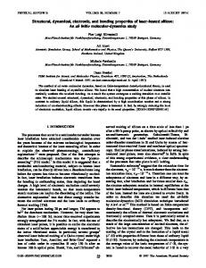
FIG. 1. C K-edge XANES spectra of unchlorinated/chlorinated N-CNTs and reference HOPG. The inset highlights the * region.
with e2u 共an antibonding state whose wave functions are antisymmetric兲 and b2g 共a bonding state whose wave functions are symmetric兲 symmetry.11 The latter is observed at ⬃289.2 eV in the * region for N-CNTs:Cl, and its intensity increases with the Cl-treatment time. This feature is attributable to the transition of 1s → *共b2g兲 similar to that of the pyridine structure with the symmetric bonding state,11 and/or C–Cl bonds, as observed by Unger et al. at 289.0 eV in x-ray photoelectron analysis of Cl-functionalized multiwall CNTs.12 Previous works of carbon systems revealed a similar double feature in the C 共1s兲 XANES spectra.13 For N-CNTs, a small shoulder observed at ⬃289 eV 共indicated by a vertical arrow兲 was attributed to the presence of interlayer graphite states, which was correlated with the calculated electronic state of dual-layer graphite sheets.14 In the * region, the centers of the maximum features of N-CNTs and N-CNTs:Cl appear at ⬃293.2 and 293.6 eV, respectively, similar to those of the pyridine structure.11 Interestingly, the intensities of 1s → *共e2u兲 共286.4/ 286.7 eV兲 and * 共293.2/ 293.6 eV兲 features decrease, while that of 1s → *共b2g兲 feature 共indicated by the arrow兲 increases with the increase of Cltreatment time, as clearly shown in the inset of Fig. 1. This trend suggests the formation of a more symmetric bonding state in the N-CNTs than in the pyridine structure. To identify clearly these features, the p-projected conduction-band partial densities of states 共PDOSs兲 of pure CNTs, N-CNTs, and N-CNTs:Cl are calculated using the CASTEP code,15 which is a plane-wave pseudopotential program based on the density functional theory and local density approximation, and are shown in Fig. 2. The benzene, pyridine, and pyridine-Cl cluster models are employed to represent local
Appl. Phys. Lett. 90, 192107 共2007兲
FIG. 3. N K-edge XANES spectra of unchlorinated/chlorinated N-CNTs. The inset highlights the * region.
bonding configurations of CNTs, N-CNTs, and N-CNTs:Cl, respectively. The hydrogen atoms in these models saturate the dangling bonds of carbon atoms, so that these carbon atoms mimic those in CNTs, N-CNTs, and N-CNTs:Cl. The insets 共a兲–共c兲 in Fig. 2 present the bonding configurations of these three CNTs; blue and green colors in the bonding structures indicate the attachment of N and Cl atoms in pure CNTs, respectively. Details of the calculations for the cluster models of these CNTs with various geometries can be found elsewhere.16 In Fig. 2, the feature located at ⬃2.6 eV between * and * for the cluster model of pyridine-Cl may correspond to the extra feature indicated by the arrow between * and * features in the inset of Fig. 1 and can be attributed to the C–Cl bond. Figure 3 displays the N K-edge XANES spectra of the N-CNTs and N-CNTs:Cl samples. The two main features centered at ⬃403.2 and ⬃409.5 eV are associated with transitions into unoccupied * and * orbitals, respectively. As stated above, the pyridine structure has two unfilled * orbitals with e2u and b2g symmetries, which are typically observed in the N K-edge XANES spectra at ⬃400.0 and ⬃403.7 eV, respectively, for nitrogenated carbon films.11 The features in the N K-edge XANES spectra in Fig. 3 are similar for all N-CNTs. The prominent feature centered at ⬃403.2 eV in the * resonance has the b2g symmetry of the pyridine structure.11 Jimènez et al.17 also observed this feature at 403.5 eV for sp2-hybridized nitrogenated carbon films, for which the * region can be resolved into four peaks. The broad feature centered at ⬃409.5 eV in the * region is associated with the C–N bond and is identical to that of the pyridine structure.11,17 The inset in Fig. 3 reveals that the intensity of the * features in the N K-edge XANES spectra decreases with the Cl-treatment time consistent with the trend of the C K-edge XANES spectra shown in the inset of Fig. 1, which consistently indicate that Cl treatment increases the occupation of the b2g symmetry states or enhances the pyridinelike C–N bonding. Figure 4 displays spatially resolved valence-band photoemission spectra of N-CNTs and N-CNTs:Cl with corresponding C 1s SPEM cross-sectional images. The bright area in the SPEM image corresponds to the N-CNTs and N-CNTs:Cl with a maximum C 1s intensity. The spectra presented in Fig. 4 exhibit photoelectron yields from the bright regions S0 共N-CNTs兲, S1 共1 min Cl treated兲, and S5 共5 min Cl treated兲, which correspond to the sidewalls of the respective N-CNTs, N-CNTs:Cl 共1 min兲, and CNTs:Cl 共5 min兲.
FIG. 2. 共Color online兲 PDOSs of various CNTs. Insets 共a兲, 共b兲, and 共c兲 show the cluster models, which represent the local bonding configurations of CNTs, N-CNTs, and N-CNTs:Cl, respectively. Blue- and light-green colored balls indicate N and Cl atoms, respectively. Downloaded 07 Jan 2010 to 193.61.144.141. Redistribution subject to AIP license or copyright; see http://apl.aip.org/apl/copyright.jsp
192107-3
Appl. Phys. Lett. 90, 192107 共2007兲
Ray et al.
FIG. 4. Valence-band photoemission spectra obtained from selected brightspot S0, S1, and S5 of C 1s SPEM cross-sectional images of unchlorinated/ chlorinated N-CNTs at the excitation photon energy of 388 eV. The inset shows difference between N-CNTs:Cl and N-CNTs spectra.
Fig. 2兲 in the calculated conduction-band PDOSs are a strong evidence of the contribution of C–Cl bonds in N-CNTs:Cl. The decrease of the intensity of the * feature 共shown in the inset of Fig. 3兲 and the increase of the intensity of the feature at ⬃289.2 eV 共shown in the inset of Fig. 1兲 with the increase of the Cl-treatment time in the N K-edge and C K-edge XANES spectra, respectively, indicate the formation of sp2 C–N bonded N-CNTs with more symmetric pyridinelike structures. In the case of SPEM shown in Fig. 4, chlorine treatment of N-CNTs broadens the p- bond associated with chlorine-derived states and markedly increases the intensity of the new feature at ⬃26 eV, which is attributable to N 2s states.22 This unusually sensitive N signal may indicate that N is on the surface. These observations in conjunction with the theoretical calculation suggest that the formation of pyridine, the increased symmetry of the N-CNTs states, and the possible formation of mixed C–Cl, N–Cl, and sp2 C–N bonds are due to chlorination. 1
The spectra contain two weak structures at binding energies of ⬃3.5 and 8.2 eV 共shown by down arrows兲 associated with the C 2p and bonds, respectively.18–20 The spectra of N-CNTs:Cl 共1 min兲 and CNTs:Cl 共5 min兲 show that chlorine treatment of N-CNTs broadens the feature of the bond and increases its intensity. The change in the -bond feature may be associated with chlorine-derived states and can be caused by the formation of C–Cl and/or N–Cl bonds. However, the physical origin of this feature remains uncertain. Features observed at ⬃15 eV 共mixed s and p characters of the C–N bond兲 and ⬃19 eV 共C 2s兲 are typically observed in nitrogenated carbon films with a graphitic structure.21 The intensities of these two features 共⬃15 and 19 eV兲 decrease with the chlorine treatment, while the intensity of another new feature in the 24– 30 eV range 共centered at ⬃26 eV兲, which is attributable to N 2s states, increases with the chlorine treatment. This result reflects the increase and decrease of the numbers of C–N and C–C bonds, respectively, and the formation of a pyridine structure in nitrogenated carbon films, as described by Bhattacharyya et al. for nitrogenated carbon films.21 The N 2s peak is very prominent, despite the fact that the N/C at. % ratio is only 0.033 共for N-CNTs兲 and 0.054 共for N-CNTs:Cl兲, which may suggest that N 2s orbital has a larger effective cross-section area or transition probability than those of C valence orbitals for the 388 eV photon. This feature is observed in various nitrogen-based materials, and its intensity increases with the nitrogen concentration.22 The difference between N-CNTs:Cl and N-CNTs spectra, as shown in the lower inset of Fig. 4, illustrates the effect of the treatment of N-CNTs with chlorine. The difference spectra contain two positive features in the and regions 共within the range of 0 – 9 eV兲 and another feature 共in the range of 24– 30 eV兲 centered at ⬃26 eV. A negative feature centered at ⬃18 eV is a signature of the decrease in the number of C 2s bonds, which is consistent with an increase of the intensity of the feature at ⬃26 eV and the formation of the C–N bond. It may also be associated with the formation of either C–Cl and/or N–Cl bonds by the substitution of C–C bonds. The increase of the intensity of the feature between * and * in the C K-edge XANES spectra 共at ⬃289.2 eV in the inset of Fig. 1 and the C–Cl bond peak 共at ⬃2.6 eV shown in
M. S. Dresselhaus, G. Dresselhaus, and P. C. Eklund, Science of Fullerenes and Carbon Nanotubes 共Academic, San Diego, 1996兲. 2 S. H. Lim, H. I. Elim, X. Y. Gao, A. T. S. Wee, W. Ji, J. Y. Lee, and J. Lin, Phys. Rev. B 73, 045402 共2006兲. 3 K. N. Kudin, H. F. Bettinger, and G. E. Scuseria, Phys. Rev. B 63, 045413 共2001兲. 4 E. T. Mickelson, I. W. Chiang, J. L. Zimmerman, P. J. Boul, J. Lozano, J. Liu, R. E. Smalley, R. H. Hauge, and J. L. Margrave, J. Phys. Chem. B 103, 4318 共1999兲. 5 A. M. Rao, P. C. Eklund, S. Bandow, A. Thess, and R. E. Smalley, Nature 共London兲 388, 257 共1997兲. 6 C. L. Yueh, J. C. Jan, J. W. Chiou, W. F. Pong, M.-H. Tsai, Y. K. Chang, Y. Y. Chen, Y. F. Lee, P. K. Tseng, S. L. Wei, C. Y. Wen, L. C. Chen, and K. H. Chen, Appl. Phys. Lett. 79, 3179 共2001兲. 7 J. W. Chiou, C. L. Yueh, J. C. Jan, H. M. Tsai, W. F. Pong, I.-H. Hong, R. Klauser, M.-H. Tsai, Y. K. Chang, Y. Y. Chen, C. T. Wu, K. H. Chen, S. L. Wei, C. Y. Wen, L. C. Chen, and T. J. Chuang, Appl. Phys. Lett. 81, 4189 共2002兲. 8 S. C. Ray, J. W. Chiou, W. F. Pong, and M.-H. Tsai, Crit. Rev. Solid State Mater. Sci. 31, 91 共2006兲. 9 L. C. Chen, C. Y. Wen, C. H. Liang, W. K. Hong, K. J. Chen, H. C. Cheng, C. S. Shen, C. T. Wu, and K. H. Chen, Adv. Funct. Mater. 12, 687 共2002兲. 10 S. Anders, J. Díaz, J. W. Ager III, R. Y. Lo, and D. B. Bogy, Appl. Phys. Lett. 71, 3367 共1997兲. 11 S. Bhattacharyya, M. Lübbe, and F. Richter, J. Appl. Phys. 88, 5043 共2000兲. 12 E. Unger, A. Graham, F. Kreupl, M. Liebau, and W. Hoenlein, Curr. Appl. Phys. 2, 107 共2002兲. 13 M. Imamura, H. Shimada, N. Matsubayashi, M. Yumura, K. Uchida, S. Oshima, Y. Kuriki, Y. Yoshimura, T. Sato, and A. Nishijima, Jpn. J. Appl. Phys., Part 2 33, L1016, 共1994兲. 14 D. A. Fischer, R. M. Wentzcovitch, R. G. Carr, A. Continenza, and A. J. Freeman, Phys. Rev. B 44, 1427 共1991兲. 15 M. C. Pyane, M. Teter, D. C. Allan, and J. D. Joannopoulos, Rev. Mod. Phys. 64, 1045 共1992兲. 16 C.-W. Chen and M.-H. Lee, Nanotechnology 15, 480 共2004兲. 17 I. Jimènez, W. M. Tong, D. K. Shuh, B. C. Holloway, M. A. Kelly, P. Pianetta, L. J. Terminello, and F. J. Himpsel, Appl. Phys. Lett. 74, 2620 共1999兲. 18 H. Ago, T. Kugler, F. Cacialli, W. R. Salaneck, M. S. P. Shaffer, A. H. Windle, and R. H. Friend, J. Phys. Chem. B 103, 8116 共1999兲. 19 S. Suzuki, Y. Watanabe, T. Kiyokura, K. G. Nath, T. Ogino, S. Heun, W. Zhu, C. Bower, and O. Zhou, Phys. Rev. B 63, 245418 共2001兲. 20 S. C. Ray, C. W. Bao, H. M. Tsai, J. W. Chiou, J. C. Jan, K. P. Krishna Kumar, W. F. Pong, M.-H. Tsai, W.-J. Wang, C.-J. Hsu, T. I. T. Okpalugo, P. Papakonstantinou, and J. A. McLaughlin, Appl. Phys. Lett. 85, 4022 共2004兲. 21 S. Bhattacharyya, C. Spaeth, and F. Richter, J. Appl. Phys. 89, 2414 共2001兲. 22 G. Beamson and D. Briggs, High Resolution XPS of Organic Polymers 共Wiley, New York, 1992兲.
Downloaded 07 Jan 2010 to 193.61.144.141. Redistribution subject to AIP license or copyright; see http://apl.aip.org/apl/copyright.jsp