Electronic Supplementary Material (ESI) for ChemComm. This journal is © The Royal Society of Chemistry 2014
Electronic Supplementary Information One-Step Synthesis of Patterned Photocatalytic Microcontact Printing
Polymer
Brushes
by
Friederike Kettling,a Benjamin Vonhörena, Jennifer A. Kringsa, Susumu Saitob and Bart Jan Ravooa,* a
Organic Chemistry Institute, Westfälische Wilhelms-Universität Münster, Corrensstrasse 40, 48149 Münster, Germany. Graduate School of Science and Institute for Advanced Research, Nagoya University, Chikusa, Nagoya 464-8602, Japan.
b
Email:
[email protected]
I.
General Information ........................................................................................................................ 2
II. Sample Preparation.......................................................................................................................... 3 I.I. Preparation of PDMS stamps......................................................................................................... 3 I.II. Preparation of 11-(trichlorosilyl)undecan-1-ol SAMs ................................................................. 3 I.III. Preparation of TiO2 nanoparticles ............................................................................................... 4 I.IV. Preparation of cellulose surfaces ................................................................................................ 4 III.
Microcontact Printing .................................................................................................................. 4
IV.
Additional Experiments ............................................................................................................... 5
Immobilization of anionic SNPs on polymer brushes ......................................................................... 5 V. Additional Analysis ......................................................................................................................... 5
ESI 1
I.
General Information
A Leica DMRE with a TCS SL scanning unit (Leica Microsystems Heidelberg GmbH, Mannheim, Germany) was used for light microscopy. Surface UV-irradiation was performed with four P8D2 High Power 364 nm UV LEDs (λ = 365 nm, Seoul Optodevice, Ansan, Gyeonggido, Korea). Dynamic light scattering (DLS) measurements were carried out on a Nano ZS Zetasizer (Malvern Instruments Ltd., Worcestershire, UK) in disposable semi-micro PMMA cuvettes (BRAND GmbH & Co. KG, Wertheim, Germany) with a path length of 1 cm. AFM measurements were performed using a Nano Wizard (JPK Instruments AG) in tapping mode under air on silicon surfaces. XPS spectra were measured on an Axis Ultra (Kratos). Analysis of the AFM data was done with the software Casa XPS (Version 2.3.15). All chemicals were purchased from Acros Organics, Fischer Scientific GmbH, Schwerte, Germany Aldrich, Sigma-Aldrich Chemie GmbH, Taufkirchen, Germany and Alfa Aesar, Alfa Aesar GmbH & Co KG, Karlsruhe, Germany and used without further purification.
ESI 2
II.
Sample Preparation
I.I. Preparation of PDMS stamps
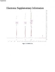
PDMS stamps were prepared from Sylgard 184, Dow Corning by mixing poly(dimethylsiloxane) and curing agent 10:1, pouring this mixture on a patterned silicon master and curing the combination at 60 °C over night. The stamps were removed from the silicon master, cut out with a scalpel and oxidized with a UV ozonizer (PSD-UV,Novascan Technologies Inc.) for 55 min before to use. If not used immediately, the PDMS stamps were stored in distilled water.
Figure 1: left: PDMS plate removed from silicon master, right: single PDMS stamp
I.II. Preparation of 11-(trichlorosilyl)undecan-1-ol SAMs Glass or silicon slides were cut into pieces of around 1.4x1.0 cm, sonicated in pentane, EtOHabs. and milliQ water for 5 min each and dried in a stream of argon. Afterwards they were immersed in a freshly prepared solution of piranha (H2SO4/H2O2 3:1) for 30 min, washed extensively with H2Odest., dried and put in a fresh solution of 11-(trichlorosilyl)undecyl acetate for 1 h. Subsequently the surfaces were sonicated in EtOHabs. for 5 min, dried and given in a 2.5 M solution of HCl and stirred for 2 h at 85 °C to deprotect the alcohol function. Directly prior to use the surfaces were cooled to room temperature, washed with milliQ water and dried in a stream of argon.
ESI 3
I.III. Preparation of TiO2 nanoparticles A 100 mL three neck flask was filled with 60 mL of diethyleneglycol under argon atmosphere was heated under stirring to 60 °C. 2 mL of TiCl4 were added by pipette and the temperature raised to 75 °C. After addition of 1 mL of H2O the mixture was stirred for 3 h at 160 °C. After cooling to room temperature the stirring was stopped and the flask was allowed to stand overnight. The suspension was transferred to a beaker, 100 mL of acetone were added and the whole mixture centrifuged at 3500 turns/min for 10 min. The white precipitate was washed at least three times with acetone and the remaining solvent was removed under vacuum. DLS measurements showed a diameter of 4-5 nm.
I.IV. Preparation of cellulose surfaces 2.5 g of cellulose were mixed in a 250 mL round bottom flask with 100 mL of dimethylacetamide. The mixture was stirred at 130 °C for 2 h. After cooling of the slurry to 100 °C 5 g of LiCl were added and the stirring continued over night and the cellulose dissolved completely. Glass or silicon slides were cut into pieces of around 1.4x1.0 cm, sonicated in pentane, EtOHabs. and milliQ water for 5 min each and dried in a stream of argon. Afterwards they were immersed in a freshly prepared solution of piranha (H2SO4/H2O2 3:1) for 30 min and washed extensively with H2Odest.. 150 µL of cellulose solution were dropped on the surface and spin coated at 7500 turns/min for 150 s. The surfaces were subsequently carefully washed with a mixture of isopropanol/ H2O 6:4 to remove LiCl and dried over night in an oven at 120 °C.
III.
Microcontact Printing
A freshly oxidized PDMS stamp was covered with 30 µL of fresh ink solution (5 mg TiO2 nanoparticles in 1 mL of MeOH were sonicated for 10 min, 3 µL of ethanolamine were added and sonicated again for 1 min) and incubated for 1 min. The stamp was dried in a stream of argon, placed carefully on a freshly prepared 11-(trichlorosilyl)undecan-1-ol SAM surface and irradiated with a 365 nm high power UV LED, which was placed around 2 cm above the stamp, for 30 min. After removing of the stamp, the surface was sonicated in DCM, EtOHabs. and milliQ water to remove all molecules, which are not covalently bond to the surface. After sonication the surface was dried in a stream of argon and left under vacuum overnight. ESI 4
IV.
Additional Experiments
Immobilization of anionic SNPs on polymer brushes A few drops of a 1 mg/mL solution of SNPs in EtOH were added to a dry patterned polymer brush surface and left till the EtOH was evaporated. To reach the half-filled polymer brushes, the surface was tilted by a few degrees during evaporation.
V.
Additional Analysis
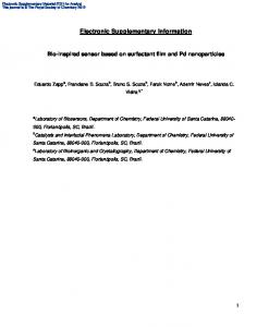
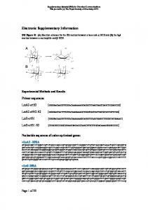
Figure S1: AFM measurements after different irradiation times and correlation of irradiation time to average profile height (different feature width due to the use of different patterned stamps).
ESI 5
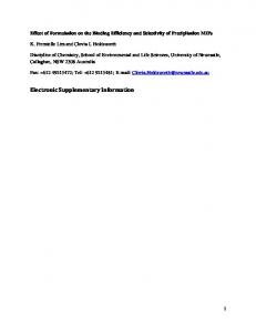
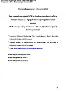
Figure S2: Profilometry measurements of cellulose coated surface.
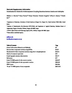
Figure S3: XPS analysis of PEI on cellulose coated surfaces.
ESI 6