677095 research-article2017
CDPXXX10.1177/0963721416677095Juan et al.Neural Correlates of Visuospatial Working Memory
Elucidating and Modulating the Neural Correlates of Visuospatial Working Memory via Noninvasive Brain Stimulation
Current Directions in Psychological Science 2017, Vol. 26(2) 165–173 © The Author(s) 2017 Reprints and permissions: sagepub.com/journalsPermissions.nav https://doi.org/10.1177/0963721416677095 DOI: 10.1177/0963721416677095 www.psychologicalscience.org/CDPS
Chi-Hung Juan1, Philip Tseng2,3,4, and Tzu-Yu Hsu3,4,5
1 Institute of Cognitive Neuroscience, National Central University; 2Graduate Institute of Humanities in Medicine, Taipei Medical University; 3TMU - Research Center for Brain and Consciousness, Taipei Medical University; 4 Shuang-Ho Hospital, Taipei Medical University; and 5Graduate Institute of Health and Biotechnology Law, Taipei Medical University
Abstract Visuospatial working memory refers to the short-term memory mechanism that enables humans to remember visual information across visual blackout periods such as eyeblinks or eye movements. In recent years, neuroscientific studies have made great progress in uncovering the brain regions that support visuospatial working memory. In this review, we focus on the role of the posterior parietal cortex in forming and maintaining visual information, and use it as an example to highlight how noninvasive brain-stimulation techniques, particularly transcranial magnetic, direct current, and alternating current stimulation, can shed light on this topic because of their unique strengths in modulating brain activities. Keywords visual short-term memory, visual working memory, TMS, tDCS, tACS
Anyone who has played children’s favorite game of “spot the difference” can agree that the game is not as easy as we would think. This is because during blackout periods when our eyes move from one image to another, or even when we blink, the eyes are practically blind (Bridgeman, Hendry, & Stark, 1975). Therefore, a game like “spot the difference” is actually an ultimate test of human abilities in attending to and remembering the right information. This highlights the intricate relationship between visual attention and memory and gave rise to the term visuospatial working memory (VWM) in the field of cognitive psychology to emphasize such active interaction instead of passive memory storage (Baddeley, 2003). VWM is an important psychological construct because it keeps just enough traces of our visual world that our perception of the world does not fall apart when vision is turned off momentarily during blackout periods like those mentioned above. As such, VWM ability has been shown to be positively correlated with fluid intelligence (Fukuda, Vogel, Mayr, & Awh, 2010; Kane & Engle, 2002). Decades of psychological research, however, have repeatedly demonstrated that people’s attention and memory can be surprisingly poor (Chabris & Simons, 2011; Simons
& Rensink, 2005). Behavioral studies using computerized psychological tasks that test people’s VWM by presenting multiple simple colors or shapes for less than a second have mostly concluded that people’s VWM performance usually averages around three to four items (Bays & Husain, 2008; Luck & Vogel, 2013). Neuroimaging studies using similar tasks have also pointed to several brain regions involving VWM processing, including the occipital cortex (Harrison & Tong, 2009; Serences, Ester, Vogel, & Awh, 2009; Silvanto & Cattaneo, 2010), the prefrontal cortex (PFC; D’Esposito, Postle, & Rypma, 2000; Feredoes, Heinen, Weiskopf, Ruff, & Driver, 2011; Fregni et al., 2005; Gazzaley, Cooney, McEvoy, Knight, & D’Esposito, 2005; Higo, Mars, Boorman, Buch, & Rushworth, 2011; Mulquiney, Hoy, Daskalakis, & Fitzgerald, 2011; Pope, Brenton, & Miall, 2015; Zaehle, Sandmann, Thorne, Jäncke, & Herrmann, 2011; Zanto, Rubens, Thangavel, & Corresponding Author: Chi-Hung Juan, Institute of Cognitive Neuroscience, National Central University, 300 Jhongda Rd., Jhongli District, Taoyuan City 320, Taiwan E-mails:
[email protected]
166 Gazzaley, 2011), and the posterior parietal cortex (PPC; Jones & Berryhill, 2012; Tseng et al., 2012; Tseng et al., 2013; Xu & Chun, 2006). Across these regions, the activation of the PPC seems to be one of the major sources to why we can only remember so little (Ikkai & Curtis, 2011), and it is therefore the focus of this brief review. Using fMRI, which traces the level of oxygenated blood in the brain, Todd and Marois (2004) found that PPC activity increased as people tried to remember more in VWM but eventually plateaued as people’s behavioral performance also plateaued around three to four items. Using electroencephalogram (EEG), which measures the brain’s electrophysiological activity, Vogel and Machizawa (2004) found similar findings: Brain-wave waveforms near the PPC area showed larger amplitudes (i.e., more activity) as people tried to remember more, and such amplitude differences can be used to predict who has good versus bad VWM. Together, converging behavioral and neuroimaging evidence has so far concluded that (a) our VWM is worse than what we think it is, and (b) the cause of such poor VWM seems to be limited activity in the PPC.
Types of NIBS Before diving into the application of noninvasive brain stimulation (NIBS) in investigating the neural mechanisms of VWM, we first introduce the tools that are often used for stimulation, as well as their strengths and weaknesses. Currently, the field of cognitive neuroscience has mostly used transcranial magnetic stimulation (TMS) and transcranial electric stimulation (TES) because they are noninvasive, in the sense that no surgery is involved at any stage of the experiment. TMS, by definition, uses magnetic forces to achieve its goal. It does so by generating a magnetic field via a coil placed over the participant’s skull, thereby inducing electric activity in the neurons beneath the skull (interested readers can look into the physics behind electromagnetism). TMS is relatively expensive, and less portable (though portable versions are starting to appear on the market), but its high spatial and temporal resolution makes it an irreplaceable tool for studies that require high levels of precision. In terms of spatial resolution, the focal point of a figure-ofeight coil is about a quarter of the size of a penny, allowing it to target smaller regions on the cortex. Timing-wise, a TMS pulse can be delivered in less than a millisecond, so it can be used to target a specific point in time during a cognitive task (e.g., stimulating the speech area right before someone is about to speak). Better yet, the number of TMS pulses can be customized by the experimenter. For example, delivering one TMS pulse every 25 milliseconds would result in a total of 40 pulses per second (aka 40 Hz), which matches the gamma-band range
Juan et al. (25–100 Hz) of neural activity. Therefore, by carefully specifying the amplitude and frequency of the pulses, TMS is also suitable for inducing neural entrainment in cortical areas, forcing the neurons to fire in a certain pattern that mimics the rhythmic activities (e.g., EEG) seen during the processing of the desired cognitive tasks (Huang, Edwards, Rounis, Bhatia, & Rothwell, 2005; Juan & Muggleton, 2012; Miniussi & Thut, 2010). Several devices that use this TMS-induced form of neural entrainment are now approved by the FDA for the treatment of refractory depression (George, Taylor, & Short, 2013; Li et al., 2014). TES uses electric current to stimulate the brain, and its earliest usage dates back to 1960 (Utz, Dimova, Oppenländer, & Kerkhoff, 2010). Unlike TMS, TES is inexpensive and very portable (roughly the size of an external hard drive). However, TES involves larger electrodes roughly the size of a large bandage (though there has been an emergence of penny-sized “HD” tDCS), and the duration of stimulation is usually 1 to 20 minutes, which is far from the millisecond advantage of TMS. There is one major advantage of TES, though, and that is due to the direct (tDCS) and alternating (tACS) natures of electric current. When stimulating with direct current, tDCS involves positively charged (anodal) and negatively charged (cathodal) direct current, and the two polarities have been suggested to increase and decrease neuronal activities, respectively (Nitsche & Paulus, 2000). Although this simple, dichotomous anodal-increase/cathodal-decrease neuronal view has changed in recent years (Fertonani & Miniussi, 2016; Jacobson, Koslowsky, & Lavidor, 2012; for a review in the context of VWM, see Hsu, Juan, & Tseng, 2016), the effect of anodal tDCS in elevating neural activities has been less controversial in the literature. This ability to increase neural activity, and thereby possibly elevate cognitive ability, has been a major advantage and explains why tDCS has been regaining momentum in cognitive neuroscience. Like TMS, TES can be used for neural entrainment if it is used in its alternating-current format, which is composed of sinusoidal waves with user-specified amplitude, phase, and frequency. So far, the number of tACS studies in the field is, although currently small, growing very rapidly. In conclusion, TMS and TES both have their advantages and weaknesses. Whereas TMS enjoys high spatial (to the millimeter) and temporal (to the millisecond) precision, TES enjoys the advantages of elevating neural activity (anodal tDCS) and customizable waveforms (tACS). With these differences in mind, it is also important to note two important similarities. First, both techniques can stimulate only the surface level of the brain (i.e., the cortex), as their stimulating strength is not strong enough to affect subcortical structures, and second, stimulating one area of the cortex can create a rippling effect,
Neural Correlates of Visuospatial Working Memory something neuroscientists call “functional connectivity” with other coactivating brain regions, both deep and cortical (see Yu, Tseng, Hung, Wu, & Juan, 2015, for a tDCSfMRI study; Liang et al., 2014, for a tDCS-EEG study; Ellison et al., 2014), that work in concert with the stimulated region to support a particular cognitive task or function. In the case of VWM, the dorsolateral prefrontal cortex, the frontal eye fields, areas within the medial temporal lobe, and visual cortices in the occipital lobe have all been implicated to work in synchrony with the PPC to support memory formation, maintenance, and retrieval. Although areas of the medial temporal lobe are too deep in the brain to be stimulated via NIBS, it is possible to alter their coactivation by applying NIBS over areas that are located on the cortex, and such alterations to a brain region’s coactivation can be very informative in shedding light on how different regions “functionally connect,” or interact with each other, to support VWM. Therefore, the effect of TMS and TES can be widespread within the brain network beyond just a single cortical region, and the mechanisms behind this rippling effect are currently under rigorous investigation.
TMS in VWM Since the discovery of PPC involvement in VWM via fMRI and EEG, several TMS studies have been done to test whether PPC activity is really causally related to VWM, and if so, at what point in time during the memory processes PPC matters the most. The question of causality is important because neuroimaging evidence is correlational, and correlations can often occur just by random chance. Additionally, a correlation between Factor A and Factor B can sometimes be real, but actually controlled by an unknown X factor (e.g., a city’s number of churches and its number of crime cases are highly correlated, but that is mediated by population). Therefore, cause-andeffect experiments are crucial before one can make inferences based on neuroimaging data. This question was tackled by Beck, Muggleton, Walsh, and Lavie (2006), who applied TMS pulses over the PPC of either the left or the right hemisphere. The logic was that applying TMS pulses while people were trying to remember something would create unintended neuronal noise in the PPC and therefore disrupt memory performance if PPC activity is really causal to VWM. Interestingly, these authors found that TMS over the right, but not the left, PPC was able to impair people’s VWM performance. This study suggested not only that PPC is indeed casually related to VWM processing, but also that only the right PPC (rPPC) is crucial to such visuospatial memory processes—something fMRI and EEG studies were not able to pick up. Beyond causality, the temporally precise nature of TMS is also able to answer the question of “when”—that is,
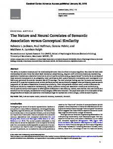
167 when is rPPC activity important? Is it when people are soaking in information (i.e., encoding), when they try to maintain information in memory (i.e., retention), or when they recall information (i.e., retrieval)? With this question in mind, Tseng and colleagues (2010) tweaked Beck et al.’s TMS design, and, instead of disruptively applying TMS pulses during the entire memory task, applied them only in the early or late memory stage. The rationale was simple: If people’s VWM performance plummets during the early stage, then rPPC activity likely reflects early encoding processes, and if it plummets during the later retrieval stage, then rPPC activity likely reflects retrieval processes. These authors found that early TMS impaired people’s VWM performance the most, with weaker impairment during the later stage. This suggests that the rPPC is doing most of its work early, when people are trying to take in as much information as they can (Fig. 1a).
tDCS in VWM With converging evidence from fMRI, EEG, and especially TMS studies, it was clear that rPPC activity had a direct cause-and-effect relationship with people’s VWM performance. The next natural question, then, became whether it was possible to help people improve their VWM by pushing their rPPC activity up a notch. This is where the unique advantage of anodal tDCS in increasing neural excitability comes in. In two experiments, Tseng and colleagues (2012) applied anodal tDCS over the rPPC and found that people below the three- to four-item average, namely those who could remember only three items or fewer, could indeed benefit from anodal tDCS and improve their memory to reach the four-item average (see also Heimrath, Sandmann, Becke, Müller, & Zaehle, 2012). However, those participants with good VWM (i.e., above four items) already, without tDCS, could not benefit from anodal tDCS. In this group of “high performers,” anodal tDCS did not help or hurt their VWM performance (Fig. 1b). This behavioral dichotomy was also reflected in people’s brain-wave data, where event-related potentials near rPPC during both the attention and memory stages showed large changes after tDCS in low performers but not high performers (Fig. 2a). One subsequent study by Hsu, Tseng, Liang, Cheng, and Juan (2014) also found that these low and high performers differed remarkably in terms of their brain-wave data and reactivity to tDCS: Low performers’ EEG signals exhibited high alpha amplitude in the parieto-occipital regions, something that has been associated with distracted attention, and anodal tDCS was able to tame it down and improve their VWM performance (Fig. 2b). As in the 2012 study, high performers did not show any strong alpha amplitude to begin with, and that remained unaffected by anodal
Juan et al.
168
a
TMS Study 2
Retrieval Stage
Memory Stage
*
*
1.6
+
1.2
d′
+
0.8 0.4
200 ms
Early TMS (10 Hz, 3 pulses)
b
200 ms
100-ms Retention Interval
0
No TMS
Late TMS (10 Hz, 3 pulses)
Sham
tDCS Study Memory Stage
4
Anodal
***
5
Retrieval Stage
Early TMS Late TMS
*
K
3 2 1 200 ms
900 ms Retention Interval
2,200 ms
0
Low
High
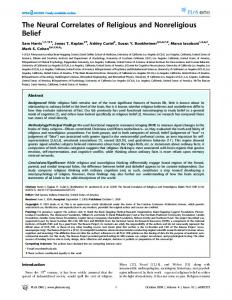
Fig. 1. Procedures and results from the (a) 2010 transcranial magnetic stimulation (TMS) study (Tseng et al., 2010) and (b) the 2012 transcranial direct current stimulation (tDCS) study (Tseng et al., 2012). Panel (a) depicts the design of the change-detection task. Participants had to remember four faces in the first array (i.e., memory stage) and try to detect any change in the second array (i.e., retrieval stage). (Note that the black bars over the faces are used here for publication but were not used in the actual experiment.) Meanwhile, to delineate the timing of right posterior parietal cortex (rPPC) involvement, TMS was applied over the rPPC either during the memory stage (i.e., early TMS) or the retrieval stage (i.e., late TMS). Panel (b) depicts the results, showing that although rPPC TMS was effective in both stages, the effect of early-memorystage TMS was by far the largest. This suggests that the rPPC is especially critical to the processes of memory encoding. Panel (b) shows the change-detection task with color blocks. The task requirement is similar to that in (a), where participants were to judge whether the first and second arrays were the same or not. The results showed that low performers who could only remember three color blocks or less were able to improve from sham to anodal tDCS. tDCS did not work for high performers who already scored above the average of three blocks to begin with. This suggests that, among all the factors that may contribute to the complex nature of tDCS effects, individual capabilities may be one of them. One possible way to help the high performers to improve is to optimize the parameters of tDCS to better excite their brain activities. This possibility should be addressed by future tDCS studies.
tDCS. Furthermore, these authors found that, in low performers, alpha amplitude before the onset of the task (i.e., before the participants saw anything on the computer screen to remember) was particularly useful in categorizing their subsequent VWM performance 2 or 3 seconds later. This observation of alpha amplitude taking place before the task is interesting, as it implies a critical involvement of visual attention throughout the processes of VWM (Tsubomi, Fukuda, Watanabe, & Vogel, 2013),
which echoes the earlier notion that attention and shortterm memory come hand in hand in most experimental settings (see Shipstead, Lindsey, Marshall, & Engle, 2014, for an insightful discussion). Here it is important to note that different variations of experimental design, such as task difficulty ( Jones & Berryhill, 2012; Wu et al., 2014; Wu et al., 2016) and the ways in which researchers categorize low and high performers, may cause different patterns of results (cf. Jones & Berryhill, 2012). Therefore,
Neural Correlates of Visuospatial Working Memory
a
169
b
Event‐Related Potentials μV
Alpha Power Distribution
SPCN
N2pc 6
Sham
Anodal
Low Performers
4 2 0 –100
0
100
200
300
400
500
600
–2
μV
–4
Low Performers
6
High Performers
4 2 0 –100
0
100
200
300
400
500
600
–2 –4
Sham_contra
Sham_lpsi
Anodal_contra
Anodal_lpsi
High Performers
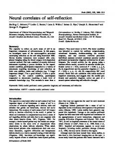
Fig. 2. Transcranial direct current stimulation– (tDCS-) induced changes in low and high performers’ brain waves. Panel (a) shows results from Tseng et al. (2012). Low performers’ behavioral improvement can be traced to their brain-wave changes after tDCS application. Here, the readers should look for the gap between the dotted and the solid lines: Bigger separation between the two indicates a bigger tDCS effect. In low performers (top panel), there is no separation between the two lines at all in the sham condition; the separation emerges only in the anodal-tDCS condition. This tDCS effect is particularly noticeable at the 300-ms and 400- to 600-ms time windows (gray areas), which are known as the N2pc and SPCN components and represent visual attention and memory processes, respectively. In high performers, note that the gap between the dotted and the solid line was already apparent even without tDCS, and applying tDCS did not help enlarge the gap, either, which coincides with the high performers’ behavioral performance quite well. Panel (b) shows similar results observed when we broke down the brain-wave signal into different frequency components in Hsu, Tseng, Liang, Cheng, and Juan (2014). In low performers, alpha wave was particularly strong in the parietal and occipital regions (sham), which is indicated by red, orange, and yellow on the chart. Alpha power is mostly used to index unfocused attention, which is in line with our behavioral observations. The strong alpha power was drastically reduced after tDCS application over the rPPC (anodal). In high performers, similar to the 2012 study, alpha power was already low in the parietal and occipital regions even without tDCS (sham), and therefore applying tDCS did not help reduce alpha power any further.
further work is necessary to clarify the interaction of task characteristics, intrinsic individual differences, and tDCS effects (for a more systematic investigation, see Hsu et al., 2016).
tACS in VWM tACS is a relatively newer form of TES but has been gaining momentum in the field of VWM because of its effectiveness in inducing neural entrainment via alternating
current (its underlying mechanism is quite different from that of TMS; see Herrmann, Rach, Neuling, & Strüber, 2013). Benchmarking studies have reported a relatively smaller effect in tACS than tDCS (Paulus, 2011), which is reasonable since tACS can be viewed as a more precise version of TES. In VWM, one notable study was conducted by Jaušovec and Jaušovec (2014). These authors applied theta-wave tACS (roughly 6 Hz), which was chosen based on persistent EEG observations of theta-gamma coupling in working memory formation (Sauseng,
Juan et al.
170 Griesmayr, Freunberger, & Klimesch, 2010), over either the left frontal or parietal cortex, and found enhanced VWM performance only when theta tACS was applied over the PPC. The behavioral improvement was also coupled with stronger brain-wave signals that indicate memory recognition. Similarly, Tseng, Chang, Liang, Chang, and Juan (2016) used 40-Hz gamma tACS over the temporal-parietal pathway and also found that gamma entrainment was critical to certain types of VWM processing. In the context of tACS, it is usually interpreted that the two electrodes of tACS are oscillating in rhythm (6 times per second, in this case), forcing the two brain regions underneath the electrodes to communicate in the same fashion. You can imagine this as a band playing, where each band member (i.e., brain region) needs to coordinate with the others in order to achieve meaningful synchronization (i.e., neural communication). This network-connectivity nature of tACS can be a useful tool once each node within the network has been identified (Battleday, Muller, Clayton, & Cohen Kadosh, 2014). In the Jaušovec and Jaušovec study, however, the authors placed the return electrode over the right eyebrow instead of over a brain region that has been identified as a node within the VWM network, and they still observed an enhancement effect even though the PPC was not communicating with other regions. Therefore, our traditional understanding of the mechanisms behind tACS and their applications may need to be updated in order to account for the increasing number of new tACS findings in VWM. This conceptual update will likely include factors such as individual difference and variability (Hsu et al., 2016), inter-electrode distance (e.g., Moliadze, Antal, & Paulus, 2010), inter-electrode phase differences (Polanía et al., 2012; Tseng et al., 2016), and functional connectivity that we have mentioned earlier (Ellison et al., 2014; Yu et al., 2015). On this note, it is exciting to see that studies have already begun to combine neuroimaging techniques with NIBS, which has several unique strengths, as it allows researchers to (a) look deeper into the mechanisms behind NIBS in the brain and, most important, (b) investigate the distal network-like effect of NIBS and therefore map out the participating nodes, as well as how they interact with each other, in order to give us a closer look at the neuronal “teamwork” that’s happening behind the scenes. One excellent example of such a study was carried out by Ellison and colleagues (2014), who applied cathodal tDCS over the rPPC (which should decrease activity in the PPC) and monitored changes in activity in other participating brain regions such as the frontal eye fields in the prefrontal cortex. These authors observed decreased frontal eye field activity even though the cathodal electrode was placed over the rPPC, which suggests functional communication between the PPC and the frontal eye fields, such that activity in the frontal eye
fields can be altered when information flow from the PPC is disrupted. Similar activity changes can also be observed in deeper brain regions that are otherwise hard to stimulate with NIBS (e.g., Yu et al., 2015). An interesting direction for follow-up research would be the use of tACS in different frequencies to test the “channel” in which parietal-frontal communication occurs. Together, a NIBS-fMRI approach like this provides a unique opportunity to investigate regional networks in the brain that were not available to researchers before. For this reason, we expect to see an increase in NIBS-neuroimaging studies in the coming years.
Concluding Remarks Here we have argued for a unique place for NIBS in cognitive neuroscience because of its advantages in establishing causality, pinpointing temporal windows, boosting excitability, and assessing network connectivity. It is important to reiterate that although NIBS is used to stimulate only one or two brain regions, from there its effect can propagate toward deeper regions that are otherwise impossible to stimulate, and the field of VWM has benefited much from these attributes. In this review we have also focused on NIBS studies that helped clarify the roles of the PPC in supporting VWM. Because VWM is a collective process that involves different aspects of cognitive processing (attention, encoding, maintenance, etc.), it is important to note that the PPC should not be taken as a do-it-all region that is self-sufficient in sustaining VWM. Rather, the network behind VWM involves contributions from the PFC, the frontal eye fields, the PPC, and regions within the occipital cortex. It is also important to note that PPC activation is by no means exclusive to VWM processing. In fact, other NIBS studies of phenomena that are not related to VWM, such as mathematical processing or time perception, have also reported a critical role for PPC activity in their respective domains (for a review, see Bueti & Walsh, 2009). Therefore, as it is with almost all brain regions, the function of the PPC likely lies in something much more fundamental across all cognitive domains. In this light, Walsh (2003) has proposed “a theory of magnitude” that views the PPC as a magnitude processor whose job is to track different aspects of magnitudes in the world, be they of space, time, number, or maybe even music (Kadosh et al., 2008)! Although this theory assumes that the human brain represents space, time, and numbers all in some kind of common spatialcoordinate systems (unbeknownst to our conscious awareness), this assumption has actually gained a lot of empirical support in the spatial, temporal, and numerical literature (Bueti & Walsh, 2009). To put this back in the context of VWM, perhaps what’s common across all the studies reviewed above is the number of spatial locations that need to be
Neural Correlates of Visuospatial Working Memory remembered or tracked in VWM. In all VWM studies, as memory load increases, so does the number of spatial locations that need to be tracked. Indeed, one study in particular by Parra, Della Sala, Logie, and Morcom (2014) used a similar VWM task but randomized the spatial locations of every memory object so that the locations were no longer relevant to the task and should not be encoded into VWM. With such a design, these authors did not find the commonly observed activation from the rPPC, a result that lends support to the magnitude theory of PPC functioning. In this respect, although most VWM studies have pointed to the rPPC as a key player, it is possible that the PPC is actually tracking “where” and “how many” while sending such information to other regions within the network via neural entrainment and functional connectivity, which can be investigated with various kinds of NIBS such as rTMS or tACS of a specific frequency. To this end, the field of cognitive neuroscience is moving toward a combined NIBS-neuroimaging approach, as many of the reviewed studies have already done, and with that we anticipate that the number of NIBS studies and their impact will only increase as the field progresses. Recommended Reading Fertonani, A., & Miniussi, C. (2016). (See References). A review and discussion of the mechanisms and models of TES. Herrmann, C. S., Rach, S., Neuling, T., & Strüber, D. (2013). Transcranial alternating current stimulation: A review of the underlying mechanisms and modulation of cognitive processes. Frontiers in Human Neuroscience, 7. doi:10.3389/ fnhum.2013.00279. A collection of tACS findings and discussion of its possible mechanism from a network perspective. Juan, C. H., & Muggleton, N. G. (2012). (See References). A systematic review of the application of NIBS in the context of cognitive control. Luck, S. J., & Vogel, E. K. (2013). (See References). A review of behavioral and neuroimaging findings on the topic of VWM. Miniussi, C., & Thut, G. (2010). (See References). A review and discussion on the effect and mechanism of TMS from a brain-network perspective. Walsh, V. (2003). (See References). A unifying theory on the function of the PPC in cognition.
Declaration of Conflicting Interests The authors declared that they had no conflicts of interest with respect to their authorship or the publication of this article.
Funding This research was supported by grants from the Ministry of Science and Technology (MOST), Taiwan, to Chi-Hung Juan (NSC 101-2628-H-008-001-MY4, MOST 103-2410-H-008-023-MY3, MOST 104-2745-B-008-002), Philip Tseng (MOST 104-2410-H-038013-MY3; MOST 104-2420-H-038-001-MY3), and Tsu-Yu Hsu (MOST 104-2410-H-038-012-MY2) and by grants from Taipei Medical University to Philip Tseng (TMU 104-AE1-B07; 105TMUSHH-20) and Tsu-Yu Hsu (TMU 104-AE1-B08, SKH-TMU-105-05).
171 References Baddeley, A. (2003). Working memory: Looking back and looking forward. Nature Reviews Neuroscience, 4, 829–839. Battleday, R. M., Muller, T., Clayton, M. S., & Cohen Kadosh, R. (2014). Mapping the mechanisms of transcranial alternating current stimulation: A pathway from network effects to cognition. Frontiers in Psychiatry, 5, Article 162. doi:10.3389/ fpsyt.2014.00162 Bays, P. M., & Husain, M. (2008). Dynamic shifts of limited working memory resources in human vision. Science, 321, 851–854. Beck, D. M., Muggleton, N., Walsh, V., & Lavie, N. (2006). Right parietal cortex plays a critical role in change blindness. Cerebral Cortex, 16, 712–717. Bridgeman, B., Hendry, D., & Stark, L. (1975). Failure to detect displacement of the visual world during saccadic eye movements. Vision Research, 15, 719–722. Bueti, D., & Walsh, V. (2009). The parietal cortex and the representation of time, space, number and other magnitudes. Philosophical Transactions of the Royal Society B: Biological Sciences, 364, 1831–1840. Chabris, C., & Simons, D. (2011). The invisible gorilla: And other ways our intuitions deceive us. New York, NY: Broadway Books. D’Esposito, M., Postle, B. R., & Rypma, B. (2000). Prefrontal cortical contributions to working memory: Evidence from event-related fMRI studies. Experimental Brain Research, 133, 3–11. Ellison, A., Ball, K. L., Moseley, P., Dowsett, J., Smith, D. T., Weis, S., & Lane, A. R. (2014). Functional interaction between right parietal and bilateral frontal cortices during visual search tasks revealed using functional magnetic imaging and transcranial direct current stimulation. PLoS ONE, 9(4), e93767. doi:10.1371/journal.pone.0093767 Feredoes, E., Heinen, K., Weiskopf, N., Ruff, C., & Driver, J. (2011). Causal evidence for frontal involvement in memory target maintenance by posterior brain areas during distracter interference of visual working memory. Proceedings of the National Academy of Sciences, USA, 108, 17510–17515. Fertonani, A., & Miniussi, C. (2016). Transcranial electrical stimulation: What we know and do not know about mechanisms. The Neuroscientist. Advance online publication. doi:10.1177/1073858416631966 Fregni, F., Boggio, P. S., Nitsche, M., Bermpohl, F., Antal, A., Feredoes, E., . . . Pascual-Leone, A. (2005). Anodal transcranial direct current stimulation of prefrontal cortex enhances working memory. Experimental Brain Research, 166, 23–30. Fukuda, K., Vogel, E., Mayr, U., & Awh, E. (2010). Quantity, not quality: The relationship between fluid intelligence and working memory capacity. Psychonomic Bulletin & Review, 17, 673–679. Gazzaley, A., Cooney, J. W., McEvoy, K., Knight, R. T., & D’Esposito, M. (2005). Top-down enhancement and suppression of the magnitude and speed of neural activity. Journal of Cognitive Neuroscience, 17, 507–517. George, M. S., Taylor, J. J., & Short, E. B. (2013). The expanding evidence base for rTMS treatment of depression. Current Opinion in Psychiatry, 26, 13–18. doi:10.1097/ YCO.0b013e32835ab46d
172 Harrison, S. A., & Tong, F. (2009). Decoding reveals the contents of visual working memory in early visual areas. Nature, 458, 632–635. Heimrath, K., Sandmann, P., Becke, A., Müller, N. G., & Zaehle, T. (2012). Behavioral and electrophysiological effects of transcranial direct current stimulation of the parietal cortex in a visuo-spatial working memory task. Frontiers in Psychiatry, 3, Article 56. Higo, T., Mars, R. B., Boorman, E. D., Buch, E. R., & Rushworth, M. F. (2011). Distributed and causal influence of frontal operculum in task control. Proceedings of the National Academy of Sciences, USA, 108, 4230–4235. Hsu, T.-Y., Juan, C.-H., & Tseng, P. (2016). Individual differences and state-dependent responses in transcranial direct current stimulation. Frontiers in Human Neuroscience, 10, Article 643. doi:10.3389/fnhum.2016.00643 Hsu, T. Y., Tseng, P., Liang, W. K., Cheng, S. K., & Juan, C. H. (2014). Transcranial direct current stimulation over right posterior parietal cortex changes prestimulus alpha oscillation in visual short-term memory task. NeuroImage, 98, 306–313. Huang, Y. Z., Edwards, M. J., Rounis, E., Bhatia, K. P., & Rothwell, J. C. (2005). Theta burst stimulation of the human motor cortex. Neuron, 45, 201–206. Ikkai, A., & Curtis, C. E. (2011). Common neural mechanisms supporting spatial working memory, attention and motor intention. Neuropsychologia, 49, 1428–1434. Jacobson, L., Koslowsky, M., & Lavidor, M. (2012). tDCS polarity effects in motor and cognitive domains: A meta-analytical review. Experimental Brain Research, 216, 1–10. Jaušovec, N., & Jaušovec, K. (2014). Increasing working memory capacity with theta transcranial alternating current stimulation (tACS). Biological Psychology, 96, 42–47. Jones, K., & Berryhill, M. E. (2012). Parietal contributions to visual working memory depend on task difficulty. Frontiers in Psychiatry, (3), Article 81. doi:10.3389/fpsyt.2012.00081 Juan, C. H., & Muggleton, N. G. (2012). Brain stimulation and inhibitory control. Brain Stimulation, 5, 63–69. Kadosh, R. C., Brodsky, W., Levin, M., & Henik, A. (2008). Mental representation: What can pitch tell us about the distance effect? Cortex, 44, 470–477. Kane, M. J., & Engle, R. W. (2002). The role of prefrontal cortex in working-memory capacity, executive attention, and general fluid intelligence: An individual-differences perspective. Psychonomic Bulletin & Review, 9, 637–671. Li, C. T., Chen, M. H., Juan, C. H., Huang, H. H., Chen, L. F., Hsieh, J. C., . . . Su, T. P. (2014). Efficacy of prefrontal thetaburst stimulation in refractory depression: A randomized sham-controlled study. Brain, 137, 2088–2098. Liang, W. K., Lo, M. T., Yang, A. C., Peng, C. K., Cheng, S. K., Tseng, P., & Juan, C. H. (2014). Revealing the brain’s adaptability and the transcranial direct current stimulation facilitating effect in inhibitory control by multiscale entropy. NeuroImage, 90, 218–234. Luck, S. J., & Vogel, E. K. (2013). Visual working memory capacity: From psychophysics and neurobiology to individual differences. Trends in Cognitive Sciences, 17, 391–400.
Juan et al. Miniussi, C., & Thut, G. (2010). Combining TMS and EEG offers new prospects in cognitive neuroscience. Brain Topography, 22, 249–256. Moliadze, V., Antal, A., & Paulus, W. (2010). Electrode-distance dependent after-effects of transcranial direct and random noise stimulation with extracephalic reference electrodes. Clinical Neurophysiology, 121, 2165–2171. Mulquiney, P. G., Hoy, K. E., Daskalakis, Z. J., & Fitzgerald, P. B. (2011). Improving working memory: Exploring the effect of transcranial random noise stimulation and transcranial direct current stimulation on the dorsolateral prefrontal cortex. Clinical Neurophysiology, 122, 2384–2389. Nitsche, M. A., & Paulus, W. (2000). Excitability changes induced in the human motor cortex by weak transcranial direct current stimulation. The Journal of Physiology, 527, 633–639. Parra, M. A., Della Sala, S., Logie, R. H., & Morcom, A. M. (2014). Neural correlates of shape–color binding in visual working memory. Neuropsychologia, 52, 27–36. Paulus, W. (2011). Transcranial electrical stimulation (tES – tDCS; tRNS, tACS) methods. Neuropsychological Rehabilitation, 21, 602–617. Polanía, R., Nitsche, M. A., Korman, C., Batsikadze, G., & Paulus, W. (2012). The importance of timing in segregated theta phase-coupling for cognitive performance. Current Biology, 22, 1314–1318. Pope, P. A., Brenton, J. W., & Miall, R. C. (2015). Task-specific facilitation of cognition by anodal transcranial direct current stimulation of the prefrontal cortex. Cerebral Cortex, 25, 4551–4558. Sauseng, P., Griesmayr, B., Freunberger, R., & Klimesch, W. (2010). Control mechanisms in working memory: A possible function of EEG theta oscillations. Neuroscience & Biobehavioral Reviews, 34, 1015–1022. Serences, J. T., Ester, E. F., Vogel, E. K., & Awh, E. (2009). Stimulus-specific delay activity in human primary visual cortex. Psychological Science, 20, 207–214. Shipstead, Z., Lindsey, D. R., Marshall, R. L., & Engle, R. W. (2014). The mechanisms of working memory capacity: Primary memory, secondary memory, and attention control. Journal of Memory and Language, 72, 116–141. Silvanto, J., & Cattaneo, Z. (2010). Transcranial magnetic stimulation reveals the content of visual short-term memory in the visual cortex. NeuroImage, 50, 1683–1689. Simons, D. J., & Rensink, R. A. (2005). Change blindness: Past, present, and future. Trends in Cognitive Sciences, 9, 16–20. Todd, J. J., & Marois, R. (2004). Capacity limit of visual shortterm memory in human posterior parietal cortex. Nature, 428, 751–754. Tseng, P., Chang, C. F., Chiau, H. Y., Liang, W. K., Liu, C. L., Hsu, T. Y., . . . Juan, C. H. (2013). The dorsal attentional system in oculomotor learning of predictive information. Frontiers in Human Neuroscience, 7, Article 404. Tseng, P., Chang, Y. T., Liang, W. K., Chang, C. F., & Juan, C. H. (2016). The critical role of phase difference in gamma oscillation within the temporoparietal network for binding visual working memory. Scientific Reports, 6, Article 32138. doi:10.1038/srep32138
Neural Correlates of Visuospatial Working Memory Tseng, P., Hsu, T. Y., Chang, C. F., Tzeng, O. J., Hung, D. L., Muggleton, N. G., . . . Juan, C. H. (2012). Unleashing potential: Transcranial direct current stimulation over the right posterior parietal cortex improves change detection in low-performing individuals. The Journal of Neuroscience, 32, 10554–10561. Tseng, P., Hsu, T. Y., Muggleton, N. G., Tzeng, O. J., Hung, D. L., & Juan, C. H. (2010). Posterior parietal cortex mediates encoding and maintenance processes in change blindness. Neuropsychologia, 48, 1063–1070. Tsubomi, H., Fukuda, K., Watanabe, K., & Vogel, E. K. (2013). Neural limits to representing objects still within view. The Journal of Neuroscience, 33, 8257–8263. Utz, K. S., Dimova, V., Oppenländer, K., & Kerkhoff, G. (2010). Electrified minds: Transcranial direct current stimulation (tDCS) and galvanic vestibular stimulation (GVS) as methods of non-invasive brain stimulation in neuropsychology—A review of current data and future implications. Neuropsychologia, 48, 2789–2810. Vogel, E. K., & Machizawa, M. G. (2004). Neural activity predicts individual differences in visual working memory capacity. Nature, 428, 748–751. Walsh, V. (2003). A theory of magnitude: Common cortical metrics of time, space and quantity. Trends in Cognitive Sciences, 7, 483–488. Wu, Y.-J., Tseng, P., Chang, C. F., Juan, C. H., Pai, M.-C., & Lin, C.-C. (2014). Modulating the interference effect on spatial
173 working memory by applying transcranial direct current stimulation over the right dorsolateral prefrontal cortex. Brain and Cognition, 91, 87–94. Wu, Y.-J., Tseng, P., Huang, H.-W., Hu, J.-F., Juan, C.-H., Hsu, K.-S., & Lin, C.-C. (2016). The facilitative effect of transcranial direct current stimulation on visuospatial working memory in patients with diabetic polyneuropathy: A pre–post shamcontrolled study. Frontiers in Human Neuroscience,10, Article 479. doi:10.3389/fnhum.2016.00479 Xu, Y., & Chun, M. M. (2006). Dissociable neural mechanisms supporting visual short-term memory for objects. Nature, 440, 91–95. Yu, J., Tseng, P., Hung, D. L., Wu, S. W., & Juan, C. H. (2015). Brain stimulation improves cognitive control by modulating medial-frontal activity and preSMA-vmPFC functional connectivity. Human Brain Mapping, 36, 4004–4015. Zaehle, T., Sandmann, P., Thorne, J. D., Jäncke, L., & Herrmann, C. S. (2011). Transcranial direct current stimulation of the prefrontal cortex modulates working memory performance: Combined behavioural and electrophysiological evidence. BMC Neuroscience, 12, Article 2. doi:10.1186/1471-220212-2 Zanto, T. P., Rubens, M. T., Thangavel, A., & Gazzaley, A. (2011). Causal role of the prefrontal cortex in top-down modulation of visual processing and working memory. Nature Neuroscience, 14, 656–661.