with 200 μg/mL hygromycin B. 8. Change the medium daily and select transfected cells over a period of 7 d. Cells may be trypsinized and replated into new ...
Cardiomyocyte Enrichment
157
14 Cardiomyocyte Enrichment in Differentiating ES Cell Cultures: Strategies and Applications Kishore B.S. Pasumarthi and Loren J. Field 1. Introduction Advances in our understanding of cardiomyocyte cell biology have been dependent largely upon the ability to generate primary cultures from enzymatically dispersed fetal, neonatal, or adult hearts. Primary cardiomyocyte cultures recapitulate many of the physiologic and molecular attributes found in intact hearts at the corresponding developmental stage. Moreover, these cultures are readily amenable to a wide variety of physical, physiologic, and molecular analyses. Gene transfer approaches including traditional calcium phosphate and lipofection techniques, as well as viral transduction with recombinant retro-, adeno-, or adeno-associated viruses are also readily accomplished. In light of these attributes, primary cardiomyocyte cultures constitute an extremely versatile experimental system. Despite their tremendous experimental utility, primary cardiomyocytes cultures are not without weaknesses. For example, it is very difficult to generate large-scale cultures of adult cardiomyocytes which retain a differentiated phenotype. Consequently, studies designed to address issues pertaining to adult cardiac biology frequently utilize primary cultures of fetal or neonatal cardiomyocytes. Given the marked physiologic differences observed in vivo between fetal–neonatal cardiomyocytes as compared to terminally differentiated adult cells, interpretation of such experiments can be problematic. Perhaps the greatest weaknesses of primary cardiomyocyte cultures is that they are subject to strict temporal constraints. This is a result of the relatively rapid cell cycle withdrawal that cardiomyocytes undergo in culture, in contrast to the sustained proliferation observed for noncardiomyocytes (i.e., fibroblasts, vascular smooth muscle cells, etc.). Although enriched cardiomyocyte preparations can be generated, the resulting cultures typically will be contaminated with noncardiomyocytes. Treatments designed to block proliferation of the contaminating cells (e.g., exposure to Ara-C or UV irradiation) can frequently have a deleterious effect on the resident cardiomyocytes as well. Efforts to circumvent this intrinsic limitation have typically relied on enhancing cardiomyocyte proliferative capacity through the targeted expression of oncogenes or proto-oncogenes. Although this approach has yielded a number of cardiomyocyte cell lines which retained varying degrees of differentiation (reviewed in ref. 1), From: Methods in Molecular Biology, vol. 185: Embryonic Stem Cells: Methods and Protocols Edited by: K. Turksen © Humana Press Inc., Totowa, NJ
157
158
Pasumarthi and Field
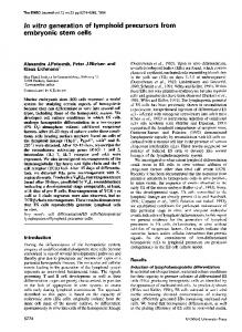
Fig. 1. Structure of the MHC-neor/pGK-hygror transgene. The transgene comprises an α-MHC-aminoglycoside phosphotransferase (MHC-neor) transcriptional unit and pGK-hygror transcriptional unit on a common pBM20 vector backbone. The α-MHC promoter consisted of 4.5 kb of 5′ flanking sequence and 1 kb of the gene encompassing exons 1 through 3 up to, but not including, the initiation codon (19). The aminoglycoside phosphotransferase (neor ) cDNA was subcloned from pMC1-neo poly(A)(Stratagene, La Jolla, CA). The pGK-hygromycin sequences were described previously (20).
sustained oncogene expression prohibits studies on many aspects related to the terminal differentiation program. In vitro differentiation of embryonic stem (ES) cells provides an alternative source of cardiomyocytes for study in tissue culture. ES cells are derived from the inner cell mass of preimplantation embryos and, when grown under appropriate conditions, can be propagated in an undifferentiated state indefinitely. Culturing ES cell in suspension and in the absence of differentiation inhibitors, such as leukemia inhibitory factor (LIF), results in the formation of multicellular structures called embryoid bodies (EBs), which reproducibly contain most if not all ectoderm-, endoderm-, and mesoderm-derived cell lineages (2,3; see also other chapters in this series). In many instances, regions of cardiomyogenesis are readily apparent, as evidenced by the presence of spontaneous contractile activity. Numerous studies have shown that cardiogenic induction in ES cells faithfully recapitulates the physical and molecular properties of developing myocardium in vivo (4–11). In addition, cell cycle withdrawal and terminal differentiation in ES-derived cardiomyocytes appears to parallel closely that which occurs during normal development (12). In this report, we describe a relatively simple genetic enrichment approach that facilitates the generation of highly enriched cultures of cardiomyocytes from differentiating ES cells (13). The approach relies on the use of two transcriptional units introduced into undifferentiated ES cells on a common vector backbone (see Fig. 1). The first transcriptional unit comprises a promoter expressed in undifferentiated ES cells linked to a marker gene suitable for enrichment of cells carrying the DNA (in our example, the phosphoglycerate kinase [PGK] promoter and a cDNA encoding resistance to hygromycin [hygror ] were used; the transcriptional unit is designated pGK-hygror). The second transcriptional unit comprises a cell lineage restricted promoter linked to a marker gene suitable for enrichment of the desired cells (in our example, the cardiomyocyte-restricted α-cardiac myosin heavy chain [α-MHC] promoter and a cDNA encoding aminoglycoside phosphotransferase were used; the transcriptional unit is designated MHC-neor ).
Cardiomyocyte Enrichment
159
Using this approach, the generation of highly enriched cardiomyocyte cultures is experimentally quite simple (Fig. 2). Undifferentiated ES cells are transfected with the MHC-neor/pGK-hygror construct. Cells incorporating the DNA are enriched based on their resistance to hygromycin. Differentiation is then induced, and once evidence of cardiomyogenesis is observed (i.e., spontaneous contractile activity), the cultures are treated with geneticin (G418). Because the α-MHC promoter is only active in cardiomyocytes, only these cells express aminoglycoside phosphotransferase and survive G418 treatment (Fig. 2). Although the example presented utilizes antibiotic resistance as the basis of the enrichment, a wide variety of analogous marker genes–enrichment protocols can readily be used (e.g., green fluorescent protein [GFP] targeted expression of cell surface markers, which could be used in conjunction with fluorescence-activated cell sorting [FACS] protocols, etc.). The genetic enrichment approach has the advantage that very long-term cultures of terminally differentiated cardiomyocytes can be generated, since noncardiomyocytes are eliminated from the culture. Moreover, the approach is easily amenable to gene transfer, either prior to differentiation or, alternatively, after the generation of terminally differentiated cells. In addition, the genetic enrichment approach is applicable to all cell lineages derived from ES cells, as well as to all multipotent stem cell systems. Here, we present a detailed description of the genetic enrichment protocols used to produce essentially pure populations of cardiomyocytes from differentiating ES cells. The Methods section is divided into subsections describing: (1) the transfection and selection of undifferentiated ES cells; (2) the “en mass” differentiation of the selected cells; (3) the generation of highly enriched cardiomyocyte cultures; and (4) the use of periodic acid Schiff’s (PAS) staining to visualize colonies of ES-derived cardiomyocytes. In addition, we provide a practical example of the use of the system to generate cardiomyocytes of sufficient purity for intracardiac engraftment (an emerging protocol aimed at restoring systolic function in diseased hearts). We also provide an example of how the system can be utilized to perform an ES-derived cardiomyocyte colony growth assay, which provides a relatively rapid throughput system to identify genes that impact on cardiomyocyte cell cycle regulation. 2. Materials 1. 2. 3. 4. 5. 6. 7. 8. 9. 10. 11. 12. 13.
ES cell line ES-D3 (obtained from the American Type Culture Collection, Rockville, MD). pMHC-neor/PGK-hygror plasmid. Restriction enzymes XhoI and HindIII. Geneclean kit (Bio 101, cat. no. 1001-400). Cell culture dishes (Corning, cat. no. 430293). ES cell grade fetal bovine serum (FBS) (Gibco, cat. no. 10439-024). Dulbecco’s modified Eagle’s medium (DMEM) (Sigma, cat. no. D-6546). ESGRO/mLIF (murine leukemia inhibitory factors) (Chemicon International, cat. no. ESG-1107). Nonessential amino acids (Gibco, cat. no. 11140-050), L-Glutamine (Gibco, cat. no. 25030-081), and Penicillin–Streptomycin solutions (Gibco, cat. no. 15070-063). 2-Mercaptoethanol (Sigma, cat. no. M-7522). Phosphate-buffered saline (PBS) (Sigma, cat. no. D-8537). Trypsin solution: 0.025% trypsin (1!250) (Gibco, cat. no. 27250-042), 1 mM EDTA and 1% chicken serum (Sigma, cat. no. 16110-082) in sterile PBS. Electroporator (Gibco, cat. no. 71600-19).
160
Pasumarthi and Field
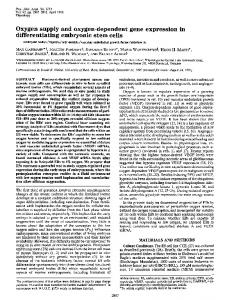
Fig. 2. (A) Schematic diagram of the genetic enrichment approach. (B) Hoechst epifluorescence of a nonselected culture of ES cells carrying the MHC-neor/pGK-hygromycin transgene 16 d after differentiation was induced. Note the high density of cells present in the field. (C) Antisarcomeric MHC immunofluorescence (green signal) of the same field depicted in panel B. Note that only a small percentage of the cells are cardiomyocytes (arrows demarcate the same group of cells in panels B and C). (D) Hoechst epifluorescence of a G418-selected culture of ES cells carrying the MHC-neor/pGK-hygromycin transgene 16 d postcardiogenic induction; note the reduction in total cell number as compared to the un-selected cultures depicted in panel B. (E) Antisarcomeric myosin immunofluorescence (green signal) of the same field depicted in panel D. Note that all of the cells present in the G418-selected culture express sarcomeric myosin (arrows demarcate the same group of cells in panels D and E). (See color plate 3, following p. 254).
Cardiomyocyte Enrichment 14. 15. 16. 17. 18. 19. 20. 21. 22. 23. 24. 25.
161
Hygromycin B (Calbiochem, cat. no. 400051). G418 (Roche, cat. no. 1464990). Falcon bacterial Petri dishes (Becton Dickinson, cat. no. 1029). ES growth medium: DMEM containing 15% heat-inactivated FBS, 0.1 mM nonessential amino acids, 2 mM glutamine, 50 U/mL penicillin, 50 µg/mL streptomycin, 0.1 mM 2-mercaptoethanol, and 103 U/mL LIF. Differentiation Medium A: DMEM containing 15% heat-inactivated FBS, 0.1 mM nonessential amino acids, 2 mM glutamine, 50 U/mL penicillin, 50 µg/mL streptomycin, and 0.1 mM 2-mercaptoethanol. Differentiation Medium B: DMEM containing 20% heat-inactivated FBS, 0.1 mM nonessential amino acids, 2 mM glutamine, 50 U/mL penicillin, 50 µg/mL streptomycin, and 0.1 mM 2-mercaptoethanol. Differentiation Medium C: DMEM containing 20% heat-inactivated FBS, -.1 mM nonessential amino acids, 2 mM glutamine, 50 U/mL penicillin, 50 µg/mL streptomycin, 0.1 mM 2-mercaptoethanol, and 200 µg/mL G418. Formaldehyde (37%) (Sigma cat. no. F-1268). Ethanol (95%). Periodic acid (Sigma, cat. no. P-7875). Schiff’s reagent (Sigma, cat. no. 395-2-016) stored at 4°C until expiration date. Sodium metabisulfite solution: 1% sodium metabisulfite (Sigma, cat. no. S-1516), 0.05 N HCl in distilled water; made fresh.
3. Methods 3.1. Transfection and Selection of Undifferentiated ES Cells 1. Prior to transfection of ES cells, digest the selection cassette (pMHC-neor/PGK-hygror) with XhoI/Hind III and isolate the 8.8-kb fragment containing the entire MHC-neor/PGKhygror sequence using a Gene cleankit. 2. ES cells are routinely maintained in an undifferentiated state by culturing them in the ES Growth Medium. 3. Dissociate cells using trypsin, count, and resuspend 4 × 10⁶ cells in 0.8 mL of ES Growth Medium. Transfer the cells into an electroporation chamber and leave on ice. 4. Mix MHC-neor/PGK-hygror DNA (1 µg) and 25 µg sonicated salmon testes DNA in a total volume of 70 µL, add this mixture to the cells, and leave the electroporation chamber on ice for 15 min. 5. Electroporate the cells (180 V, 800 µF) and leave on ice for 15 min. 6. Plate the cells in 100-mm Corning dishes (6 × 10⁵ cells/dish) in ES Growth Medium for 24 h. 7. Aspirate the medium the next day and switch the cells to ES Growth Medium supplemented with 200 µg/mL hygromycin B. 8. Change the medium daily and select transfected cells over a period of 7 d. Cells may be trypsinized and replated into new dishes if a plate becomes confluent.
3.2. “En Mass” Differentiation of Transfected ES Cells 1. After 7 d of hygromycin selection, dissociate the cells using trypsin and plate 4 × 10⁶ cells in a 100-mm bacterial Petri dish in 10 mL of Differentiation Medium A. Cells will grow in suspension under these conditions (see Note 1). 2. Supplement the cells with 5 mL of Differentiation Medium A on the next day, to facilitate EB formation.
162
Pasumarthi and Field
3. On the third day, transfer the medium containing EBs using a 10-mL pipet into a sterile 50-mL cell culture tube and allow the EBs to settle by gravity. Aspirate the medium, resuspend the EBs in 10 mL of fresh Differentiation Medium A, and plate in a new bacterial Petri dish. 4. Supplement cells with 5 mL of Differentiation Medium A on the next day (see Note 2). 5. Collect EBs on the fifth day by gravity and resuspend in 10 mL of Differentiation Medium B. Plate EBs in 100-mm Corning cell culture dishes at different dilutions (1!2, 1!5, 1!10 etc). 6. Change the medium daily; regions of cardiogenesis can be readily identified by the presence of spontaneous contractile activity within 4–6 d of EB attachment.
3.3. Selection of Cardiomyocyte Restricted Lineages 1. For enrichment of cardiomyocyte restricted lineages, cultures exhibiting spontaneous contractile activity are grown in Differentiation Medium C. 2. Cultures can be grown in Differentiation Medium C for as long as required to eliminate noncardiomyocytes (see Note 3).
3.4. Use of PAS Staining to Visualize Colonies of ES-Derived Cardiomyocytes 1. For a rapid visualization of areas of cardiogenic induction, the PAS reaction can be used. Cardiomyocytes are rich in glycogen, and the PAS reaction is based on the oxidative action of periodic acid on glycol groups present in glucose residues giving rise to aldehyde groups. These aldehyde groups react with Schiff’s reagent producing a new complex compound with a purple or magenta color. 2. Aspirate the medium, rinse cells in PBS, and fix with formyl alcohol (9!1 mixture of 95% ethanol and formaldehyde) for 15 min at room temperature. 3. Aspirate the fixative, rinse once with 95% ethanol, air-dry the plate, and rinse twice with tap water. 4. Add 1% periodic acid to the plate and incubate for 10 min at room temperature. 5. Rinse twice with tap water. 6. Add Schiff’s reagent and incubate for 10 min in the hood (see Note 4). 7. Rinse the plate with sodium metabisulfite solution for 2 min in the hood; repeat three times. 8. Rinse twice with tap water. 9. Areas with cardiogenic induction can be readily visualized by intense purple color staining (see Fig. 3).
3.5. Practial Examples Illustrating the Utility of the Technique 3.5.1. Example 1: Generation of Intracardiac Grafts with ES-Derived Cardiomyocytes
Many forms of cardiovascular disease are characterized by progressive loss of cardiomyocytes, which result in decreased systolic function. The ability to increase the number of functional cardiomyocytes in diseased hearts might have a positive impact of systolic function (14). One approach to accomplish this relies on the physical delivery of donor cardiomyocytes into the diseased heart, with the expectation that such cells can form stable functional grafts. Proof of concept studies with donor cells from mouse, dog, and rat demonstrated that fetal cardiomyocytes can form stable intracardiac grafts, and at least in the case of the mouse model (15,16), ultrastructural analyses revealed the presence of physical attributes necessary for force and action potential propagation
Cardiomyocyte Enrichment
163
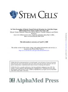
Fig. 3. PAS staining provides rapid assessment of cardiomyocyte yield in differentiating ES cells. (A) PAS staining of enzymatically dispersed cells prepared from a differentiating ES cell culture. Image shows a cardiomyocyte that stained dark purple due to its high glycogen content. In contrast, the nonmyocyte present in the same field was not stained. (B) Image of a PAS-stained cardiomyocyte colony following the genetic enrichment protocol. (C) Image of a PAS-stained tissue culture dish of ES-derived cardiomyocytes following the genetic enrichment protocol. Note the presence of many independent colonies. (See color plate 4, following p. 254).
between donor and host cardiomyocytes (i.e., fascia adherens, desmosomes, and gap junctions). While intracardiac engraftment of fetal cardiomyocytes holds great promise for functional augmentation in diseased hearts, identification of a suitable source of donor cells is quite problematic for clinical application in humans. Cardiomyocytes derived from differentiating ES cells could constitute a suitable surrogate source of donor cells for therapeutic intracardiac engraftment. Accordingly, a proof of concept experiment was initiated in mice (13). Cardiomyocytes derived from ES cells using the genetic selection approach described in this Subheading were digested with trypsin, and approx 1 × 10⁴ cells were delivered into the left ventricular free wall of dystrophic adult muscular dystrophy (mdx) recipient mice. The mdx mice harbor a mutation in the dystrophin gene and show no immune reactivity to antidystrophin antibodies. Consequently, the fate of the engrafted cardiomyocytes, which expressed a wild-type dystrophin gene, could easily be monitored by antidystrophin immune histology. Phase contrast microscopic examination of cryosections from the recipient hearts revealed that engrafted regions frequently exhibited normal myocardial topography (Fig. 4A). Immune cytologic assays with antidystrophin antibody revealed the presence of dystrophin-positive G418-selected cardiomyocytes (Fig. 4B). Comparison of phase contrast and antidystrophin images revealed the presence of myofibers in the engrafted ES-derived cardiomyocytes. Additional analyses with other antidystrophin antibodies, as well as transgenespecific polymerase chain reaction (PCR) analysis of material harvested from the graftbearing regions of the cryosections, confirmed that the dystrophic immune reactivity
164
Pasumarthi and Field
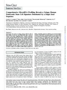
Fig. 4. Genetically enriched cardiomyocytes form stable intracardiac grafts. Phase contrast image (A) and antidystrophin immune fluorescence (B) of the same field from an mdx heart engrafted with G418-selected cardiomyocytes. Dystrophin immune reactivity appears as a green signal in panel B. (See color plate 5, following p. 254).
corresponded to engrafted ES-derived cardiomyocytes. Grafts were observed for as long as 7 wk post-implantation, which was the latest time point analyzed. Thus, the use of the genetic enrichment approach provides a suitable source of donor cardiomyocytes for intracardiac engraftment. Current research efforts are focused on enhancing graft size in experimental models of heart disease. If this approach is successful at restoring partial or complete systolic function in appropriate animal models, the recent development of human ES (17) raises the possibility that the approach can be tested therapeutically. 3.5.2. Example 2: Development of an ES-Derived Cardiomyocyte Colony Growth Assay
Cell cycle reactivation (i.e., induced proliferation) constitutes an alternative strategy through which to increase the number of functional cardiomyocytes in a diseased
Cardiomyocyte Enrichment
165
heart. The premise of the approach is that increased myocardial mass resulting from the proliferation of differentiated functional cardiomyocytes will augment systolic function. The successful application of this approach requires the identification of gene products that are capable of reactivating the cell cycle in genetically naive terminally differentiated cardiomyocytes. It has proven to be somewhat difficult to identify exploitable cardiomyocyte cell cycle regulatory genes. Although adenoviral-mediated gene transfer in primary cultures have identified some genes that promote cardiomyocyte growth, long-term analyses of the transfected cardiomyocytes in these cultures was limited because of the intrinsic proliferative capacity of the resident noncardiomyocytes. The use of transgenic mice with targeted expression in the heart or, alternatively, the use of retroviral transfection in developing avian hearts has identified several genes that are capable of inducing cardiomyocyte proliferation (1,18). However, these latter approaches are both time-consuming and can incur significant expenses related to animal husbandry charges. By incorporating a slight modification of the genetic enrichment protocol, coupled with the use of a muscle-specific cytologic stain suitable for tissue culture dishes, it should be possible to generate an ES-derived cardiomyocyte colony growth assay. Such an assay would provide a comparatively high-throughput, as compared to in vivo gene transfer approaches, and also would not be subject to the temporal constraints encountered with gene transfer approaches in primary cardiomyocyte cultures. As a proof of concept, we compared the activities of simian virus 40 (SV40) Large T-Antigen (T-Ag) vs the adenoviral E1A oncoproteins directly in the ES-derived cardiomyocyte colony growth assay. Previous studies, performed largely in transgenic mice with either wild-type or a conditionally active mutant T-Ag, have shown that expression of T-Ag gene (TAG) in the atrial or ventricular myocardium during fetal life is sufficient to induce sustained cardiomyocyte proliferation, while expression after terminal differentiation does not induce cell cycle activation. In contrast, studies utilizing adenoviral transduction of primary cardiomyocytes indicate that expression of E1A at any point of cardiac development can induce cell cycle reentry (as evidenced by the initiation of DNA synthesis), followed by a rapid apoptotic response. To compare the activities of T-Ag and E1A directly in the same system, undifferentiated ES cells were cotransfected with the MHC-neor/pGK-hygror and an MHC-T-Ag expression construct or, alternatively, with the MHC-neor/pGK-hygror and an MHCE1A expression construct. Control cultures were transfected with the MHC-neor/pGKhygror transgene only. After hygromycin selection, differentiation was induced, and cardiomyocytes were enriched as described above. Following 52 d of culture in G418 (a total of 60 d following the induction of differentiation), the dishes were rinsed, fixed, and stained with PAS as described above. The effect of oncogene expression was easily scored by simple visualization (Fig. 5). The density of cardiomyocyte colonies in the control dishes (MHC-neor/pGK-hygror transgene only) is used for reference. In good agreement with the data obtained via adenoviral transfection of primary cardiomyocytes, targeted expression of E1A in the ES-derived cardiomyocyte growth assay induced apoptosis, as evidenced by the marked reduction of cardiomyocyte colonies, as well as the presence of pronounced DNA fragmentation (Fig. 5). In contrast, targeted expression of T-Ag in the ES-derived cardiomyocyte growth assay resulted in fulminate proliferation, with the dish appearing as a nearly confluent
166
Pasumarthi and Field
Fig. 5. Use of the ES-derived cardiomyocyte colony growth assay to monitor the effects of gene transfer on cardiomyocyte proliferation. Undifferentiated ES cells were transfected with the MHC-neor/pGK-hygror alone or in combination with MHC-E1A or MHC-T-Ag expression constructs. Transfected cells were processed via the genetic enrichment protocol as described in the text, and at 60 d after differentiation, the cultures were fixed and stained with PAS. In addition, DNA fragmentation was monitored as an indirect indication for apoptosis. DNA prepared from parallel cultures was analyzed by agarose gel electrophoresis and visualized by ethidium bromide staining and UV illumination. Note the marked decrease in cardiomyocyte colonies in cells transfected with the MHC-E1A construct and the marked increase in cardiomyocyte colonies in cells transfected with the MHC-T-Ag construct as compared to the control dish. Note also the presence of extensive DNA fragmentation (as evidenced by the small molecular weight species) in DNA prepared from MHC-E1A transfected cells, but not in the control nor the MHC-T-Ag transfected cultures. (See color plate 6, following p. 254).
synchronously beating mass (Fig. 5). In agreement with results obtained in transgenic animals, T-Ag-induced proliferation was not accompanied by pronounced cardiomyocyte dedifferentiation, nor was an apoptotic response apparent (Fig. 5). Thus, the ES-derived cardiomyocyte growth assay described here faithfully recapitulates the results of gene transfer observed in other experimental systems and appears to be
Cardiomyocyte Enrichment
167
suitable for relatively high-throughput screens of the activity of cardiomyocyte cell cycle regulatory proteins.
3.6. Conclusions The genetic enrichment approach described in this chapter facilitates the generation of essentially pure cardiomyocyte cultures. Importantly, several groups have already used the approach in cardiomyocytes (13,19), attesting to both its reproducibility and relative ease of utilization. Moreover, it is clear that the enrichment protocol works in other cell lineages, as evidenced by the recent generation of relatively pure cultures of neurons (20) as well as insulin-secreting pancreatic β-cells (21). This approach should thus be useful for generation of a wide variety ES-derived cells suitable for both in vitro and in vivo applications. 4. Notes 1. Test several types/batches of bacterial Petri dishes to screen for lots that exhibit minimal EB attachment. 2. Change medium at all stages with care to avoid detachment of differentiating ES cell clusters. 3. The genetic enrichment approach described above is highly reproducible, relatively straightforward in nature, and can yield cardiomyocyte cultures in excess of 99% purity. As indicated in the Introduction, it is well established that ES-derived cardiomyocytes are highly differentiated. Similarly, molecular, immune cytologic, and ultrastructural analyses all indicate that genetically enriched cardiomyocyte cultures share these attributes (13). Moreover, cardiomyocytes from traditional murine ES cultures, as well as those from the genetically enriched cultures, follow cell cycle withdrawal and terminal differentiation programs, which are temporally similar to the programs observed during mouse embryonic development (12). Finally, the cultures can be maintained in vitro for as long as 11 mo while still retaining spontaneous contractile activity, thereby eliminating most of the temporal restraints encountered with traditional primary cardiomyocyte cultures. 4. Filter Schiff’s reagent prior to use, bring it to room temperature, and take precautions not to inhale vapors. Incubations with Schiff’s reagent and subsequent washes during PAS staining should be performed in the hood.
References 1. Pasumarthi, K. B. S. and Field, L. J. (2001) Strategies to identify cardiomyocyte cell cycle regulatory genes, in Molecular Approaches to Heart Failure Therapy (Hasenfuss, G. and Marban, E., eds.), Steinkopff, Verlag, Dormstadt. 333–351. 2. Doetschman, T. C., Eistetter, H., Katz, M., Schmidt, W., and Kemler, R. (1985) The in vitro development of blastocyst-derived embryonic stem cell lines: formation of visceral yolk sac, blood islands and myocardium. J. Embryol. Exp. Morphol. 87, 27–45. 3. Risau, W., Sariola, H., Zerwes, H. G., Sasse, J., Ekblom, P., Kemler, R., and Doetschman, T. (1988) Vasculogenesis and angiogenesis in embryonic-stem-cell-derived embryoid bodies. Development 102, 471–478. 4. Sanchez, A., Jones, W. K., Gulick, J., Doetschman, T., and Robbins, J. (1991) Myosin heavy chain gene expression in mouse embryoid bodies. An in vitro developmental study. J. Biol. Chem. 266, 22,419–22,426. 5. Muthuchamy, M., Pajak, L., Howles, P. L., Doetschman, T., and Wieczorek, D. F. (1993) Developmental analysis of tropomyosin gene expression in embryonic stem cells and mouse embryos. Mol. Cell. Biol. 13, 3311–3323.
168
Pasumarthi and Field
6. Miller-Hance, W. C., LaCorbiere, M., Fuller, S. J., Evans, S. M., Lyons, G., Schmidt, C., et al. (1993) In vitro chamber specification during embryonic stem cell cardiogenesis. Expression of the ventricular myosin light chain-2 gene is independent of heart tube formation. J. Biol. Chem. 268, 25,244–25,252. 7. Ganim, J. R., Luo, W., Ponniah, S., Grupp, I., Kim, H. W., Ferguson, D. G., et al. (1992) Mouse phospholamban gene expression during development in vivo and in vitro. Circ. Res. 71, 1021–1030. 8. Boer, P. H. (1994) Activation of the gene for type-b natriuretic factor in mouse stem cell cultures induced for cardiac myogenesis. Biochem. Biophys. Res. Commun. 199, 954–961. 9. Metzger, J. M., Lin, W.-I., and Samuelson, L. C. (1994) Transition in cardiac contractile sensitivity to calcium during the in vitro differentiation of mouse embryonic stem cells. J. Cell Biol. 126, 701–711. 10. Maltsev, V. A., Rohwedel, J., Hescheler, J., and Wobus, A. M. (1993) Embryonic stem cells differentiate in vitro into cardiomyocytes representing sinusnodal, atrial and ventricular cell types. Mech. Dev. 44, 41–50. 11. Wobus, A. M., Wallukat, G., and Hescheler, J. (1991) Pluripotent mouse embryonic stem cells are able to differentiate into cardiomyocytes expressing chronotropic responses to adrenergic and cholinergic agents and Ca++ channel blockers. Differentiation 48, 173–182. 12. Klug, M. G., Soonpaa, M. H., and Field, L. J. (1995) DNA synthesis and multinucleation in embryonic stem cell-derived cardiomyocytes. Am. J. Physiol. 269, H1913–H1921. 13. Klug, M. G., Soonpaa, M. H., Koh, G. Y., and Field, L. J. (1996) Genetically selected cardiomyocytes from differentiating embryonic stem cells form stable intracardiac grafts. J. Clin. Invest. 98, 216–224. 14. Reinlib, L. and Field, L. J. (2000) Transplantation: Future Therapy for Cardiovascular Disease? An NHLBI Workshop. Circulation 101, e182–e187. 15. Soonpaa, M. H., Koh, G. Y., Klug, M. G., and Field, L. J. (1994) Formation of nascent intercalated discs between grafted fetal cardiomyocytes and host myocardium. Science 264, 98–101. 16. Koh, G. Y., Soonpaa, M. H. Klug, M. G., Pride, H. P., Zipes, D. P. Cooper, B. J., and Field, L. J. (1995) Stable fetal cardiomyocyte grafts in the hearts of dystrophic mice and dogs. J. Clin. Invset. 96, 2034–2042. 17. Thomson, J. A., Itskovitz-Eldor, J., Shapiro, S. S., Waknitz, M. A., Swiergiel, J. J., Marshall, V. S., and Jones, J. M. (1998) Embryonic stem cell lines derived from human blastocysts. Science 282, 1145–1147. 18. Franklin, M. and Field, L. J. (1997) Use of Transgenic Animals in Cardiovascular Toxicology Testing, in Comprehensive Toxicology, Vol. 6. Cardiovascular Toxicology (Bishop, S. P., McQueen, C. A., and Gandolfi, A. J., eds.), Cambridge University Press, Cambridge, U. K., pp. 201–212. 19. Minamino, T., Yujiri, T., Papst, P. J., Chan, E. D., Johnson, G. L., and Terada, N. (1999) MEKK1 suppresses oxidative stress-induced apoptosis of embryonic stem cell-derived cardiac myocytes. Proc. Natl. Acad. Sci. USA 96, 15,127–15,132. 20. Li, M., Pevny, L., Lovell-Badge, R., and Smith, A. (1998) Generation of purified neural precursors from embryonic stem cells by lineage selection. Curr. Biol. 8, 971–974. 21. Soria, B., Roche, E., Berna, G., Leon-Quinto, T., Reig, J. A., and Martin, F. (2000) Insulinsecreting cells derived from embryonic stem cells normalize glycemia in strptozotocininduced diabetic mice. Diabetes 40, 157–162.