FoxM1, Stat3, Myc, Jun, RelA, Zipro1, Lmo4, Nr3c1, and Miz1 showed a very high expression and may play essential roles that are still unexplored [26]. Finally ...
EMBRYONIC STEM CELLS Embryonic Stem–Derived Versus Somatic Neural Stem Cells: A Comparative Analysis of Their Developmental Potential and Molecular Phenotype ELENA COLOMBO,a SERENA G. GIANNELLI,a ROSSELLA GALLI,a ENRICO TAGLIAFICO,b CHIARA FORONI,a ELENA TENEDINI,b SERGIO FERRARI,b STEFANO FERRARI,b GIORGIO CORTE,c,d ANGELO VESCOVI,a GIULIO COSSU,a VANIA BROCCOLIa a
Stem Cell Research Department, San Raffaele Scientific Institute, Milan, Italy; bDepartment of Biomedical Sciences, University of Modena and Reggio Emilia, Modena, Italy; cNational Institute for Cancer Research, Genoa, Italy; dDepartment of Oncology, Biology and Genetics, Genova University Medical School, Genova, Italy Key Words. Neural stem cell • Embryonic stem cell • Neural differentiation • Self-renewal • Multipotency Transcriptional profile
ABSTRACT Reliable procedures to induce neural commitment of totipotent undifferentiated embryonic stem (ES) cells have provided new tools for investigating the molecular mechanisms underlying cell fate choices. We extensively characterized the developmental potential of ES-induced neural cells obtained using an adaptation of the multistep induction protocol. We provided evidence that ES-derived neural proliferating cells are endowed with stem cell properties such as extensive self-renewal capacity and single-cell multipotency. In differentiating conditions, cells matured exclusively into neurons, astrocytes, and oligodendrocytes. All these features have been previously described in only somatic neural stem cells (NSCs). Therefore, we consider it more appropriate to rename our cells ES-derived NSCs. These similarities between the two NSC populations induced us to carefully compare their proliferation ability and differentiation potential. Although they were very similar in
overall behavior, we scored specific differences. For instance, ES-derived NSCs proliferated at higher rate and consistently generated a higher number of neurons compared with somatic NSCs. To further investigate their relationships, we carried out a molecular analysis comparing their transcriptional profiles during proliferation. We observed a large fraction of shared expressed transcripts, including genes previously described to be critical in defining somatic NSC traits. Among the genes differently expressed, candidate genes possibly responsible for divergences between the two cell types were selected and further investigated. In particular, we showed that an enhanced MAPK (mitogen-activated protein kinase) signaling is acting in ES-induced NSCs, probably triggered by insulin-like growth factor–II. This may contribute to the high proliferation rate exhibited by these cells in culture. STEM CELLS 2006;24: 825– 834
INTRODUCTION
possible to explore in vitro the conditions by which ES totipotent cells can be induced to give rise to a specific population of tissue-restricted stem cells able to grow and differentiate. Over the years, several in vitro systems have been described for the derivation of neural progeny from ES cells. Notably, some of these allow the generation of mature cells through an intermediate step represented by progenitor cells able to grow for some time in vitro and generate differentiated progeny upon changes in culture conditions [5–7]. These protocols have been widely used to generate a variety of differentiated neural cell types such as astrocytes, oligodendrocytes, and glutamatergic, GABAergic,
Stem cells are defined by three fundamental features: the ability to self-renew, to give rise to a differentiated progeny, and to maintain these features over a long period of time [1, 2, 3]. In particular, embryonic stem (ES) cells are cell lines derived from the inner cell mass of the developing blastocyst, which generates all the different cell types of the future organism [4]. During development, the cells of the inner cell mass generate the three germinal layers of the embryos and, conceivably, will then give rise to tissue-restricted stem cells that generate the organs and tissues of the future organism. With this in mind, it may be
Correspondence: Vania Broccoli, Ph.D., Stem Cell Research Department, Dipartmento di Biotecnologie, San Raffaele Scientific Institute, Via Olgettina 58, 20132 Milan, Italy. Telephone: ⫹39 02 26434612; Fax: ⫹39 02 26434621; e-mail: broccoli.vania@ hsr.it Received July 12, 2005; accepted for publication December 5, 2005; first published online in STEM CELLS EXPRESS December 9, 2005. ©AlphaMed Press 1066-5099/2006/$20.00/0 doi: 10.1634/stemcells.2005-0313
STEM CELLS 2006;24:825– 834 www.StemCells.com
Characterization of ES-Derived Neural Stem Cells
826
and dopaminergic neurons [8, 9]. However, the nature of the in vitro– derived neural progenitors is still elusive, and a better characterization of their self-renewal features and neural potential over time is desirable. Somatic neural stem cells (NSCs) can be retrieved from the embryonic neural tissue or from the neurogenetic regions of the adult brain (subependymal layer and dentate gyrus) and can self-renew in vitro for a long period of time and differentiate into neurons, astrocytes, and oligodendrocytes [10, 11]. Their self-renewal features have been clearly characterized using clonogenic assays to evaluate single-cell potentialities [12, 13]. ES cell– derived neural progenitors, on the other hand, have not been studied as well, and their clonogenic potentials are still unknown. Moreover, a thorough comparison between ES cell– derived neural progenitors and somatic NSCs in terms of stable growth, differentiation potential, and overall gene expression is still missing. To fill this gap, we studied a population of ESderived cells that showed all the cardinal properties of stem cells and thus defined ES-derived NSCs. Moreover, we extensively compared them with NSCs isolated from embryonic neural tissue, in terms of proliferation ability, neural differentiation potential, and overall gene expression.
MATERIALS
AND
METHODS
Materials and methods are available as supplemental online data.
RESULTS Derivation of Homogenous and Stably Proliferating Neural Progenitors from ES Cells To differentiate ES cells toward a neural phenotype, we employed a protocol based on a three-step procedure established by Okabe et al. [1] and Lee et al. [5], with a number of modifications. Briefly, whole undifferentiated ES cell colonies were isolated from the fibroblast feeder layers by dispase treatment and re-plated in bacteriological plates containing ES cell medium with 20% serum replacement to obtain embryoid bodies (EBs). After 4 days, EBs were transferred in matrigel-coated plates and cultured in a serum-free medium enriched in insulin (0.025 mg/ml) and transferrin (100 ug/ml) and completed with fibroblast growth factor (FGF-2) (10 ng/ml) and epidermal growth factor (EGF) (20 ng/ml) (see Materials and Methods). This medium is normally used for selection and maintenance of NSCs isolated from embryonic and adult brains [12, 14]. Furthermore, matrigel-coated dishes allowed a better cell attachment and spreading than did gelatin, used in previous studies. Within 5 days, a homogenous population of cells expressing the neural precursor markers Nestin and Vimentin had grown [15, 16] (Fig. 1C–1E). The overall procedure required 8 days, thus shortening the period of time needed to obtain neural commitment as compared with the protocol of Okabe et al. After the first selection phase, we were able to maintain stable cell growth of undifferentiated neural progenitors in NSC culture medium for several months. To monitor neural induction in our cells, we tested a number of neural-specific genes by reverse transcription–polymerase chain reactions (RT-PCRs). Interestingly, the expression of molecular markers typical of totipotent ES cells such as Oct4, Cripto, and Nanog was not detected in the neural progenitor
Figure 1. Establishment of homogenous cultures of ES cell– derived NPs. (A): Outline of the overall protocol based on three major steps: I, embryoid body formation; II, plating in matrigel-coated dishes and cultured with NSC medium; and III, disaggregation after amplification. (B): Determination of the cell growth in a window of 1 month. (C): Phase-bright microphotograph showing the general morphology of the ES-derived NSCs in their undifferentiated state. Immunofluorescence for nestin (D) and vimentin (E) on proliferating ES-derived cells. (F): Normal karyotype (2n ⫽ 40) isolated from P17 cell cultures. (G): Reverse transcription–polymerase chain reaction analysis of a series of molecular markers of naı¨ve ES cells (Oct4) and forebrain neural stem cells (Sox2, Emx2, Pax6, Lhx2, and Foxg1). Abbreviations: dNP, differentiated embryonic stem cell– derived neural progenitors; EGF, epidermal growth factor; ES, embryonic stem; FGF, fibroblast growth factor; KSR, knockout serum replacement; LIF, leukemia inhibitory factor; NP, neural progenitor; NSC, neural stem cell.
cultures (Fig. 1G; data not shown), whereas genes specific to somatic NSCs, such as Sox2, and forebrain-specific genes such as Emx2, Pax6, Lhx2, and Foxg1 were activated and detectable at similar levels (Fig. 1G) [17]. Cultures of ES cell– derived neural progenitors were stable for months without undergoing either senescence or growth factor–independent proliferation. They showed a stable doubling time (approximately 24 hours) and a normal caryotype (2n ⫽ 40) as tested up to 17 passages, spanning 1 month of in vitro culture (Fig. 1B, 1F). ES cell– derived neural progenitors were differentiated by sequential removal of FGF-2 and EGF and finally switched to a hippocampal culture medium (see Materials and Methods) [18]. Neural progenitors exposed to differentiating conditions completed their maturation within 9 days. -III-Tubulin and microtubule associated protein 2 (MAP-2)–positive neurons, glial fibrillary acidic protein (GFAP)–positive astrocytes, and galactocerebrosidase (GALC)–positive oligodendrocytes were present in a ratio of 54% ⫾ 4%, 34% ⫾ 5%, and 12% ⫾ 2% (200 total cells counted per field, n ⫽ 3), respectively (Fig. 2C). Most of the neurons presented a bipolar shape, whereas a minor fraction showed a multipolar morphology (Fig. 2D, 2E). Differentiated cultures contained different types of neurons, including glutamatergic (Fig. 2J), GABAergic (Fig. 2H), dopaminergic (Fig. 2K), and cholinergic neurons (Fig. 2L) in a ratio of 53% ⫾ 6%, 37 ⫾ 5%, 6% ⫾ 4%, and 4% ⫾ 2% (200 cells, n ⫽ 3),
Colombo, Giannelli, Galli et al.
827
cell lines (TVB2, R1, YC5, and D3), indicating a widespread applicability of this protocol to ES cells [19]. To evaluate the in vivo differentiation potential of ES cell– derived precursors, we took advantage of the YC5 ES cell line that constitutively expresses enhanced green fluorescent protein (eGFP) [20]. Neural precursors derived from this line showed stable growth and multilineage differentiation in vitro. We transplanted the cells into the lateral ventricles of embryonic day 14.5 (E14.5) mouse embryos, and results were analyzed between P1 and P5. A diffuse integration of eGFP cells was normally observed in the neural parenchyma of different regions, depending on the area of graft integration (supplemental online Fig. 2). Generally, the lateral cortex, the olfactory bulbs, and the ventral forebrain showed many integrated eGFP cells. In particular, in the lateral cortex, many GFP cells were detected in all the layers and spread along the dorsal-ventral axis (supplemental online Fig. 2). eGFP integrated cells expressed markers of differentiated neurons and astrocytes as assessed by -III-tubulin or NeuN and GFAP immunofluorescence, respectively (supplemental online Fig. 2). In all the experiments analyzed (n ⫽ 3), neurons, astrocytes, and oligodendrocytes were scored, but the ratio among the different cell types was highly variable, depending on the injection site. Figure 2. Differentiation protocol and phenotypes of embryonic stem (ES)-derived neural cells. (A, B): Low magnification of differentiated cell cultures stained with -III-tubulin (neurons) and GFAP (astrocytes). (C): Quantitative analysis of neurons, glia, and oligodendroglia in the differentiated cells. (D, E): Morphological characterization of the ES cell– derived neurons stained with NF160 and MAP2. (F): Quantitative analysis of the different types of neurons according to their neurotransmitter content. (G, Gⴕ): Mature oligodendrocyte highlighted in the ES cell– derived cultures by GalC and O4 antibody stainings, respectively. (H, J, K, L): Immunocytochemistry for neuron subtype–specific markers in the differentiated cell cultures using antibodies against GABA (H), glutamate (J), TH (K), and ChAT (L). (I): Double staining for synaptophysin (green) and -III-tubulin (red) to assess the formation of synaptic buttons in neurites of ES cell– derived neurons. Abbreviations: IIItub, -III-tubulin; ChAT, choline acetyltransferase; GABA, ␥-aminobutyric acid; GalC, galactocerebroside; GFAP, glial fibrillary acidic protein; MAP2, microtubule associated protein 2; TH, tyrosine hydroxylase.
respectively (Fig. 2F). Interestingly, we noted the widespread formation of many synaptic contacts among neurons as assessed by synapsin-I immunofluorescence (Fig. 2I). This molecule reveals the organization of neural synapses in mature differentiated neurons. Furthermore, we asked whether ES cell– derived neural progenitor differentiation could be manipulated adding specific growth factors or cytokines both in early cultures. Remarkably, retinoic acid (RA) supplementation (5 M) drastically strengthened the overall neuronal differentiation, reducing astrocyte formation, whereas a combination of RA (0.2 M) and Sonic Hedgehog (Shh-N, 300 nM) led to a detectable increase in the amount of cholinergic neurons (from 5% ⫾ 2% to 13% ⫾ 2%) (120 cells, n ⫽ 3) (supplemental online Fig. 1). Similar results were obtained differentiating ES cell– derived cultures from late passages (P16). Thus, long-term cultures of the ES cell– derived neural progenitors did not lose their ability to respond to external cues, modifying their differentiation potential accordingly. Notably, isolation and differentiation of ES-derived NSCs were accomplished using four different ES www.StemCells.com
ES Cell–Derived Neural Progenitors Are Clonogenic and Multipotent Clonal analysis is a stringent paradigm for testing multipotency at the single-cell level. Although it has been extensively used in the NSC field, it has rarely been applied to ES cell– derived neural cultures. Individual ES-derived cells from different passages (P3, P7, and P11) were transferred into four-well chamber slides (1 cell/1 well) pre-coated with matrigel. In the presence of FGF-II/EGF, approximately 5% of these single cells proliferated, giving rise to a clone in approximately 3 weeks (Fig. 3A–3D). Upon differentiation, cells were tested for -III-tubulin, GFAP, and GalC immunofluorescence (Fig. 3E–3H). In all cases (n ⫽ 12), immunostainings revealed neural tri-lineage commitment, confirming the multipotent nature of the clone founder cells. The percentages of mature cell progenies (neurons, astrocytes, and oligodendrocytes) over the whole differentiated population derived from either a single clone or the sister uncloned culture were very similar (Fig. 3I). This indicated that each individual clonal culture maintained its differentiation potentialities with no alteration. Finally, we tested the growth rate of the three cultures derived by single-cell expansion (no. 35 from P3, no. 102 from P7, and no. 155 from passage P11) in comparison with their sister bulk cultures. The four cultures exhibited a similar growth rate when measured for a period of 4 weeks (Fig. 3J). These results suggest that the ES cell– derived progenitors exhibit self-renewal features as tested with stringent criteria. Taken together, these data demonstrate that ES cell– derived neural progenitors are self-renewing, multipotent, and clonogenic and maintain these potentialities for a long period of time in vitro. In conclusion, neural precursors derived from ES cells are endowed with all the cardinal features of stem cell lines and thus represent bona fide NSCs. We thus renamed them ESderived NSCs. This finding cleared the way for a comparison of the cellular and molecular phenotypes of these two NSC lines in similar experimental conditions.
828
Characterization of ES-Derived Neural Stem Cells
Figure 3. Clonogenic assays and analysis of bulk and cloned cell cultures. (A–D): Bright-field pictures of a growing clone. (E–H): Immunoreactivity for markers of neuronal (-III-tubulin [E, G]), astroglial (GFAP [F, G]), and oligodendroglial (GalC [H]) fate of a clone derived by single-cell expansion. (I): Characterization of the mature phenotypes observed in both bulk and cloned cell cultures (nos. 35, 102, and 135). (J): Compared analysis of the growth rate of the bulk and three cloned cell cultures over a period of 3 weeks. Abbreviations: IIITub, -III-tubulin; GalC, galactocerebroside; GFAP, glial fibrillary acidic protein.
ES-Derived NSCs and Somatic NSCs Compared with In Vitro Phenotypes We isolated somatic NSCs from forebrain regions of E14.5 mouse embryos to compare in vitro growth and differentiation potential of somatic and ES-derived NSCs. Therefore, we maintained both cell lines in the same culture medium (see Materials and Methods) and compared cells at the same passages (between P8 and P14). The two cell lines grew in different ways. Whereas somatic NSCs proliferated better as clusters of cells floating freely in the medium, generally called neurospheres, the ES-derived NSCs grew optimally when adhering to a substrate (supplemental online Fig. 3). In fact, ES-derived NSCs were unable to form growing “spheres,” and their growth was impaired when unable to attach to a substrate (data not shown). Under optimal conditions for each of the cultures and using the same culture medium, cells showed a different proliferation rate at similar passage numbers. In fact, ES-derived NSCs showed a two-time log 10 increase with respect to embryonic somatic NSCs after 6 passages, spanning less that 1 month of in vitro culture (supplemental online Fig. 3). Then, we analyzed their differentiation potential. Both cell lines were plated on laminin-coated dishes and exposed to similar differentiation conditions (see Materials and Methods). After 8 days, cells were processed for immunofluorescence to detect neurons (-III-tubulin), astroglia
(GFAP), and oligodendroglia (GalC). In all cases analyzed (n ⫽ 18), the proportion of neuronal and glial cells was different between the two cell lines. In fact, differentiated progenies derived from either ES-derived or somatic NSCs contained 54% ⫾ 4% and 40% ⫾ 3% of neurons, 36% ⫾ 2% and 52% ⫾ 3% of astrocytes, and 10% ⫾ 3% and 8% ⫾ 3% of oligodendrocytes, respectively (200 cells, n ⫽ 5) (Fig. 4). Furthermore, we assessed the differentiation potential of NSCs derived from adult brains in similar conditions and we observed a further general decrease in neuronal cell production (28% ⫾ 4%) and a corresponding enrichment in astrocytes (64% ⫾ 5%) (200 cells, n ⫽ 5) (Fig. 4C, 4F, 4H). Overall, ES-derived NSCs displayed a higher efficiency in neuronal differentiation compared with somatic NSCs. We then assessed the neuronal subtype composition of the mature neurons in embryonic NSCs and ES-derived NSCs. In both cases, glutamatergic neurons were the most abundant phenotype (69% ⫾ 6% and 53% ⫾ 4%) (200 cells, n ⫽ 5), whereas GABAergic neurons represented 29% ⫾ 3% and 37% ⫾ 3% of the entire differentiated cell population, respectively (n ⫽ 5) (supplemental online Fig. 4). However, only in the mature progeny derived by ES-derived NSCs was it possible to observe a portion of dopaminergic (6% ⫾ 4%) and cholinergic (4% ⫾ 3%) neurons (200 cells, n ⫽ 5) (Fig. 2I; supplemental online Fig. 4).
Colombo, Giannelli, Galli et al.
Figure 4. Composition and morphology of differentiated cell cultures derived from embryonic somatic NSCs (A, D), adult somatic NSCs (B, E), and ES-derived NSCs (C, F, G). (A–C): Double stainings for -III-tubulin and GFAP to visualize the ratio of neurons/astrocytes in the overall differentiated cultures. (D–G): Neural morphology visualized by -III-tubulin immunostaining of neurons derived from either somatic embryonic (D) and adult (E) NSCs or ES-derived NSCs (F, G) with nuclei labeled with 4⬘,6-diamidino-2-phenylindole. (H): Quantitative analysis of the neural, glial, and oligodendroglial phenotypes obtained by differentiation of somatic and ES-derived cell cultures. ⴱ, p ⬍ .05 versus ES cell– derived neurons; ⴱⴱ, p ⬍ .05 versus ES cell– derived astrocytes. Abbreviations: IIITub, -III-tubulin; ES, embryonic stem; GFAP, glial fibrillary acidic protein; NSC, neural stem cell.
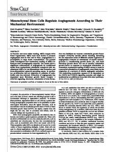
ES-Derived NSCs and Somatic NSCs Compared with Transcription Profiles To test the molecular heterogeneity between ES-derived NSCs and somatic NSCs and to score genes differently expressed in the two cell populations, we performed a compared global transcriptional profile by means of gene chip analysis comparing cells between passages P9 and P10. Briefly, fluorescentlabeled cRNAs derived from ESC-derived NSCs and E14.5 embryonic striatal NSCs (between P8 and P9) were hybridized to the Affymetrix Mouse Expression 430 set (Santa Clara, CA, http://www.affymetrix.com). This platform consists of two GeneChip probe arrays (MOE430A and MOE430B) containing more than 45,000 probe sets representing more than 39,000 transcripts and variants, including more than 34,000 well substantiated mouse genes. As shown in the scatter plot in Figure 5A, both NSC lines expressed a large number of common sequences. In this category, 15,031 sequences were not changed in the two cell populations. Although 3,158 and 2,482 showed an increased expression in ES-derived NSCs and somatic NSCs, respectively, as identified by Affymetrix MAS 5.0 absolute and comparative analyses, the number of sequences, which can be considered specifically expressed in either cell population, on the basis of a ratio equal to or higher than threefold (i.e., a signal-log ratio grater than or equal to 1.5 or less than or equal to ⫺1.5), was 276 in ES-derived NSCs (among them, 185 present only ES-derived NSCs) and 372 in somatic NSCs (among them, 201 present only in somatic NSCs). Thus, less than 2% of the studied transcripts were found significantly changed, indicating a substantial transcriptional homogeneity of the two cell populations. www.StemCells.com
829
Figure 5. Compared global transcriptional profile of somatic and ES-derived NSCs. (A): Scatter plot showing the distribution of hybridization signals of somatic (E14-NSCs) on the y-axis and ES-derived (ES-NSCs) NSCs on the x-axis. Transcripts more abundant in E14NSCs are shown in red, transcripts more abundant in ES-NSCs are shown in green, and unchanged transcripts are shown in yellow. (B): Probe sets considered as positive controls for the microarray experiment: Xist, H2-K, H2-D1, and H13. (C): Microarray data of selected genes chosen for their proven or suggested role played in somatic NSCs. (D–F): Signaling pathways whose main components show equal expression in both ES-derived and somatic NSCs. (E): Wnt--catenin schematic transduction pathway and Western blotting showing Wnt5A and -catenin protein levels in both NSC cell lines. (C, D): Signals are shown as color-coded cells (black for “absent” or “marginal” transcripts and blue to red for “present” transcripts: blue for signals ⬍100; yellow for signals between 101 and 300; light orange for signals between 301 and 600; dark orange for signals between 601 and 1,000; and red for signals ⬎1,001). The fold change is also shown as SLR. (F): Notch transduction pathway and protein level analysis of Notch1 and Hes1. (G): Hh schematic molecular pathway and analysis of Dhh and Smo protein levels in both stem cell lines. Abbreviations: Bmpr1a, bone morphogenetic protein receptor 1a; Bmpr2, bone morphogenetic protein receptor 1; Ccnb1, cyclinb1; Ccnnd1, cyclind1; Ccnd2, cyclind2; Dhh, desert hedgehog; Disp1, Dispatched1; Dll1, Delta1; Dnclc1, dynein light chain 1; Dvl1, Dishevelled 1; E14, embryonic day 14; Egfr1, epidermal growth factor 1; ES-NSC, embryonic stem–neural stem cell; Fgfr1, fibroblast growth factor receptor 1; Fz2/9, Frizzled receptor 2, 9; Fu, fused; Hdac, histone deacetylase; Hdgf, hepathoma-derived growth factor; Hh, Hedgehog; Jag1, Jagged1; Nes, nestin; Nic, nicastrin, NISD, notch intracellular domain; Nr3c1, nuclear receptor 3c1; Pdgfa, plateletderived growth factor A; PS1, presenilin1; Ptch1, Patched1; SLR, signal log 2 ratio; Smo, Smoothened; Stmn1, Stathmin1; ThrA, thyroid receptor A; Vegfb, vascular endothelial growth factor B; Vim, vimentin.
Importantly, the Xist gene resulted as one of the most widely differentially expressed (Fig. 5B). This gene is exclusively expressed in the female lineage from the blastocyst stage onward because it produces a main molecular switch that inactivates one of the two X chromosomes. In fact, the ES cell lines used for the transcriptional profile were all male (XY), and thus the Xist gene was not expressed. Conversely, the NSCs were
830
derived by a pool of both male and female E14.5 embryos, and thus Xist gene expression was clearly detectable in the screening. Another expected difference between the two cell lines was represented by the low expression of some genes coding for major histocompatibility complex (MHC) class I proteins in ES-derived NSCs (H2-K, H2-D1, and H13) (Fig. 5C), as already stated in previous reports describing the very low level of MHC class I gene expression in mouse and human ES cells with respect to many somatic cell lines [20, 21]. Among the 15,031 genes equally expressed in the two cell lines, several genes encode for a variety of proteins already known to play a critical role in somatic NSCs, such as the cytoskeletal components nestin [15] and vimentin [16] and the cytosolic proteins stathmin [22], Pten [23], and ShcA [24] (Fig. 5D). Much evidence has been reported recently on the key functions, such as self-renewal and cell fate choices, played by some transcription factors in controlling the cardinal properties of somatic NSCs. Most of these transcription factors are expressed in both cell types at very similar levels, as detected for Sox2 [25], Emx2 [14], and Olig1,2 [17]. Among many other transcription factors equally expressed in both cell types, FoxM1, Stat3, Myc, Jun, RelA, Zipro1, Lmo4, Nr3c1, and Miz1 showed a very high expression and may play essential roles that are still unexplored [26]. Finally, chromatin re-modeling proteins such as histone deacetylases Hdac-1, -2, and -3 [27] and Hmga1, Hmgb1, and Hmgb3 [28] were highly expressed in both cell lines, as expected with their function in modulating cell cycle progression in a variety of cells (Fig. 5D). We scored a similar presence not only of single genes but also of entire transduction pathways implicated in regulating somatic NSC behavior. For instance, three transduction pathways highly related to proliferation and self-renewal activity of somatic NSCs, such as the Wnt–-catenin (-cat), Notch, and Hedgehog (Hh) pathways, were similarly expressed in ESderived NSCs [28]. In particular, many single components of the -cat pathway were detected in both cell types, including the Wnt receptors Frizzled-1, -2, and -9 (Fzd-1, -2, and -9), the transduction components Dishevelled1 (Dvl1), Glicogen sinthase kinase 3 (GSK3-), APC, Axin, as well as many protein kinases (Csnk1e, Csnk1a1, and Csnk2a2) and phosphatases (Ppp2cb, Ppp2r1a, and Ppp2r5c), which are known to regulate the activity of the Wnt pathway. Finally, -cat and its associated transcription factors Tcf4 and (to a lesser extent) Tcf3 were equally detected (Fig. 5E). Both cell lines expressed Wnt family members and in particular Wnt5a and Wnt7b, suggesting the existence of a functional Wnt autocrine loop, which might contribute to NSC maintenance. In particular, we detected the presence of Wnt5A and -cat proteins, suggesting the functional activity of this pathway in ES-derived NSCs (Fig. 5E). Another main transduction pathway implicated in somatic NSC proliferation activity is the Notch pathway [29]. Both Notch-1 and -2, their ligands Delta-1 to -3 (Dll-1 to -3), and Jagged1 (Jag1) are highly expressed (Fig. 5F). The downstream effectors of this pathway, Hes1 and Hes5, are also detected in the microarray. Furthermore, some other essential components of the pathway such as RBP-Jk, Presenilin-1 (PS1), Nicastrin (Nic), and Lunatic and Radical Fringe were enriched in both populations. Supporting some functional involvement of this pathway, we identified the presence of both Notch1 and Hes1 proteins (Fig. 5F) by Western blotting. Finally, we considered the Hh
Characterization of ES-Derived Neural Stem Cells pathway, which has been recently found to be involved in supporting somatic NSC proliferation both in vitro and in vivo [30, 31]. Both the Hh receptor Patched1 (Ptch1) and its coreceptor Smoothened (Smo) as well as the downstream effectors Gli2-Gli3 were found expressed in both cell lines (Fig. 5G). Interestingly, we found only Desert Hedgehog (Dhh), out of the whole Hh ligand family, to be expressed in both cell types. Indeed, we confirmed these results by Western blot analysis, which indicated the presence of both Dhh and Ptch1 proteins (Fig. 5G). Taken together, these data show that somatic and ES-derived NSCs share a great part of their molecular phenotype, including important molecular components and specific signal transduction pathways, suggesting the similar nature and potentials of the two cell lines. Furthermore, this study identified 245 sequences specifically enriched in either of the two NSC lines (supplemental online Table 1). We identified three groups of genes among them that may unravel the molecular pathways at the basis of the differences of the two cell lines. Some of the genes most differently expressed coded for proteins of the extracellular matrix (ECM) such as collagen proteins such as Col1a1, Col3a1, and Col1a2 (Fig. 6A). Furthermore, other important components of the ECM, such as fibronectin (Fn) and fibrillins (Fbn-1 and -2), were upregulated in ES-derived NSCs [32]. To independently confirm the reproducibility of the array data, RT-PCRs were performed. Semiquantitative amplification reactions for Col1a1, Fn1, and Fbn1 confirmed gene expression diversity in the two cell populations and showed even greater differences as in the case of Fbn1 (6.5-fold increase in ES-derived NSCs vs. a 3.5-fold increase in the gene-chip analysis) (Fig. 6A). The enhanced expression of extracellular components in ES-derived cells may explain why these cells show a higher ability to attach and grow over many types of surfaces in respect to somatic NSCs, which typically cannot grow on a naı¨ve substrate. Among the 75 sequences found enriched in the ES-derived NSCs and corresponding unambiguously to known genes, eight (Nkx2.2, Irx2, En1, Hoxa1, Hoxa2, Hoxa3, Hoxa4, and Hoxa5) were represented by genes coding for transcription factors mostly or exclusively active along the caudal neural tube. During embryonic development, a series of transcription factors regionalize the neural tube along the anterior-posterior (A-P) axis. Growing evidence supports the hypothesis that these molecules are producing a molecular identity that specifies the different regions of the neural tube such as forebrain, midbrain, and hindbrain. Many of these transcription factors were expressed in both cell lines; however, we found a general enrichment in ES-derived NSCs of mRNAs of transcription factors that act on the spinal cord. Nkx2.2, Irx2, and En1 are genes expressed starting from the mid-hindbrain region all along the ventral spinal cord, whereas the Hox genes are active in the most caudal regions of the body. We confirmed these findings by means of semiquantitative RT-PCRs (Fig. 6B) and widened this data set by analysis other Hox genes not included in the microarray platform, such as Hoxa9, Hoxa10, and Hoxa13 (Fig. 6B; data not shown). In all cases, these genes were expressed in ES-derived NSCs but were represented at a low or undetectable level in somatic NSCs. These data strongly suggested that ES-derived NSCs displayed the positional molecular information of the whole A-P axis of the developing neural tube.
Colombo, Giannelli, Galli et al.
831
ingly, all these intracellular mediators of the IGF pathway were highly expressed in ES-derived NSCs at an equivalent level to those detected in somatic NSCs, as shown by gene-chip and RT-PCR analyses (Fig. 6C). Also, the IGF binding proteins 2 and 4, which have been implicated in regulating the overall actions of the IGF molecules [36], were expressed at comparable levels (Fig. 6C). These data prompted us to determine whether the IGF signaling was effectively upregulated in the ES-derived NSCs and to correlate this molecular diversity with a particular aspect of the in vitro behavior of these cells.
The Enhanced IGF Signaling Is Responsible for the High Proliferation Rate of ES-Derived NSCs In Vitro To verify whether the IGF-II enhanced expression resulted in the functional activation of the downstream targets, we evaluated the relative amounts of either phosphorylated MAPK (mitogen-activated protein kinase) and phophorylated Akt, two key molecules of the Ras-Raf-MAPK and PI3-Kinase (PI3K) pathways, respectively. Indeed, we found an increase in the phosphorylated forms of MAPK and Akt, in the ES-derived in respect to the somatic NSCs, without any clear increase of the total amount of these proteins (Fig. 7A). Thus, a sustained increase in IGF signaling was exhibited by ES-derived NSCs Figure 6. Classes of genes differentially expressed in ES-derived NSCs (ES-NSCs) versus somatic NSCs (E14-NSCs). (A): Microarray and semiquantitative reverse transcription-polymerase chain reaction (RT-PCR) analysis of ES-derived NSC– enriched transcripts coding for extracellular matrix proteins. (B): Gene expression analysis by GeneChip and RT-PCR of transcripts coding for transcription factors contributing to caudal neural tube identity. (C): Microarray and RT-PCR study of genes coding for IGF signaling pathway. In tables, signals are shown as color-coded cells (black for “absent” transcripts and blue to red for “present” transcripts, as in Figure 5). The fold change is also shown as SLR. Abbreviations: aNSC, adult neural stem cell; Col1a1, collagen 1a1; Col3a1, collagen 3a1, Col1a2, collagen 1a2; Col6a3, collagen 6a3; E14-NSC, embryonic day 14 –neural stem cell; ES-NSC, embryonic stem–neural stem cell; Fn1, fibronectin 1; Fbn1, fibrillin 1; Fbn2, fibrillin 2; Itga9, Integrin a9; SLR, signal log 2 ratio.
Conversely, somatic NSCs showed a general low expression or absence of members of the Hox gene family and other transcription factors of the Nkx and Irx classes, which are critical for the establishment and shaping of the spinal cord. Among the 75 known transcripts enriched in the ES-derived NSCs, insulin-like growth factor (IGF)-II is coding for the growth factor most expressed in a relative and absolute manner in these cells. IGF-II belongs to the IGF family and shares many pleiotropic activities in neural cells, such as supporting cell proliferation, migration, and survival, with the other members of the family, IGF-I and IGF-III [33, 34]. IGF receptors are expressed in the germinal zone of the developing neural tissue as well as in the subventricular zone (SVZ) of adult mouse brain. Targeted mutations of members of this family lead to deficits in both body and brain development [35]. IGF signals are transduced through the two IGF receptors (IGF-1R and -2R), which are receptor tyrosine kinases closely related to the insulin receptor. Activation of these receptors results in tyrosine phosporylation of cytoplasmic insulin receptor substrates (IRSs) that in turn can activate the Ras-MAP kinase or the PI3 kinase (phosphatidylinositide-3⬘ kinase)-Akt pathways [35]. Interestwww.StemCells.com
Figure 7. Active IGF signaling is enhanced in ES-derived NSCs with respect to somatic NSCs. (A): Western blotting analysis of IGF-II and active forms of key components of the pathway. p42/p44 (Erk1/2) and Akt phosphorylated forms are enriched in ES-derived NSCs. Cell lysates were normalized with respect to their -actin protein levels. (B): IGF growth factors sustained ES cell– derived neural proliferation in culture in the absence of insulin. (C): Functional arrest of IGFR-I function by a blocking antibody strongly decreased IGF-II– dependent cell proliferation. Suramin is an inhibitor of both IGF and epidermal growth factor. (D): Cell proliferation elicited by IGF-II is mediated mostly by activation of the mitogen-activated protein kinase signal transduction pathway. Abbreviations: aNSC, adult neural stem cell; DIV, days in vitro; ES-NSC, embryonic stem–neural stem cell; IGF, insulin-like growth factor; IGFR-I, insulin-like growth factor receptor I.
832
during their proliferating stage. Previous studies revealed an essential action of IGF-I in maintaining proliferation of somatic NSCs in cultures [37]. Indeed, all the media used for NSC cultures include insulin at a concentration of 5–25 g/ml. Its withdrawal in ES-derived NSC cultures led to a rapid block in cell proliferation, which is followed by a widespread cell death even in the presence of the growth factors FGF-II and EGF (Fig. 7B). Similar behavior was described for adult NSCs in a medium not supplemented with insulin growth factors [38]. In addition, a culture medium deprived of insulin, but supplemented with IGF-I or IGF-II (100 nM), sustained the growth of ES-derived NSCs. Interestingly, the effect of IGF-I on proliferation was higher than that of IGF-II, although the latter still had a significant effect. To identify the receptor through which IGF-II promoted cell proliferation, we added blocking antibodies for either IGF receptor I (IGF-IR) or IGF receptor II (IGF-IIR) in conditions in which cell growth was sustained by IGF-II (see Materials and Methods). Only cultures treated with anti–IGF-IR, but not antiIGF-IIR, exhibited a dramatic decrease in cell proliferation (Fig. 7C). Accordingly, we found a reduction in MAPK phosporylation in cultures exposed to IGF-RI blocking antibody (Fig. 7C). These findings suggest that IGF-II elicits cell growth mainly by signaling via the IGF-IR. Finally, we investigated the intracellular pathway involved in mediating IGF action. Specific inhibitors of the MAPK or the PI3K pathways were used, in particular inhibitors of the ERK kinases such as PD098059, U0126, and LY294002, to block PI3-kinase [37]. Addition of the MAPKK inhibitor PD098059 to IGF-II treated cultures abolished the effect of the growth factor on the increased proliferation in a dose-dependent manner (Fig. 7C). Conversely, a minor effect was noted on the IGF-II–induced proliferation upon the addition of the PI3-K inhibitor, LY294002 (Fig. 7C). These results suggest that IGF-II–induced proliferation is mediated mainly by the activation of the MAPK signaling and only marginally by the PI3K molecular component pathway.
DISCUSSION ES Cell–Derived Neural Progenitors Exhibited Main Properties of Stem Cells Several protocols suitable for ESC differentiation toward a neural cell lineage have been established over the years. In particular, the work originally described by Okabe et al. [1] and further elaborated by Lee et al. [5] allowed the induction of neuroepithelial precursors that can be maintained in vitro and induced to differentiate upon growth factor withdrawal. We opted for a revision of the general protocol, which included some important changes in culture mediums and cell-substrate choices. In our view, these modifications accelerate the entire procedure and probably maintained the ES cell– derived neural precursors in a prolonged and stable state of proliferation and multipotent behavior. Furthermore, we employed a series of experiments, including clonogenic assays and long-term growth-rate determination, that enabled us to clearly define the stem cell features of ES cell– derived neural progenitors. In fact, we showed that at different cell passages (P3, P7, and P11), and therefore at different times in culture, ES-derived neural proliferating cells exhibited clonogenic potential and multipotency. Such functional features are critical attributes of cultured stem
Characterization of ES-Derived Neural Stem Cells cells and are probably a consequence of their ability to selfrenew. Therefore, these cells displayed all cardinal properties of stem cells. Recently, Barberi et al. provided a different in vitro method based on a coculture system in which stromal feeder cells were able to prompt a neural commitment of naı¨ve ES cells [7]. A series of clonogenic assays allowed the authors to emphasize the stem cell nature of the induced ES cells. All these results indicate that different strategies may be applied to derive NSCs from more primitive totipotent cells. This suggests that further studies are required to understand the differences among various NSC-inducing systems and the developmental potential of derived cells.
Cellular and Molecular Phenotypes of Somatic and ES-Derived NSCs The stem cell features of these ES cell– derived populations prompted us to investigate their relationships with somatic NSCs. We used embryonic NSCs derived from the forebrain regions as a basis of comparison with the ES-derived NSCs, although in some circumstances adult NSCs isolated from the subependymal zone were added in the analysis. ES-derived NSCs showed an in vitro behavior completely different from that displayed by somatic NSCs. In fact, the former grew attached to a substrate, whereas the latter formed clonal aggregates known as neurospheres. Our large-scale gene transcription analysis, based on GeneChip technology, provided some possible explanations for these dissimilar behaviors. In fact, we found ES-derived NSCs to highly express the genes coding for Fibronectin, Fibrillin-1 and -2, and various forms of Collagens. All of them are important components of the extracellular matrix; Fibronectin, in particular, allows the cells to strongly adhere, linking the substrate to the cell membrane and to the intracellular cytoskeleton via ␣-5--1 integrin. Undifferentiated ES cells show a highly organized extracellular matrix, forming focal adhesions in specific circumstances [39]. Interestingly, naı¨ve ES cells and ES-induced NSCs exhibit a very similar behavior on different cell substrates (data not shown). Therefore, it is tempting to speculate that some specific cellular features of ES cells are maintained during their conversion to a neural fate and that extracellular matrix organization may be one of those. On the other hand, these data provide evidence that stem cell potential is partially independent from the physical composition of the extracellular matrix. Despite this diversity, ES-derived and somatic NSCs exhibited a large variety of common features. In fact, they share the main properties of stem cells—self-renewal and multipotency activities—as tested by clonogenic assays. Furthermore, we showed here that their transcriptional profiles are highly overlapping with only a few hundred genes enriched in either one of the two cell populations. Many genes known to play a pivotal role in somatic NSC are also present in ES-derived NSCs with a similar transcriptional level. We should emphasize that this analysis offers some clues for the identification of novel genes with an unexplored role in NSCs. Indeed, genes commonly expressed in both proliferating cell populations, but not in their differentiated progenes and in ES cells, may represent good candidates. For instance, FoxM1 is among the strongest expressed transcription factors in both cell lines. FoxM1 has been implicated in regulating the cell cycle in hepatocytes [40], but its expression was reported also in proliferating NSCs [41]. Interestingly, the gene is overexpressed at
Colombo, Giannelli, Galli et al. various degrees in different forms of astrocytoma and/or glioblastoma [42]. Among the chromatin architectural proteins, the Hmgb proteins may reveal unsuspected functions in NSCs. In fact, it has been reported that Hmgb1 protein acts in specific cellular contexts as a secreted cytokine [43] and that it is able to trigger proliferation and migration of vessel-associated stem cells (mesoangioblasts) [44]. Thus, a similar function may be supposed in NSCs, where it shows a high level of expression.
ES-Derived NSCs Exhibit a High Proliferative Index and Sustained IGF Signaling ES-derived NSCs displayed a higher proliferation rate in comparison with both embryonic and adult somatic NSCs. Seeking the molecular basis of this behavior, we used the microarray analysis to identify the enriched expression of IGF-II in the ES-derived cell population. IGFs are widely expressed molecules that regulate proliferation, survival, and differentiation. In particular, several studies have revealed their essential role in supporting proliferation of NSCs either in culture or during neural development [36, 38]. Thus, IGF signaling could represent a good candidate to explain the different proliferation rates between the two cell populations. Indeed, in addition to the increased IGF-II expression detected both by microarray and semiquantitative RT-PCRs, Western blotting analysis contributed to show an effective upregulation of the entire IGF pathway by scoring the amount of activated forms of key components of
REFERENCES 1
2
Okabe S, Forsberg-Nilsson K, Spiro AC et al. Development of neuronal precursor cells and functional postmitotic neurons from embryonic stem cells in vitro. Mech Dev 1996;59:89 –102. Smith AG. Embryo-derived stem cells: Of mice and men. Annu Rev Cell Dev Biol 2001;17:435– 462.
833
the intracellular transduction pathway, represented by phosphorylated MAPK and AKT proteins. Importantly, we showed that IGF factors modulate ES-derived NSC growth, in particular, through activation of the MAPK pathway. Interestingly, recent data described that somatic NSC cultures also need an activated MAPK signaling to sustain proliferation and neurosphere formation [45]. Thus, these and other data suggest that MAPK acts as a primary signaling in regulating proliferation in ES-derived and somatic NSCs, reinforcing the view of a general homogeneity between the two NSC cell populations. Taken together, these findings represent an initial step toward the molecular characterization of different types of NSCs, highlighting specific features for each cell line. Furthermore, these characterizations will reveal new molecules able to control cell differentiation and cell lineage choices.
ACKNOWLEDGMENTS We thank L. Cornaghi and T. Veneroso for expert technical assistance, Drs. A. Faedo and A. Bulfone for initial support in the microarray analysis, and Dr. A. Nagy for providing YC5-ES cells. This research was supported by Istituto Superiore di Sanita´ grant CS-71 to V.B.
DISCLOSURES The authors indicate no potential conflicts of interest.
13 Doetsch F, Caille I, Lim DA et al. Subventricular zone astrocytes are neural stem cells in the adult mammalian brain. Cell 1999;97:703–716. 14 Galli R, Fiocco R, De Filippis L et al. Emx2 regulates the proliferation of stem cells of the adult mammalian central nervous system. Development 2002;129:1633–1644. 15 Lendahl U, Zimmerman LB, McKay RD. CNS stem cells express a new class of intermediate filament protein. Cell 1990;60:585–595.
3
Lagasse E, Shizuru JA, Uchida N et al. Toward regenerative medicine. Immunity 2001;14:425– 436.
4
Loebel DA, Watson CM, De Young RA et al. Lineage choice and differentiation in mouse embryos and embryonic stem cells. Dev Biol 2003;264:1–14.
16 Suzuki SO, Goldman JE. Multiple cell populations in the early postnatal subventricular zone take distinct migratory pathways: A dynamic study of glial and neuronal progenitor migration. J Neurosci 2003;15:4240 – 4250.
5
Lee S-H, Lumelsky N, Studer L et al. Efficient generation of midbrain and hindbrain neurons from mouse embryonic stem cells. Nat Biotechnol 2000;18:675– 679.
17 Hack MA, Sugimori M, Lundberg C et al. Regionalization and fate specification in neurospheres: The role of Olig2 and Pax6. Mol Cell Neurosci 2004;25:664 – 678.
6
Ying Q-L, Stavridis M, Griffiths D et al. Conversion of embryonic stem cells into neuroectodermal precursors in adherent monoculture. Nat Biotechnol 2003;21:183–186.
18 Brewer GJ, Cotman CW. Survival and growth of hippocampal neurons in defined medium at low density: Advantages of a sandwich culture technique or low oxygen. Brain Res 1989;494:65–74.
7
Barberi T, Klivenyi P, Calingasan NY et al. Neural subtype specification of fertilization and nuclear transfer embryonic stem cells and application in parkinsonian mice. Nat Biotechnol 2003;21:1200 –1207.
19 Hadjantonakis AK, Gertsenstein M, Ikawa M et al. Generating green fluorescent mice by germline transmission of green fluorescent ES cells. Mech Dev 1998;76:79 –90.
8
Brustle O, Jones KN, Learish RD et al. Embryonic stem cell-derived glial precursors: A source of myelinating transplants. Science 1999;285:754 – 756.
20 Fandrich F, Lin X, Chai GX et al. Preimplantation-stage stem cells induce long-term allogeic graft acceptance without supplementary host conditioning. Nat Med 2002;8:171–178.
9
Kim J-H, Auerbach JM, Rodriguez-Gomez JA et al. Dopamine neurons derived from embryonic stem cells function in an animal model of Parkinson’s disease. Nature 2002;418:50 –56.
21 Drukker M, Katz G, Urbach A et al. Characterization of the expression of MHC proteins in human embryonic stem cells. Proc Natl Acad Sci U S A 2002;99:9864 –9869.
10 Morshead CM, van der Kooy D. Disguising adult neural stem cells. Curr Opin Neurobiol 2004;14:125–131.
22 Jin K, Mao XO, Cottrell B et al. Proteomic and immunochemical characterization of a role for stathmin in adult neurogenesis. FASEB J 2004;18:287–299.
11 Doetsch F. The glial identity of neural stem cells. Nat Neurosci 2003; 11:1127–1134. 12 Gritti A, Frolichsthal-Schoeller P, Galli R et al. Epidermal and fibroblast growth factors behave as mitogenic regulators for a single multipotent stem cell/like population from the subventricular region of the adult mouse forebrain. J Neurosci 1999;19:3287–3297.
www.StemCells.com
23 Groszer M, Erickson R, Scripture-Adams DD et al. Negative regulation of neural stem/progenitor cell proliferation by the Pten tumor suppressor gene in vivo. Science 2001;294:2186 –2189. 24 Conti L, Sipione S, Magrassi L et al. Shc signaling in differentiating neural progenitor cells. Nat Neurosci 2001;4:579 –586.
Characterization of ES-Derived Neural Stem Cells
834
25 Zappone MV, Galli R, Catena R et al. Sox2 regulatory sequences direct expression of a (beta)-geo transgene to telencephalic neural stem cells and precursors of the mouse embryo, revealing regionalization of gene expression in CNS stem cells. Development 2000;127:2367–2382. 26 Ramaldho-Santos M, Soonsang Y, Matsuzaki Y et al. “Stemness”: Transcriptional profiling of embryonic and adult stem cells. Science 2002; 298:597– 600. 27 Muller S, Scaffidi P, Degryse B et al. The double life of HMGB1 chromatin protein: Architectural factor and extracellular signal. EMBO J 2001;20:4337– 4340. 28 Temple S. The development of neural stem cells. Nature 2001;414:112– 117. 29 Hitoshi S, Alexson T, Tropepe V et al. Notch pathway molecules are essential for the maintenance, but not the generation, of mammalian neural stem cells. Genes Dev 2002;16:846 – 858. 30 Palma V, Lim DA, Dahmane N et al. Sonic hedgehog controls stem cell behavior in the postnatal and adult brain. Development 2005;132:335– 344. 31 Machold R, Hayashi S, Rutlin M et al. Sonic hedgehog is required for progenitor cell maintenance in telencephalic stem cell niches. Neuron 2003;39:937–950. 32 Zamir E, Geiger B. Molecular complexity and dynamics of cell-matrix adhesions. J Cell Sci 2001;114:3583–3590. 33 Stewart CEH, Rotwein P. Growth, differentiation and survival: Multiplephysiological functions for insulin-like growth factors. Physiol Rev 1996;76:1005–1026. 34 de Pablo F, de la Rosa EJ. The developing CNS: A scenario for the action of proinsulin, insulin and insulin-like growth factors. Trends Neurosci 1995;18:143–150. 35 Blume-Jensen P, Hunter T. Oncogenic kinase signalling. Nature 2001; 411:355–365.
36 Aberg MAI, Aberg ND, Palmer TD et al. IGF-I has a direct proliferative effect in adult hippocampal progenitor cells. Mol Cell Neurosci 2003; 24:23– 40. 37 Arsenijevic Y, Weiss S. Insulin-like growth factor-I is a differentiation factor for post mitotic CNS stem cell derived neuronal precursors: Distinct actions from those of brain derived neurotrophic factor. J Neurosci 1998;18:2118 –2128. 38 Arsenijevic Y, Weiss S, Schneider B et al. Insulin-like growth factor-I is necessary for neural stem cell proliferation and demonstrates distinct actions of epidermal growth factor and fibroblast growth factor-2. J Neurosci 2001;21:7194 –7202. 39 Priddle H, Hemmings L, Monkley S et al. Disruption of the talin gene compromises focal adhesion assembly in undifferentiated but not differentiated embryonic stem cell. J Cell Biol 1998;142:1121–1133. 40 Wang X, Kiyokawa H, Dennewitz MB et al. The Forkhead Box m1b transcription factor is essential for hepatocyte DNA replication and mitosis during mouse liver regeneration. Proc Natl Acad Sci U S A 2002;99:16881–16886. 41 Karsten SL, Kudo LC, Jackson R et al. Global analysis of gene expression in neural progenitors reveals specific cell-cycle, signaling, and metabolic networks. Dev Biol 2003;261:165–182. 42 van den Boom J, Wolter M, Kuick R et al. Characterization of gene expression profiles associated with glioma progression using oligonucleotide-based microarray analysis and real-time reverse transcription-polymerase chain reaction. Am J Pathol 2003;163:1033–1043. 43 Scaffidi P, Misteli T, Bianchi ME. Release of chromatin protein HMGB1 by necrotic cells triggers inflammation. Nature 2002;418:191–195. 44 Palumbo R, Sampaolesi M, De Marchis F et al. Extracellular HMGB1, a signal of tissue damage, induces mesoangioblast migration and proliferation. J Cell Biol 2004;164:441– 449. 45 Campos LS, Leone DP, Relvas JB et al. Beta1 integrins activate a MAPK signalling pathway in neural stem cells that contributes to their maintenance. Development 2004;131:3433–3444.
See www.StemCells.com for supplemental material available online.