Aug 11, 1993 - RICHARD L. KENDALL AND KENNETH A. THOMAS. Department ... The publication costs of this article were defrayed in part by page charge.
Proc. Natl. Acad. Sci. USA Vol. 90, pp. 10705-10709, November 1993
Biochemistry
Inhibition of vascular endothelial cell growth factor activity by an endogenously encoded soluble receptor (endothelial ceils/mitogenic inhibitor/alternative transcription/angiogenesis)
RICHARD L. KENDALL AND KENNETH A. THOMAS Department of Biochemistry, Merck Research Laboratories, Rahway, NJ 07065
Communicated by P. Roy Vagelos, August 11, 1993
ABSTRACT Vascular endothelial cell growth factor, a mitogen selective for vascular endothelial cells in vitro that promotes angiogenesis in vivo, functions through distinct membrane-spanning tyrosine kinase receptors. The cDNA encoding a soluble truncated form ofone such receptor, fms-like tyrosine kinase receptor, has been cloned from a human vascular endothelial cell library. The mRNA coding region distinctive to this cDNA has been confirmed to be present in vascular endothelial cells. Soluble fms-like tyrosine kinase receptor mRNA, generated by alternative splicing of the same premRNA used to produce the full-length membrane-spanning receptor, encodes the six N-terminal immunoglobulin-like extracellular ligand-binding domains but does not encode the last such domain, transmembrane-spanning region, and intracellular tyrosine kinase domains. The recombinant soluble human receptor binds vascular endothelial cell growth factor with high affinity and inhibits its mitogenic activity for vascular endothelial cells; thus this soluble receptor could act as an efficient specific antagonist of vascular endothelial cell growth factor in vivo.
Angiogenesis, the formation of new blood vessels, is critical for embryonic development, subsequent growth, and tissue repair. Neoangiogenesis is associated with multiple diseases such as rheumatoid arthritis, psoriasis, diabetic retinopathy, and cancer (1). Vascular endothelial cell growth factor (VEGF)* is a particularly selective mitogen for vascular endothelial cells, promotes angiogenesis (2-5) and, under some circumstances, induces vascular permeability (6). This growth factor has been isolated from several tumor cell lines and implicated as a tumor angiogenesis factor in some human gliomas (7). VEGF expression has also been found to be induced by hypoxia (8), so it could be elevated in a wide variety of hypoxic solid tumors and function as a general tumor angiogenesis factor. Inhibition of VEGF function by anti-VEGF monoclonal antibodies recently has been shown to inhibit tumor growth in immune-deficient animals (9). VEGF is a homodimeric glycoprotein of two 23-kDa subunits exhibiting sequence homology with platelet-derived growth factor A and B chains (5, 6, 10, 11) and placenta growth factor (12). The homologous tyrosine kinase receptors fms-like tyrosine kinase receptor (FLT) and kinase insert domain-containing receptor (KDR) function as high-affinity VEGF receptors (13, 14). KDR (15) and, presumably, FLT are selectively expressed by vascular endothelial cells. Both FLT and KDR are membrane-spanning receptors that each contain seven immunoglobulin-like domains in the extracellular ligand-binding region and cytoplasmic tyrosine kinase domains. We report the identification of a vascular endothelial cell-derived cDNA sequence encoding a secreted soluble form of the VEGF FLT receptor. Recombinant human sol-
uble FLT (sFLT), purified on the basis of its avid binding to immobilized heparin, specifically binds VEGF with high affinity and inhibits its mitogenic activity for vascular endothelial cells in culture. Therefore, this endogenously encoded soluble receptor functions as a specific high-affinity antagonist of VEGF function.
EXPERIMENTAL PROCEDURES sFLT Cloning. A 587-bp DNA probe for flt (16) was
obtained by the PCR amplification of a human umbilical vein endothelial cell (HUVEC) AgtlO cDNA phage library (Clontech) using the primers flt-i (nt 1808-1827; 5'-GCACCTTGGTTGTGGCTGAC-3') and flt-2 (nt 2377-2394; 5'-TGGAATTCGTGCTGCTTCCTGGTCC-3'). The resulting DNA fragment was cloned into pGEM3Z as a Xba I-EcoRI fragment. The probe was prepared by the Megaprime system (Amersham) at a specific activity of 1 x 10⁷ cpm/ng. The HUVEC cDNA library was plated, and 1 x 10⁶ plaques were screened by hybridization as described (17). Inserts were cloned into pGEM3Z (Promega) for DNA sequence analysis. The 5' end of the flt coding sequence was also cloned from this cDNA library by PCR using the 5' primer flt-6 (nt 240-258; 5'GGAATTCCGCGCTCACCATGGTCAGC-3') and the 3' primer flt-7 (nt 711-728; 5'-TTTGAATTCACCCGGCAGGGAATGACG-3'). The PCR fragment produced with this set of primers was cloned into the unique Sac I site of the sFLT A cDNA clone as an EcoRI-Sac I fragment to generate the full-length coding sequence of sFLT. Presence of the entire contiguous 2.3-kb coding sequence of sFLT mRNA in HUVEC was confirmed by using reverse transcriptase (RT)PCR. Poly(A)+-selected RNA was isolated from confluent HUVEC using the PolyATtract system 1000 kit (Promega), and 100 ng was used to generate single-strand cDNA with the SuperScript preamplification system (GIBCO/BRL). RTPCR was done as follows: 50 ng of the primers flt-6 and sflt-3 (nt 2537-2554; 5'-CAACAAACACAGAGAAGG-3') was added; the mixture was incubated first at 94°C for 5 min followed by 30 cycles of 94°C for 1 min, 58°C for 2 min, and 72°C for 4 min. Splice-Site Analysis. The site of sequence divergence between membrane-spanning FLT and sFLT was analyzed by RT-PCR of HUVEC mRNA, as described above, except that the 72°C extension time was shortened to 2 min. The primers flt-i, flt-2, sflt-3, sflt-4 (nt 2223-2240; 5'-GCACTGCAACAAAAAGGC-3'), and flt-5 (nt 2162-2180; 5'-CCAGGAATGTATACACAGG-3'), targeted toward FLT and sFLT seAbbreviations: VEGF, vascular endothelial cell growth factor; FLT, fms-like tyrosine kinase receptor; sFLT, soluble FLT; KDR, kinase insert domain-containing receptor; HUVEC, human umbilical vein endothelial cell(s); Sf9, Spodoptera frugiperda clone 9; FGF, fibroblast growth factor; IL, interleukin; RT, reverse transcriptase. *The C-terminally truncated sequence of human soluble fms-like tyrosine kinase receptor reported in this paper has been deposited in the GenBank data base (accession no. U01134).
The publication costs of this article were defrayed in part by page charge payment. This article must therefore be hereby marked "advertisement" in accordance with 18 U.S.C. §1734 solely to indicate this fact.
10705
10706
Biochemistry: Kendall and Thomas
quences on either side of the splice site, were used. The corresponding region of human genomic DNA (300 ng) was also analyzed with this PCR amplification protocol and some of these same primers. The identity of all PCR products spanning the FLT/sFLT splice site was confirmed by either restriction-site analysis or direct DNA sequencing. Expression. The full-length sequence encoding sFLT was cloned as an EcoRI-BamHI fragment into pGEM3Z. The EcoRI site was then modified to a BamHI site and cloned into pBlueBac III (Invitrogen) downstream from the polyhedrin promoter. This plasmid was transfected into Spodoptera frugiperda clone 9 (Sf9) insect cells using a liposome transfection system (Invitrogen). Virus was plaque-purified in soft agar containing 5-bromo-4-chloro-3-indolyl-,-D-galactoside at 150 ,g per ml, and recombinant plaques were identified by blue color. High-titer viral stocks were prepared as instructed in the Invitrogen baculovirus manual. Protein expression was accomplished by infecting Sf9 cells at a density of 2 x 10⁶ cells per ml with the high-titer viral stock at a multiplicity of infection of 5-10. Cells were changed to serum-free SF9001I medium (GIBCO/BRL) 24 hr after infection, incubated for an additional 48 hr, and removed by centrifugation; the conditioned medium was then collected. Protein Purification. The sFLT-containing Sf9-conditioned culture medium (50 ml) was loaded onto a 0.7 x 5 cm heparin-Sepharose CL-6B column (Pharmacia) equilibrated in 10 mM sodium phosphate buffer, pH 6.2/0.15 M NaCl. The column was washed with the same buffer containing 0.6 M NaCl, and sFLT was eluted as a single peak with 1.0 M NaCl in phosphate buffer. sFLT protein was analyzed by SDS/ 12.5% PAGE and visualized by staining with Coomassie blue R250. lodination of VEGF. Purified recombinant human VEGF was radiolabeled with Na125I (Amersham) by the chloramine-T method (18) to a specific activity of 0.5-1 x 10⁶ cpm/ng. Labeled VEGF was separated from free 125I by gel filtration on a 0.7 x 15 cm Sephadex G-25 column equilibrated in phosphate-buffered saline containing gelatin at 1 mg/ml. Covalent Crosslinking of VEGF-sFLT Complexes. Human 1251-labeled VEGF (0.2 ng) was incubated for 3 hr at 23°C in binding buffer [Dulbecco's modified Eagle's medium (DMEM)/25 mM Hepes, pH 7.5/0.3% gelatin] with 10 ng of purified sFLT. Complexes were covalently crosslinked with 1 mM bis(sulfosuccinimidyl)suberate (Pierce) for 15 min at 23°C, the reaction was terminated with 3 ,ul of 0.1 M Tris HCl, pH 7.4, the products were separated by SDS/7.5% PAGE, and labeled bands were visualized with a phospholmager (Molecular Dynamics). Saturation Binding. Binding of VEGF to sFLT was done essentially as described by Duan et al. (19). Purified sFLT (=200 ng) was diluted to 10 ml in 25 mM Tris, pH 7.4/100 mM NaCl/20 mM NH4HCO3. Aliquots (100 ,l) were absorbed to the surface of 96-well plates (Dynatech) for 18 hr at 4°C. The plates were then washed twice with blocking buffer (DMEM/25 mM Hepes, pH 7.5/0.5% gelatin), nonspecific sites were blocked in the same buffer for 2 hr at 23°C, and the plates were washed twice in DMEM/Hepes/gelatin binding buffer used in the crosslinking experiment. Increased amounts of 125I-labeled VEGF were added to each well and incubated for 3 hr at 23°C. The wells were washed three times with 100 ,ul of binding buffer, the bound protein was solubilized with 100 ,l of 1% SDS/0.5% bovine serum albumin per well, and radioactivity was counted in a y counter. sFLT Inhibition of VEGF Activity. HUVEC (Clonetics, San Diego) were plated in DMEM/10% fetal calf serum (GIBCO/ BRL) on gelatin-coated 96-well plates at a density of 4000 cells per well. After 16 hr the appropriate wells were pretreated for 15 min with various amounts of sFLT (1.4-50 ng/ml) followed by addition of VEGF at 10 ng/ml. After an
Proc. Natl. Acad Sci. USA 90 (1993)
additional 24 hr [methyl-3H]thymidine at 0.64 ,uCi per well (20 Ci/mmol, Amersham; 1 Ci = 37 GBq) was added; the cells were incubated for 72 hr at 37°C, washed, and solubilized with 100 ,l4 of lysis buffer (0.2% Na2CO3/0.1 M NaOH); and [3H]DNA was quantified by scintillation counting.
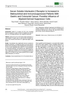
RESULTS Cloning and Structure. We have isolated and characterized four independent cDNA clones of the FLT VEGF receptor from a human vascular endothelial-cell cDNA library identified by hybridization with a DNA probe derived from the extracellular domains of this receptor (16). On the basis of sequence analysis, two of these clones appear to be derived from the previously identified membrane-spanning receptor, whereas the other two clones encode a C-terminally truncated form of the FLT VEGF receptor. The two truncated FLT clones are also independent of each other because they terminate at different 5' and 3' sites. The shorter 1.8-kb clone begins at bp 803, whereas the longer 2.6-kb clone starts at bp 597 of the knownflt coding sequence. The longer clone spans the coding sequence from the second immunoglobulin-like domain to the 3' noncoding region. The cDNA coding sequences of both the truncated flt clones are identical to full-length flt up to bp 2218, after which they encode a distinctive hydrophilic C-terminal 31-aa residue sequence, containing two cysteine residues but not resembling an immunoglobulin-like domain, before the open reading frame is terminated by TAA (Fig. 1A). The presence of HUVEC-derived mRNA containing the entire 2.3-kb coding sequence of sFLT, including the secretory leader sequence and first immunoglobulin-like domain, was established by RT-PCR and confirmed by restriction A 687
651
700
FLT WKEIT EA PYLLRNLSDH TVAISSSTTL DCHANGVPEP QITWFKNNHK sFLT KEITIItQEHC NKKAVFSRIS KFKSTRNDCT TQSNVKH*
FLT AUC AGA GAU CAG GAA AGA GGU GAG CAC
SFLT AUC
Apparent splice site
B Extracellular
Intracellular kinase domains
ligand-binding domains N
L5 -
5
-S
5-5
5-5
(1338 aa) Signal eptide (687 aa) Unique 31 amino acid C-terminu
p)lasma
Meembrane
FIG. 1. Deduced protein sequence of human sFLT. (A) Alignment of aa 651-700 of membrane-spanning FLT and C-terminal aa 651-687 of sFLT. Sequences N terminal to aa 651 are identical, as are those in box. *, Translational stop site. mRNA sequences spanning the site of divergence are shown below the corresponding protein sequences. The characteristic donor AG and acceptor G/A sequences flanking the apparent splice site of FLT and the full donor splice-site sequence of sFLT are bracketed. (B) Domain structures of membrane-spanning FLT and sFLT are schematically illustrated.
Immunoglobulin-like domains in the extracellular ligand-binding regions are numbered from the N termini with the characteristic immunoglobulin-fold disulfide-bonded cysteine-residue sulfur atoms (S) marked.
Biochemistry: Kendall and Thomas
Proc. Natl. Acad. Sci. USA 90 (1993)
mapping of the PCR product. Therefore, the entire 687-aatruncated receptor is composed of the secretory leader sequence and the N-terminal six extracellular immunoglobulinlike domains present in the previously described membranespanning receptor fused to the specific C-terminal polypeptide sequence. The receptor is missing the membrane-proximal seventh immunoglobulin-like domain, the transmembrane-spanning sequence, and the kinase domains (Fig. 1B) and, thus, is a soluble form of FLT. Altemative Splicing. The different 3' sequences encoding membrane-spanning and sFLT arise by alternative mRNA splicing. The base pairs flanking the apparent splice site (Fig. 1A) incorporate the characteristic splice-site donor (AG) and acceptor (G>A) nucleotides (20). PCR analysis confirmed that the soluble receptor is generated by an alternatively spliced message. RT-PCR reactions using HUVEC poly(A)+selected RNA as a template were done with a primer derived from the extracellular domain of FLT (flt-i) common to both forms of the receptor and primers specific for either the membrane-spanning form (flt-2) or the soluble form (sflt-3) of FLT (Fig. 2A). The expected 587-bp band (Fig. 2B, lane 1) was produced using the primers for the membrane-spanning form of FLT. The PCR reaction done with primers that flank the putative splice site of the soluble receptor generates the predicted 747-bp fragment (lane 2), and primers specific for the soluble receptor (sflt-3 and sflt-4) produce the expected 332-bp band (lane 3). These PCR products were not detected in control reactions without RT (data not shown), eliminating the possibility of contamination by genomic DNA.
A 1700
2500 _ fIt-2 -_ sfit-3 sfit-4 I-sflt-3 fit-5 _-sflt-3 2219
fit-1 _.-
FLT
fit-1 _
sFLT
B
bp
bp
1198 >
747 587
676 > 517 w 396 > 222
w 393 < 332
1
2
j
4
FIG. 2. Alternative splicing of FLT mRNA. (A) Coding region from nt 1700-2500 spanning the splice site preceding nt 2219. Relative locations of PCR primers used to amplify sequences encoding membrane-spanning FLT and sFLT are labeled. Nucleotide sequences at left of vertical dashed line encode amino acid sequences common to both membrane-spanning FLT and sFLT, whereas those at right of this dashed line are specific to either one or the other sequence. Primers that hybridize with the coding regions either common to FLT and sFLT or unique to FLT are denoted flt, whereas those that hybridize to nucleotide sequences unique to sFLT are designated sflt. (B) With these primers, RT-PCR products of the expected size were generated, spanning the splice site encoding membrane-spanning FLT (flt-1/flt-2, 587 bp, lane 1) and sFLT (flt-1/flt-3, 747 bp, lane 2) along with the sFLT primers both 3' of the splice site (flt-4/flt-3, 332 bp, lane 3). PCR products of human genomic DNA designed to amplify the sequence around the putative splice site of membrane-spanning FLT (flt-5/flt-2, 233 bp, lane 4) and sFLT (flt-5/sflt-3, 393 bp, lane 5) are shown. Positions of DNA fragment standards and PCR product sizes in bp are labeled at left and right, respectively.
10707
PCR analysis ofRNase-treated human genomic DNA using flt primers is also shown in Fig. 2B (lanes 4 and 5). The reaction with flt-5, a primer common to both receptor forms located 38 bp 5' to the putative splice site, and flt-2, a primer specific for the membrane-spanning receptor, does not produce the predicted 233-bp fragment (lane 4), inferring the existence of an intervening intron. In contrast, the reaction with flt-5 and the sFLT-specific primer sflt-3 generates the expected 395-bp fragment (lane 5), suggesting that sFLT mRNA is generated by direct read-through "splice skipping" at this site. Therefore, either membrane-spanning FLT or sFLT mRNAs are generated, depending on whether either intron excision or read-through occurs at this position. Expression and Purification. To analyze the ligand-binding and biological properties of this truncated form of the receptor, the sFLT protein was generated by using a baculovirus expression system. Recombinant human sFLT present in conditioned medium of Sf9 insect cells infected with baculovirus encoding sFLT binds to heparin-Sepharose and is step-eluted from 0.6 to 1.0 M NaCl. The eluted protein migrates on SDS/PAGE as a single major band of 85-90 kDa (Fig. 3). The polypeptide mass of mature sFLT, generated by cleavage of the secretory leader sequence, is deduced from the cDNA sequence to be 75 kDa, which is presumably increased by carbohydrate attachment to some or all of the 12 N-linked glycosylation-site consensus sequences. The somewhat broadened band width of this putative glycoprotein could be attributable to the heterogeneity of its constituent oligosaccharides. Amino acid sequence analysis (data not shown) reveals a single N terminus starting at Ser-27 of the full-length translation product consistent with the signalpeptide-cleavage site predicted by the method of von Heijne (21). VEGF Binding and Inhibition of Mitogenic Activity. Initially, complex formation between purified recombinant human 125I-labeled VEGF and sFLT in unfractionated culture medium from the baculovirus-infected Sf9 insect cells was shown by the appearance of large radiolabeled complexes that eluted at the void volume of a Sephacryl S200 gel filtration column (data not shown). To determine the masses of these complexes, 125I-labeled VEGF and purified sFLT were covalently crosslinked with bis(sulfosuccinimidyl)suberate and characterized by SDS/PAGE and autoradiography (Fig. 4, lane 1). A broad band ranging from 115 to 145 kDa and a narrower band of 220 kDa are seen. The broad band is presumably composed of complexes containing one molecule of sFLT crosslinked to single 23-kDa VEGF subunits and 46-kDa homodimers. The mass of the 220-kDa band corresponds to a VEGF dimer linked to two soluble receptor molecules. This higher mass complex indicates that each subunit of dimeric VEGF probably can bind one molecule of sFLT, perhaps promoting soluble receptor dimerization. These bands are eliminated by excess unlabeled VEGF (lane kDa
*.