TECHNIQUE AND APPLICATION
Endoscopic Approach to the Infratemporal Fossa: Anatomic Study Philip V. Theodosopoulos, MD Department of Neurosurgery, University of Cincinnati Neuroscience Institute and College of Medicine; Mayfield Clinic, Cincinnati, Ohio
Bharat Guthikonda, MD Department of Neurosurgery, University of Cincinnati College of Medicine, Cincinnati, Ohio
Aaron Brescia, MD Department of Otolaryngology, University of Cincinnati Neuroscience Institute and College of Medicine, Cincinnati, Ohio
Jeffrey T. Keller, PhD Department of Neurosurgery, University of Cincinnati Neuroscience Institute and College of Medicine; Mayfield Clinic, Cincinnati, Ohio
Lee A. Zimmer, MD, PhD Department of Otolaryngology-Head and Neck Surgery, University of Cincinnati Neuroscience Institute and College of Medicine, Cincinnati, Ohio Reprint requests: Philip V. Theodosopoulos, MD, c/o Editorial Office, Department of Neurosurgery, University of Cincinnati College of Medicine, PO Box 670515, Cincinnati, OH 45267-0515. Email:
[email protected] Received, August 22, 2008. Accepted, July 29, 2009. Copyright © 2010 by the Congress of Neurological Surgeons
OBJECTIVE: Classic surgical exposures of the infratemporal fossa region, including the adjacent intracranial space, temporal bone, and sinonasal region, require the extensive exposure associated with the transcranial, transfacial, and transmandibular approaches with their inherent neurological and cosmetic morbidities. In this study, we evaluated the feasibility and exposure afforded by combining 2 endoscopic transmaxillary approaches, endonasal and Caldwell-Luc supplement, to the infratemporal fossa. METHODS: Endoscopic transmaxillary dissection was performed in 4 formalin-fixed cadaver heads (8 sides). We quantified the extent of exposure achieved within the pterygopalatine and infratemporal fossae after our initial dissection, which was endonasal with a medial antrostomy, and after addition of a Caldwell-Luc incision with an anterior antrostomy. Complementing this anatomic study, we report on a patient in whom this endoscopic transmaxillary approach combining the endonasal and Caldwell-Luc approaches was used for resection of a trigeminal schwannoma in the infratemporal fossa. RESULTS: The combination of these 2 endoscopic transmaxillary approaches enabled visualization of the entire region of the pterygopalatine fossa and anteromedial aspect of the infratemporal fossa. Additional posterolateral exposure of the infratemporal fossa requires significant traumatic traction on the nose. Addition of the Caldwell-Luc transmaxillary approach exposed the remainder of the infratemporal fossa, including the mandibular nerve and branches, middle meningeal artery, and even the distal cervical portion of the internal carotid artery. CONCLUSION: Endoscopic exposure of the infratemporal fossa is feasible. Using the combination of the endonasal and Caldwell-Luc approaches for direct transmaxillary access significantly extended exposure, allowing safe and effective resection of infratemporal fossa lesions. KEY WORDS: Caldwell-Luc, Endoscopy, Infratemporal fossa Neurosurgery 66:196-203, 2010
T
DOI: 10.1227/01.NEU.0000359224.75185.43
he infratemporal fossa is a retromaxillary space in which neoplasms can arise primarily or from secondary spread from nearby regions (e.g., middle cranial fossa, maxillary sinus, orbit). As such, this is an area of interest for multiple subspecialties including neurosurgery, otolaryngology, ophthalmology, maxillofacial surgery, and radiation oncology. Access to the infratemporal fossa has traditionally been through the transfacial or transmandibular anterior corridor and transcranial lateral and posterolateral corridors.1-8 Surgical interest in the infratemporal fossa dates back to the 1850s when exposure was necessary to treat sphenopalatine neuralgia.9-11 The initial transmaxillary (transantral) approach was described in 1928 by Sewall 12 to expose the
196 | VOLUME 66 | NUMBER 1 | JANUARY 2010
www.neurosurgery- online.com
sphenopalatine ganglion in the pterygopalatine fossa. More recently, Conley,13 Crockett,14 and DelGaudio15 renewed interest in this region, using more extensive surgical exposures. Ugo Fisch,16,17 often considered the father of infratemporal fossa surgery, and Fisch et al3 popularized a series of surgical approaches, types A through C, to access a progressively larger area of the infratemporal fossa. Although the exposure provided was excellent, these approaches carried the increased risks of hearing loss, facial weakness and hypoesthesia, mastication difficulty, and cerebrospinal fluid leakage. Minimally invasive endoscopic approaches to the midline cranial base, from the planum sphenoidale to the clivus and even as caudal to the
www.neurosurgery-online.com
ENDOSCOPIC APPROACH TO THE INFRATEMPORAL FOSSA
A
B
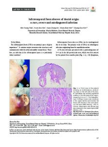
FIGURE 1. Endonasal approach to pterygopalatine and infratemporal fossae. A, cadaveric exposure of the sphenopalatine artery (SPA), the largest distal branch of the internal maxillary artery, in the pterygopalatine fossa. Maxillary antrostomy is made in the medial wall of the maxillary sinus. Elevation of the mucosa of the lateral wall of the nasal cavity over the crista ethmoidalis (CE) exposes the SPA and posterior antrum. B, illustration of the SPA and posterior nasal artery (PNA) emerging from the sphenopalatine foramen. C, posterior antrostomy is made to expose the sphenopalatine ganglion
odontoid, have been described.18-26 However, although numerous reports describe the surgical exposure of the pterygopalatine fossa,15,23,27-38 a dearth of literature exists on the endoscopic approaches to the lateral cranial base. In fact, some have argued that the location of the infratemporal fossa makes it inaccessible for endoscopic evaluation.40 Because we found no detailed cadaveric study in our review of the literature that defined the endoscopic anatomy or quantification of exposure of the infratemporal fossa, our study attempted to understand the potential effectiveness of endoscopy to explore this anatomic region, with special attention to the associated exocranial anatomy. We provide an anatomic study and case report of a trigeminal schwannoma to assess the effectiveness of an endoscopic transmaxillary approach that combines the endonasal and Caldwell-Luc approaches. Our case represents the second report in the literature that deals solely with endoscopic resection of an infratemporal trigeminal schwannoma.
MATERIALS AND METHODS Four formalin-fixed cadaveric skull specimens used for dissection were injected with colored latex to accentuate the vasculature according to the technique described by Sanan et al.41 Each side of each specimen was dissected, leading to endoscopic exposure of 8 infratemporal fossae. The extent of endoscopic exposure was identified for stage 1 of the dissection via an endonasal approach with a medial wall antrostomy and for stage 2 via a Caldwell-Luc incision with an anterior wall antrostomy. A computed tomographic scan was obtained before and after each stage of dissection to assess exposure and for qualitative comparison. We discuss the lessons learned from our anatomic findings in a clinical application, reporting on a patient in whom an endoscopic transmaxillary approach combining these 2 approaches, endonasal and Caldwell-Luc supplement, was used.
NEUROSURGERY
C
(SPG), vidian nerve, greater palatine nerve, and maxillary division of the trigeminal nerve (V2) and infraorbital nerve (IN). Shown transparently are the internal maxillary artery and its branches, that is, the descending palatine artery, PNA, and SPA. SOF, superior orbital fissure. (Reprinted with permission of the Mayfield Clinic.)
RESULTS Cadaveric Dissection Stage 1: Endonasal, Transmaxillary Exposure During stage 1 of the cadaveric dissection, we assessed the extent of exposure possible via an endonasal approach. After the middle turbinate was removed using a microdébrider (Medtronic Xomed Inc., Jacksonville, FL), the ethmoid bulla was identified lateral to the middle turbinate. A large maxillary antrostomy was then performed to allow access to the maxillary sinus. The ethmoid bulla was removed to expose the anterior ethmoid sinuses and increase intranasal exposure. The inferior turbinate was removed. The sphenopalatine artery (Fig. 1), the largest distal branch of the internal maxillary artery, was then identified. The mucosa of the lateral wall of the nasal cavity over the crista ethmoidalis was elevated, exposing the sphenopalatine artery. The course of the artery and its branches including the descending pharyngeal artery were variable. Division of the vessel was performed after standard application of vascular hemoclips in an effort to reproduce the operative environment. The pterygopalatine ganglion was exposed posterior to the sphenopalatine artery, which could be traced medially to identify the vidian nerve and pterygoid canal. Further extension of the exposure along the course of the pterygoid canal leads to the medial border of the distal petrous segment (C2) of the internal carotid artery. The infraorbital nerve, a branch of the trigeminal nerve (V2), was also identified originating lateral to the sphenopalatine ganglion and coursing anteriorly and superiorly toward the floor of the orbit. At this stage, the root of the pterygoid plate was located at the posterolateral extent of the exposure. The foramen ovale was located just lateral to the root of the lateral pterygoid plate. The
VOLUME 66 | NUMBER 1 | JANUARY 2010 | 197
THEODOSOPOULOS ET AL.
A
B
FIGURE 2. Addition of a Caldwell-Luc incision to the endonasal transmaxillary approach. Illustration of a lateral sublabial incision (A) and intraoperative view ( B) of an anterior maxillotomy providing 2 simultaneous access routes, endonasal and transmaxillary, to the infratemporal fossa. (Reprinted with permission of the Mayfield Clinic.)
pterygoid plate, which is a thick bony strut, was then drilled posteriorly until both the lateral and medial pterygoid plates were identified. The lateral pterygoid plate was the lateral boundary that could be easily exposed via a purely endonasal approach. Further lateral and posterior dissection was limited by the nasal bony pyramid and the nasolacrimal canal; a Caldwell-Luc approach was used to provide direct access to lateral structures and eliminated the use of angled endoscopes and instrumentation. Cadaveric Dissection Stage 2: Endonasal Transmaxillary Exposure via the Caldwell-Luc Approach During stage 2, we added a Caldwell-Luc incision (Fig. 2). After exposure of the anterior face of the maxilla, an anterior maxillo-
198 | VOLUME 66 | NUMBER 1 | JANUARY 2010
FIGURE 3. Further exposure of the infratemporal fossa is achieved by combining the endonasal-transmaxillary and Caldwell-Luc transmaxillary approaches. The lateral pterygoid plate has been removed. The lateral pterygoid muscle (LPtM) can be traced back to its insertion on the mandibular condyle. Medial to the mandibular nerve (V3) is the levator veli palatini muscle (LVP), which overlies deep to the cartilaginous portion of the eustachian tube. IMAX, internal maxillary artery. (Reprinted with permission of the Mayfield Clinic.)
tomy was performed. The endonasal and Caldwell-Luc transmaxillary approaches were used simultaneously as working channels. Removal of the lateral pterygoid plate flush to the cranial base allowed exposure of the foramen ovale and the exiting mandibular nerve (V3) just lateral and posterior to the root of the lateral pterygoid plate. The lateral pterygoid muscle could be followed all the way to its insertion point at the mandibular condyle. Dissecting medial to the mandibular nerve allowed exposure of the levator veli palatini muscle with the cartilaginous eustachian tube lying deep to the muscle (Fig. 3). During this stage of dissection, one of our main goals was to assess whether an endoscopic route could expose the mandibular nerve, middle meningeal artery, and upper portion of the cervical carotid artery. After identifying the foramen ovale and mandibular nerve, we continued our dissection, exposing the middle meningeal artery anterolaterally and, through a generous fat pad, the upper cervical carotid artery posterolaterally. Approximately 1.5 to 2 cm of the upper cervical carotid artery was exposed via this approach (Figs. 4 and 5). Illustrative Case A 36-year-old woman presented with headache and progressive symptoms of a left-sided facial dysesthesia in the trigeminal nerve and mandibular nerve distributions. Clinical examination revealed decreased pinprick sensation in the trigeminal nerve distribution and an intact corneal reflex and symmetrical bite. Radiographic evaluation revealed an enhancing lesion that extended from the inferior posterior cavernous sinus through a widened foramen ovale into the infratemporal fossa, consistent with a trigeminal nerve tumor (Fig. 6). After discussion regarding treatment options, she underwent an endoscopic subtotal resection of the lesion
www.neurosurgery-online.com
ENDOSCOPIC APPROACH TO THE INFRATEMPORAL FOSSA
A
B
C
D
FIGURE 4. Exocranial view of the inferior skull base in a
dry skull demonstrating the relationship of important foramina. The foramen spinosum is posterior and lateral to the foramen ovale. The cervical carotid artery enters the carotid canal posterior to the foramen spinosum and foramen ovale. (Reprinted with permission of the Mayfield Clinic.)
FIGURE 6. Gadolinium-enhanced magnetic resonance imaging scans.
Preoperatively, axial (A) and coronal (B) planes showing a lesion (arrow) extending from a widened foramen ovale into the infratemporal fossa. Postoperatively, axial (C) and coronal (B) planes showing near-total resection (arrow). (Reprinted with permission of the Mayfield Clinic.)
FIGURE 5. Overview of important neurovascular structures exposed in the infratemporal fossa. The mandibular nerve (V3) and its branches, the upper cervical carotid artery, and the middle meningeal artery could all easily be identified. (Reprinted with permission of the Mayfield Clinic.)
through a combined endonasal transmaxillary corridor that was preceded by embolization of an external carotid branch (Fig. 7). We used a 4-handed technique through 2 access points (endonasal and CaldwellLuc) with frameless stereotactic guidance. The lesion was removed in a piecemeal manner, with a small remnant at the level of the foramen ovale (Fig. 8). Pathological evaluation identified this tumor as a trigeminal schwannoma, World Health Organization grade 1. The patient was discharged on the second postoperative day without any evidence of trigeminal dysfunction.
DISCUSSION Endoscopic access to exocranial and endocranial lesions of the midline anterior cranial base has become an important part of the
NEUROSURGERY
armamentarium of cranial base surgeons. This approach is often minimally invasive, well tolerated by the patient, and effective in the exposure and treatment of certain pathologies of the sella, clivus, and anterior cranial base. However, lateral cranial base lesions continue to be approached via more traditional open approaches. In this anatomic study, we demonstrated the extent and limits of endoscopic exposure of the infratemporal fossa and documented the successful application of this technique in a patient who underwent endoscopic resection of a trigeminal schwannoma. Several pathological conditions could benefit from an endoscopic exposure of the infratemporal fossa. These include extensive juvenile nasopharyngeal angiofibromas involving the pterygopalatine and infratemporal fossae, trigeminal schwannomas and meningiomas with an extracranial extension into the infratemporal fossa via the foramen ovale42,43 or schwannomas that originate primarily from the extracranial portion of the mandibular nerve,44 and lymphoproliferative disorders that present as isolated infratemporal fossa lesions. Traditional approaches to these lesions include the preauricular infratemporal approach, a transmandibular approach, and a Fisch type C approach. Each of these approaches is effective in exposing lesions of the infratemporal fossa but carries the risk of cranial nerve morbidity and extensive soft-tissue disruption.
VOLUME 66 | NUMBER 1 | JANUARY 2010 | 199
THEODOSOPOULOS ET AL.
A
B
FIGURE 8. Intraoperative photograph showing the sur-
gical exposure obtained by combining the endonasal transmaxillary and Caldwell-Luc transmaxillary approaches. (Reprinted with permission of the Mayfield Clinic.).
FIGURE 7. Endoscopic approach to the infratemporal
fossa. Extent of exposure is shown through the 2 corridors: first, an endonasal approach with a medial wall antrostomy (A) and second, a Caldwell-Luc incision with an anterior wall antrostomy (B). (Reprinted with permission of the Mayfield Clinic.).
Based on our results, we propose a restructuring of the treatment paradigm for lesions of the infratemporal fossa. The indolent nature of most benign lesions in this area results in little morbidity from the natural progression of the disease. Additionally, small remnants often remain on the trigeminal ganglion and cavernous sinus, even when extensive approaches are used to avoid significant neurovascular injury. Considering this indolent nature and residuals, one may argue for a more minimally invasive alternative in the treatment of such lesions. We propose that the combined endoscopic transmaxillary approaches via an endonasal approach and Caldwell-Luc supplement is a feasible alternative that not only obviates the need for skin incisions but does not compromise the extent of exposure necessary in the treatment of infratemporal fossa lesions. Combination of these 2 approaches allows easy dissection within the entire width and depth of the infratemporal fossa and provides adequate exposure for vascular control of the internal maxillary, sphenopalatine, and middle meningeal arteries. This combined approach also allows safe dissection around the high cervical internal carotid
200 | VOLUME 66 | NUMBER 1 | JANUARY 2010
artery, adequate exposure of the foramen ovale, and, with the frequent widening of the foramen by most lesions in this region, easier dissection in and around the mandibular nerve at its origin in the cranial base and along its entire course. Our case illustrates the efficacy of this technique in the treatment of a trigeminal schwannoma that extended into the infratemporal fossa and provided a number of clinical lessons for our surgical team. Although the Caldwell-Luc approach was considered an adjunct to the endonasal approach used in our anatomic study, it proved to be a necessary step for allowing a significant extent of tumor dissection and control of intraoperative bleeding in our patient. The 2-port access, 4-hand technical variation proposed as a new technique is essential for adequate tumor exposure and effective dissection within this space, which is quite difficult to access (Fig. 9). The Denker approach can be considered an alternative to the Caldwell-Luc approach; yet this approach requires the removal of supporting structures such as the maxilla at the inferior lateral piriform aperture, and therefore its limitation is the resulting cosmetic deformity of the lateral nasal ala. Removal of the inferior turbinate would improve visualization inferiorly if the tumor removal required such exposure. However, we prefer preservation of this turbinate when possible to preserve nasal function. Appropriate instrumentation, particularly with respect to tumor removal, is still limited for effective application through this corridor. Intraoperative navigation remains essential for the safety and maximal effectiveness of this approach as it does on all endoscopic cranial base procedures, particularly given the often distorted local anatomy of these primarily slow-growing lesions. Finally, the rostral limitation of this exposure around the area of a widened foramen ovale precludes at this point the safe approach to lesions with significant intracranial extension via a purely endoscopic approach.
www.neurosurgery-online.com
ENDOSCOPIC APPROACH TO THE INFRATEMPORAL FOSSA
A
B
FIGURE 9. Axial computed tomography images of the cadaver dissection after
a stage 1 endonasal approach (A) and after a stage 2 endonasal approach combined with a supplemental Caldwell-Luc approach (B). A, endonasal dissection alone exposed the pterygopalatine fossa and medial infratemporal fossa, limited by the lateral pterygoid plate (yellow area). B, Caldwell-Luc exposure increased the exposure gained, specifically the high cervical internal carotid artery and mandibular condyle (orange area). (Reprinted with permission of the Mayfield Clinic.)
CONCLUSION Endoscopic exposure of the infratemporal fossa by combining the endonasal and Caldwell-Luc approaches is feasible and safe. Limits of the exposure are the ramus of the mandible laterally, the osseous skull base and foramen ovale superiorly, and the distal upper cervical internal carotid artery posteriorly. This less invasive technique may prove to be most beneficial in the surgical management of juvenile nasopharyngeal angiofibroma and trigeminal schwannoma with extracranial extension and in the biopsy of malignant lesions that extend into the infratemporal fossa. In light of the complex anatomy of the region and the technical instrumentation limitations, we suggest a stepwise progression for the adoption of this approach into clinical practice, starting with the surgical exposure of the sella turcica, proceeding with the application of the same techniques to the clivus and anterior cranial base, and finally exploring more lateral regions (e.g., the pterygopalatine and infratemporal fossae). A multidisciplinary approach, with both an endoscopic sinus surgeon and neurosurgeon with cranial base oncology experience, is recommended. Disclosure The authors have no personal financial or institutional interest in any of the drugs, materials, or devices described in this article.
REFERENCES 1. Browne JD, Jacob SL. Temporal approach for resection of juvenile nasopharyngeal angiofibromas. Laryngoscope. 2000;110(8):1287-1293. 2. Cass SP, Hirsch BE, Stechison MT. Evolution and advances of the lateral surgical approaches to cranial base neoplasms. J Neurooncol. 1994;20(3):337-361. 3. Fisch U, Fagan P, Valavanis A. The infratemporal fossa approach for the lateral skull base. Otolaryngol Clin North Am. 1984;17(3):513-552.
NEUROSURGERY
4. Fukushima T, Day JD, Hirahara K. Extradural total petrous apex resection with trigeminal translocation for improved exposure of the posterior cavernous sinus and petroclival region. Skull Base Surg. 1996;6(2):95-103. 5. Hadley KS, Shelton C. Infratemporal fossa approach to the hypoglossal canal: Practical landmarks for elusive anatomy. Laryngoscope. 2004;114(9):1648-1651. 6. Hegazy HM, Carrau RL, Snyderman CH, Kassam A, Zweig J. Transnasal endoscopic repair of cerebrospinal fluid rhinorrhea: A meta-analysis. Laryngoscope. 2000; 110(7):1166-1172. 7. Sennaroglu L, Slattery WH 3 rd. Petrous anatomy for middle fossa approach. Laryngoscope. 2003;113(2):332-342. 8. Zhang M, Garvis W, Linder T, Fisch U. Update on the infratemporal fossa approaches to nasopharyngeal angiofibroma. Laryngoscope. 1998;108(11 Pt 1):1717–1723. 9. Carnochan J. Excision of the trunk of the second branch of the fifth nerve. Am J Med Soc. 1858;1:134. 10. Segmond P. De la reaction du nerf maxillaire superieur et du ganglion sphenopalatin dans le fente pterygomaxillaire par la voie temporale [in French]. Rev Chirurg.1890;173-197. 11. Sluder G. Etiology, diagnosis, prognosis and treatment of sphenopalatine ganglion neuralgia. JAMA. 1913;60(16):1202-1205. 12. Sewall E. An operation for the removal of the sphenopalatine ganglion. Ann Otol Rhinol Laryngol. 1926:967-969. 13. Conley J. The surgical approach to the pterygoid area. Ann Surg. 1956;144(1):39-43. 14. Crockett DJ. Surgical approach to the back of the maxilla. Br J Surg. 1963;50:819821. 15. DelGaudio JM. Endoscopic transnasal approach to the pterygopalatine fossa. Arch Otolaryngol Head Neck Surg. 2003;129(4):441-446. 16. Fisch U. Infratemporal fossa approach for glomus tumors of the temporal bone. Ann Otol Rhinol Laryngol. 1982;91(5 Pt 1):474-479. 17. Fisch U. Infratemporal fossa approach for lesions in the temporal bone and base of the skull. Adv Otorhinolaryngol. 1984;34:254-266. 18. Cappabianca P, Alfieri A, de Divitiis E. Endoscopic endonasal transsphenoidal approach to the sella: Towards functional endoscopic pituitary surgery (FEPS). Minim Invasive Neurosurg. 1998;41(2):66-73. 19. Cappabianca P, Frank G, Pasquini E, de Divitiis O, Calbucci F. Extended endoscopic endonasal transsphenoidal approaches to the suprasellar region, planum sphenoidale & clivus, in de Divitiis E, Cappabianca P (eds): Endoscopic Endonasal Transsphenoidal Surgery. New York, Springer-Verlag, 2003: pp 176-187. 20. Carrau RL, Jho HD, Ko Y. Transnasal-transsphenoidal endoscopic surgery of the pituitary gland. Laryngoscope. 1996;106(7):914-918. 21. Carrau RL, Kassam AB, Snyderman CH. Pituitary surgery. Otolaryngol Clin N Am. 2001;34(6):1143-1155, ix. 22. Cavallo LM, Cappabianca P, Galzio R, Iaconetta G, de Divitiis E, Tschabitscher M. Endoscopic transnasal approach to the cavernous sinus versus transcranial route: Anatomic study. Neurosurgery. 2005;56(2 suppl):379-389. 23. Cavallo LM, Messina A, Gardner P, et al. Extended endoscopic endonasal approach to the pterygopalatine fossa: Anatomical study and clinical considerations. Neurosurg Focus. 2005;19(1):E5. 24. Jho HD, Carrau RL. Endoscopic endonasal transsphenoidal surgery: Experience with 50 patients. J Neurosurg. 1997;87(1):44-51. 25. Jho HD, Carrau RL, McLaughlin MR, Somaza SC. Endoscopic transsphenoidal resection of a large chordoma in the posterior fossa. Acta Neurochir (Wien). 1997;139(4):343-348. 26. Kassam AB, Gardner P, Snyderman C, Mintz A, Carrau R. Expanded endonasal approach: Fully endoscopic, completely transnasal approach to the middle third of the clivus, petrous bone, middle cranial fossa, and infratemporal fossa. Neurosurg Focus. 2005;19(1):E6. 27. Al-Nashar IS, Carrau RL, Herrera A, Snyderman CH. Endoscopic transnasal transpterygopalatine fossa approach to the lateral recess of the sphenoid sinus. Laryngoscope. 2004;114(3):528-532. 28. Alfieri A, Jho HD, Schettino R, Tschabitscher M. Endoscopic endonasal approach to the pterygopalatine fossa: Anatomic study. Neurosurgery. 2003;52(2):374-380. 29. Bolger W, Osenbach R. Endoscopic transpterygoid approach to the lateral sphenoid recess. Ear Nose Throat J. 1999;78:36-46. 30. Bolger WE. Endoscopic transpterygoid approach to the lateral sphenoid recess: Surgical approach and clinical experience. Otolaryngol Head Neck Surg. 2005;133(1):2026. 31. Har-El G. Combined endoscopic transmaxillary-transnasal approach to the pterygoid region, lateral sphenoid sinus, and retrobulbar orbit. Ann Otol Rhinol Laryngol. 2005;114(6):439-442.
VOLUME 66 | NUMBER 1 | JANUARY 2010 | 201
THEODOSOPOULOS ET AL.
32. Klossek JM, Ferrie JC, Goujon JM, Fontanel JP. Endoscopic approach of the pterygopalatine fossa: Report of one case. Rhinology. 1994;32(4):208-210. 33. Mitskavich MT, Carrau RL, Snyderman CH, Weissman JL, Fagan JJ. Intranasal endoscopic excision of a juvenile angiofibroma. Auris Nasus Larynx. 1998;25(1):3944. 34. Ong BC, Gore PA, Donnellan MB, Kertesz T, Teo C. Endoscopic sublabial transmaxillary approach to the rostral middle fossa. Neurosurgery. 2008;62(3 Suppl 1):30-37. 35. Pasquini E, Sciarretta V, Farneti G, Ippolito A, Mazzatenta D, Frank G. Endoscopic endonasal approach for the treatment of benign schwannoma of the sinonasal tract and pterygopalatine fossa. Am J Rhinol. 2002;16(2):113-118. 36. Pasquini E, Sciarretta V, Farneti G, Mazzatenta D, Modugno GC, Frank G. Endoscopic treatment of encephaloceles of the lateral wall of the sphenoid sinus. Minim Invasive Neurosurg. 2004;47(4):209-213. 37. Schwartz TH, Fraser JF, Brown S, Tabaee A, Kacker A, Anand VK. Endoscopic cranial base surgery: Classification of operative approaches. Neurosurgery. 2008;62(5):991-1005. 38. Sekhar LN, Schramm VL Jr, Jones NF. Subtemporal-preauricular infratemporal fossa approach to large lateral and posterior cranial base neoplasms. J Neurosurg. 1987;67(4):488-499. 39. Statham M, Tami T. Endoscopic anatomy of the pterygopalatine fossa. Oper Tech Otolaryngol Head Neck Surg. 2006;17:197-200. 40. Tiwari R, Quak J, Egeler S, et al. Tumors of the infratemporal fossa. Skull Base Surg. 2000;10(1):1–9. 41. Sanan A, Abdel Aziz KM, Janjua RM, van Loveren HR, Keller JT. Colored silicone injection for use in neurosurgical dissections: Anatomic technical note. Neurosurgery. 1999;45(5):1267-1274. 42. Kafadar AM, Tanriverdi T, Canbaz B, Kuday C. Trigeminal neuroma with extracranial extension: The 31st case. Minim Invasive Neurosurg. 2006;49(4):230-233. 43. Lunardi P, Missori P, Gagliardi FM, Fraioli B. Trigeminal schwannoma with infratemporal extension. Case report. J Neurosurg Sci. 1989;33(3):293-295. 44. Roh JL. Removal of infratemporal fossa schwannoma via a transmandibular transpterygoid approach. Eur Arch Otorhinolaryngol. 2005;262(5):428-431.
COMMENTS
T
heodosopoulos et al. present an anatomical study on a purely endoscopic approach to the infratemporal fossa, which combines an endonasal and a sublabial transmaxillary approach. They also present a case of an extracranial schwannoma of the mandibular nerve (V3) that was successfully operated on with this approach. This article is a contribution to the recent literature on endoscopic approaches to the skull base and, in particular, the infratemporal fossa. The sublabial transmaxillary approach, a modification of the Caldwell-Luc maxillotomy is a welldescribed approach to the cavernous sinus and infratemporal fossa. The advantage of using the endoscope in this relatively deep and narrow approach is intuitive, but only recently have some authors suggested its use during this approach (1) and its combination with an endonasal approach for a wider working space (2), as Theodosopoulos et al. also report. Other experienced cranial base endoscopic neurosurgeons have presented the endoscopic endonasal exposure of the infratemporal fossa as well as the pterygopalatine (3, 4). Future studies will need to address the indications for these approaches, when the combination of the 2 is needed and their possible complications. Francesco Doglietto Fred Gentili Rome, Italy
T
heodosopoulos et al. report on the use of the endonasal approach combined with a Cauldwell-Luc incision to reach the pterygopalatine and infratemporal fossa. We have used the endnasal approach alone to reach these same areas without the use of the Cauldwell-Luc incision. In fact, in our first case, we did add the Cauldwel-Luc and found that
202 | VOLUME 66 | NUMBER 1 | JANUARY 2010
it was not needed and did not add any additional exposure. In the figure provided by the authors, they imply the cut on the computed tomographic scan that shows the additional exposure provided by the Cauldwell-Luc is more inferior than the cut showing the reach of the endonasal approach by itself. This is misleading. I would encourage them to try the endonasal approach by itself without the Cauldwell-Luc incision and determine if it is needed, because it can always be added later in the operation. Theodore H. Schwartz New York, New York
I
n this article, the authors have demonstrated once again the possibility to apply for an extracranial route to access such deep located area of the cranial base, namely the infratemporal fossa, and deal with pathologies involving either the extracranial and the intracranial and intradural compartments. They report both a detailed anatomical study and its clinical/in vivo application. It has been highlighted the contribution of the endoscopic technique, which has tremendously boosted the development of endonasal surgery, affording its extension among neurosurgeons on one hand and ear, nose, and throat and maxillofacial surgeons on the other in past decades (1, 2). Even though the anatomy of this area has been reported in the pertinent literature more than once, it has to be said that authors have underlined the surgically-oriented relevance of the different anatomical details. Though, the most interesting considerations concern the adjunct of Caldwell-Luc approach to allow a safer dissection of tumor and control the intraoperative bleeding. Hence, this should be kept in mind, especially when approaching through the endonasal route lesions with lateral extent. As a matter of fact, the combination of an antrostomy via a transoral sublabial access provides the availability to achieve a wider overview of the targeted area with a minimal supplemental approach. In the case of clinical applications, the relevant role played by image-guidance system, in providing surgeon with correct orientation should not be underestimated. Luigi M. Cavallo Paolo Cappabianca Naples, Italy
1. Fortes FS, Sennes LU, Carrau RL, Brito R, Ribas GC, Yasuda A, Rodrigues AJ Jr, Snyderman CH, Kassam AB: Endoscopic anatomy of the pterygopalatine fossa and the transpterygoid approach: development of a surgical instruction model. Laryngoscope 118:44–49, 2008. 2. Kassam AB, Vescan AD, Carrau RL, Prevedello DM, Gardner P, Mintz AH, Snyderman CH, Rhoton AL: Expanded endonasal approach: vidian canal as a landmark to the petrous internal carotid artery. J Neurosurg 108:177–183, 2008.
T
he endoscopic sublabial transmaxillary approach presented by Theodosopoulos et al. is a novel approach to access to the rostral middle fossa. Meningiomas, dumbbell schwannomas, and head and neck tumors that extend to the infratemporal and middle fossa are prototypical tumors of this region that could be addressed by the endoscopic sublabial transmaxillary approach. The authors deeply debate on advantages and limits of the technique, comparing it with the more traditional approaches (subtemporal, pterional, orbitozygomatic) and to the endoscopic endonasal approaches. Even if the argumentation of the authors is quite convincing and conclusive, I still maintain a suspicion in respect to the Caldwell-Luc approach: could it be that complications, such as
www.neurosurgery-online.com
ENDOSCOPIC APPROACH TO THE INFRATEMPORAL FOSSA
facial numbness and dysestesias due to infraorbital and alveolar nerve injury, occur rarely, but their occurrence is so troublesome and difficult to treat, that it can not be evaluated only by the percentage of incidence? Therefore, I maintain my preference for the endonasal transmaxillary approach. We utilize an endoscopic endonasal inferior turbinectomy transmaxillary approach: using a drill, we remove part of the ascending branch of the maxillary bone, making the surgical corridor more straight
and avoiding the need for an endoscope angled more than 30 degrees to gain a better access than the method described in the article. The clinical setting of these patients would answer in the future to the question about the preferable method, the sublabial or the endonasal. Giorgio Frank Bologna, Italy
Hawaiians tell the story of Pele, the goddess who found and named the island of Hawai’i. After winning a war with the fire god that lived on Kilauea with her, Pele became ruler of the island and all of Hawai’i. The people of the islands loved and respected the goddess, but after learning that Hi’iaka, her sister, and the man of her dreams fell in love with each other, Pele caused an eruption. Today, the smell of sulphur reminds the natives that Pele, the fire goddess of the volcanoes, is still there. Credit corbis.com.
NEUROSURGERY
VOLUME 66 | NUMBER 1 | JANUARY 2010 | 203