Biotechnology and Bioprocess Engineering 22: 225-230 (2017) DOI 10.1007/s12257-016-0484-y
pISSN 1226-8372 eISSN 1976-3816
RESEARCH PAPER
Engineering Chimeric Two-component System into Escherichia coli from Paracoccus denitrificans to Sense Methanol Vidhya Selvamani, Irisappan Ganesh, Murali kannan Maruthamuthu, Gyeong Tae Eom, and Soon Ho Hong
Received: 16 August 2016 / Revised: 18 October 2016 / Accepted: 16 November 2016 © The Korean Society for Biotechnology and Bioengineering and Springer 2017
Abstract Escherichia coli does not have the methanol sensing apparatus, was engineered to sense methanol by employing chimeric two-component system (TCS) strategy. A chimeric FlhS/EnvZ (FlhSZ) chimeric histidine kinase (HK) was constructed by fusing the sensing domain of Paracoccus denitrificans FlhS with the catalytic domain of E. coli EnvZ. The constructed chimeric TCS FlhSZ/OmpR could sense methanol by the expression of ompC and gfp gene regulated by ompC promoter. Real-time quantitative PCR analysis and GFP-based fluorescence analysis showed the dynamic response of the chimeric TCS to methanol. The expression of ompC and the gfp fluorescence was maximum at 0.01 and 0.5% of methanol, respectively. These results suggested that E. coli was successfully engineered to sense methanol by the introduction of chimeric HK FlhSZ. This strategy can be employed for the construction of several chimeric TCS based bacterial biosensors for the development of biochemical producing recombinant microorganisms. Keywords: methanol, chimeric two-component system, green fluorescent protein,Escherichia coli
Vidhya Selvamani, Irisappan Ganesh, Murali kannan Maruthamuthu, Soon Ho Hong* Department of Chemical Engineering, University of Ulsan, Ulsan 680749, Korea Tel: +82-52-259-1293; Fax: +82-52-259-1689 E-mail:
[email protected] Gyeong Tae Eom Research Center for Bio-based Chemistry, Korea Research Institute of Chemical Technology (KRICT), Ulsan 681-802, Korea Department of Green Chemistry and Environmental Biotechnology, Korea University of Science and Technology (UST), Daejeon 305-350, Korea
1. Introduction Methanol is one of the highest produced chemical in the world for versatile applications. Methanol is primarily used as an industrial solvent for inks, resins, adhesives and dyes. It is also used as an alternative motor fuel [1], as a solvent in the manufacture of cholesterol, streptomycin, vitamins, hormones and other pharmaceuticals [2]. According to methanol institute, about 35 million metric tons of methanol are produced per year. Considering the increasing demand for methanol and its widespread use, it is high-time to develop a high-throughput screening strategy. Two-component system (TCS) is considered as a basic stimulus and response coupled mechanism, which allows microbes to sense environmental changes. It is a method used in drug discovery, biology and chemistry to sense and monitor the process. The fundamental units of TCS include histidine kinase (HK) and a response regulator (RR) [3,4]. The HK sense the environmental change consequently induces autophosphorylation in transmitter domain, which aids as phosphor donor for the RR. The RR further activates the regulator protein and leading transcriptional regulon. It is also reported that engineering chimeric HK in E. coli can be employed for the development of powerful biosensor [5]. Paracoccus denitrificans is a nutritionally adaptable bacterium found in soil, sewage and sludge. It can grow heterotrophically on a variety of carbon sources and lithographically using hydrogen source or reduced C1 compounds such as methanol, methylamine or formate [6]. The genes involved in C1 compound metabolism are tightly regulated, one such regulatory system which presented in P. denitrificans is FlhR/FlhS TCS. These genes are located in the fgh cluster in P. denitrificans [9,10]. The regulatory proteins involved in controlling the expression of fgh gene
226
Biotechnology and Bioprocess Engineering 22: 225-230 (2017)
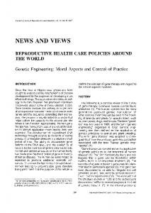
Fig. 1. Engineered E. coli to respond exogenous methanol. The P. denitrificans periplasmic histidine kinase domain of FlhS and the cytoplasmic catalytic domain of EnvZ were fused to form the FlhSZ chimeric protein, which phosphorylates the response regulator OmpR of the TCS while sensing methanol. This activates the ompC promoter, resulting in GFP expression in E. coli.
cluster are not yet identified [7]. The FlhR/FlhS deficient mutants cannot grow in methanol, methylamine, and formaldehyde environment [8]. In the present study, the FlhSZ chimeric HK was constructed by fusing the periplasmic domain of FlhS with the cytoplasmic domain of EnvZ. The resulting chimeric HK enables the E. coli to sense methanol by activating OmpR, which sequentially induce the expression of the ompC and the gfp gene regulated by the ompC promoter (Fig. 1). The expression of ompC and gfp are considered as a dynamic response of the chimeric HK FlhSZ for methanol. The expression profile of ompC gene was monitored by the real-time quantitative PCR (qRT-PCR) and GFP by a spectrofluorometer.
2. Materials and Methods 2.1. Bacterial strains and culture conditions The host strains for recombinant DNA manipulation and the expression of the recombinant proteins are E. coli XL1Blue (XB) and BL21 (DE3). The bacterial strains and plasmids used in this study are listed in Supplementary Table S1. E. coli strains were grown aerobically in LuriaBertani (LB) broth (10 g/L bacto-tryptone, 5 g/L bacto-yeast extract and 5 g/L NaCl) and in M9 minimal salts medium (Sigma) supplemented with 4 g/L of glucose, 2 mM MgSO4, 0.1 mM CaCl2 and 1% thiamine HCl at 37oC under vigorous shaking. 2.2. Molecular modeling A computational study was performed to predict the fusion site of chimeric HK (flhS/envZ). The interaction between
the two peptides was further structurally investigated by molecular modeling. The three-dimensional structure of the EnvZ protein was solved by X-ray diffraction method and it is available on Protein Data Bank (PDB ID 4CTI) [9]. The homology model of EnvZ was fused with peptide structure of FlhS modeled using Graphical user interface program of Easy Modeller 4.0 [10] using 2JIN as a template. The refined structure of the homology model was obtained by 3Drefine Protein Structure Refinement Server [11]. 2.3. Construction of chimeric FlhS/EnvZ The plasmid pFlhSZ1 contains chimeric HK FlhSZ under the control of T7 promoter. The periplasmic sensor domain of flhS (717 bp) and the cytoplasmic catalytic domain of envZ (675 bp) was amplified from the chromosomal DNA of P. denitrificans and E. coli XB by polymerase chain reaction (PCR). PCR was performed with the MJ mini Personal Thermal Cycler (BioRad Laboratories, USA) using the Expand High Fidelity PCR system (Roche Molecular Biochemicals, Mannheim, Germany). The PCR products were cloned into the low copy number plasmid pACYCDuet-1 using SacI and HindIII restriction sites to construct the pFlhSZ1 plasmid (Supplementary Fig. S1). Expression of FlhSZ chimeric HK was induced by the addition of isopropyl β-D-1-thiogalactopyranoside (IPTG) regulated by T7 promoter. 2.4. Monitoring of the ompC Gene expression by qRTPCR The transcription of the ompC gene in response to methanol was measured by qRT-PCR in E. coli cells harboring pFlhSZ1. A single colony of E. coli BL21 (DE3) (pFlhSZ1)
Engineering Chimeric Two-component System into Escherichia coli from Paracoccus denitrificans to Sense Methanol
was grown overnight in LB medium at 37oC. It was then diluted 100-fold in fresh M9 medium and incubated at 37oC in an orbital shaker at 250 rpm until the optical density at 600 nm (OD600) reached 0.5. Ten micromolar IPTG was added to the culture and the cells were grown aerobically for an additional 4.5 h at 30oC in the presence of varying concentrations of methanol (0.01 ~ 8%). After 4.5 h, cells were harvested by centrifugation for total RNA preparation using the RNeasy Mini kit according to the manufacturer’s instructions (Qiagen) followed by DNase treatment. Reverse transcription was performed with a cDNA synthesis kit (Applied Biosystems, USA) using a random hexamer primer mix according to the manufacturer’s instructions. Specific primers were designed with OLIGO software (version 5.0; Molecular Biology Insights, Cascade, CO, USA) for quantitative expression of the ompC gene and 16sRNA (Supplementary Table S2). Samples for which the RT step was omitted were used as negative controls to ensure that the extracted RNA was not contaminated with DNA. qRT-PCR reactions were performed on the Mini Opticon detection system using the SYBR Green PCR Master Mix as recommended by the manufacturer. Each qRT-PCR experiment was performed in triplicate for biological samples using separate cultures grown under aerobic and identical conditions (n = 3) and was calculated automatically by the Mini-Opticon software using 16sRNA as an internal control [12]. 2.5. Monitoring of the gfp expression by fluorescence A single colony of E. coli BL21 (DE3) (pFlhSZ1 and pOGFP1) was grown overnight at 37oC in LB medium. The overnight cultures were diluted to 100-fold in fresh M9 medium supplemented with appropriate antibiotics and incubated aerobically in an orbital shaker at 37oC at 250 rpm until OD600 reached 0.5. E. coli BL21 (DE3). 10 µM IPTG was added to the E. coli BL21 (DE3) culture medium and were cultured aerobically at 30oC to induce the expression of the chimeric TCS. The strains were screened for fluorescence with a 100 x objective on a reflected fluorescence microscope with a cooled, charge-coupled device camera (B&W SenSys, KAF1401). Emission intensity was recorded using MetaMorph image analysis software (Molecular device, Sunnyvale, CA, USA) with excitation and emission filters set optimized for EGFP imaging [13]. Cell density and fluorescence were also measured for the strains under varying concentration of methanol during 8 h of culture. Cell density was monitored by measuring the optical density at 600 nm with a spectrophotometer (Shimadzu, Japan). GFP fluorescence was measured using an RF-5301PC spectrofluorometer (Shimadzu, Japan). The excitation wavelength of the spectrofluorometer was set at 485/10 nm and the emission
227
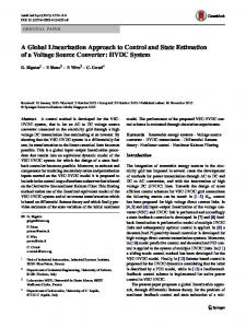
Fig. 2. The modelled structure of chimeric protein FlhS (Blue) / EnvZ (White).
wavelength was set at 515/10 nm. The specific fluorescence intensity (SFI) was defined as the raw fluorescence intensity expressed in relative fluorescence units divided by the optical density at 600 nm measured at each time point. At a minimum, triplicate measurements were obtained for each sample.
3. Results and Discussion 3.1. Construction and expression of the chimeric FlhSZ/OmpR TCS E. coli does not have the natural methanol sensing apparatus. Meanwhile, in P. denitrificans, FlhS/FlhR TCS sensed extracellular methanol. In the present work, E. coli was engineered to sense methanol by employing a chimeric TCS strategy. The chimeric HK was constructed by fusing the catalytic domain of EnvZ HK from EnvZ/OmpC with the sensing domain of P. denitrificans FlhS. The chimeric HK FlhSZ senses extracellular methanol and activates OmpR, which induces the expression of the ompC gene. Therefore, recombinant E. coli was engineered to express the ompC gene in response to methanol. 3.2. Molecular modeling and SDS-PAGE for chimeric HK Molecular modeling was performed to construct stable chimeric HK for methanol sensing. Based on the analysis, it was found that integration of FlhS at 240th methionine residue of EnvZ provides chimeric HK with a stable structure and efficient methanol sensing properties. The blue and white colour represents FlhS and EnvZ, respectively (Fig. 2). Methionine is a hydrophobic amino acid and can be classified as aliphatic amino acids. Hydrophobic amino acid prefers to be buried inside the protein core brings
228
Biotechnology and Bioprocess Engineering 22: 225-230 (2017)
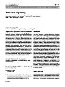
be used as methanol biosensor, high-level expression of the membrane bound FlhSZ chimera HK can result in destabilization of the cell membrane or a reduction in cellular activity. Therefore, FlhSZ was cloned into the low copy number vector, pACYCDuet-1.
Fig. 3. SDS-PAGE expression analysis of FlhS/EnvZ fusion protein extracts in the culture medium of pFlhsZ1 transformed BL21 (DE3) cells and non-induced recombinant plasmid as control. M, Molecular weight marker in KDa.
stability to the fused protein structure. The sulphur present in methionine does not interfere with the function of the fused protein it is due to the bond formation with the methyl group amino acid. The expression of chimeric HK FlhSZ was analyzed by SDS-PAGE, and the results indicated that FlhSZ was successfully expressed by IPTG induction (Fig. 3). The maximum expression of chimeric HK was observed at 0.75 mM IPTG concentration and decreased with further increase in IPTG. Though a higher output signal is better to
3.3. Monitoring the ompC gene expression by qRT-PCR The FlhSZ/OmpR chimeric TCS-mediated ompC gene expression was estimated by qRT-PCR in M9 minimal media supplemented with varying concentrations of methanol. Results of qRT-PCR showed that the transcription of the ompC gene was a maximum of 0.01% of methanol and decreased with further increase in methanol (Fig. 4). The qRT-PCR results show that the ompC gene expression started to decrease beyond 0.01% methanol concentration, which might be due to the reduced cellular activity and protein expression at a higher concentration of methanol [14]. Based on this result, it can be deduced that the chimeric FlhSZ/OmpR TCS generated an output signal strong enough to induce expression of the ompC gene. This suggests that ompC gene expression is quite precisely regulated by chimera FlhSZ in response to the low concentration of extracellular methanol. 3.4. Monitoring the gfp gene expression by fluorescence analysis Considering the qRT-PCR results, a fluorescence-based methanol biosensor system was constructed using the ompC promoter. The E. coli BL21 (DE3) strain harboring pFlhSZ1 and pOGFP1 was cultured with varying methanol concentration (0.01 ~ 4%) and the fluorescence was
Fig. 4. (A) Comparative study of transcriptional levels of the ompC gene in M9 medium after a 4.5 h exposure to methanol. After exposure, the Ct value was normalized using the 16sRNA Ct value as an internal control. The error bars indicate one standard deviation from the mean. (B) The linear correlation between the relative transcriptional levels of the ompC gene of the chimera in M9 medium at varying percentages of methanol. The data are the aggregate results from replicated experiments (n = 3).
Engineering Chimeric Two-component System into Escherichia coli from Paracoccus denitrificans to Sense Methanol
229
Fig. 5. The image of E. coli (pFhSZ1 and pOGFP1). (A) Differential interference contrast (DIC) microscopy image of the recombinant cells carrying GFP. (B) The same cells in which reflects the fluorescence in the reflected fluorescence microscope.
Fig. 6. (A) Time course of GFP fluorescence for an E. coli strain harboring pFlhSZ1 and pOGFP1 after induction with varying concentrations of methanol in M9 medium. Control (closed square); 0.001% ( ▲); 0.01% ( ○); 0.05% ( △); 0.1% ( □ ); 0.5% (◊); 1.0% ( ● ); 2.0% (▼); 4.0% (open inverted triangle); 8.0% (♦). The data are the aggregate results from replicate experiments (n = 3); (B) GFP fluorescence value of E. coli strain harboring pFlhSZ1 and pOGFP1 after induction with varying concentrations of methanol in M9 medium at 8th hour of incubation.
measured. Negligible fluorescence was observed in the control strain, harboring pACYCDuet-1 and pUC19 (Fig. 5). Though the IPTG was not added, a low level of fluorescence was observed, this might be due to the basal level of ompC promoter expression. Time profiles showed that fluorescence increased with culture time (Fig. 6). The maximum fluorescence was obtained only at 0.5% of methanol concentration and decreased at higher concentration. The fluorescence gradually increased with increasing methanol concentration, which correlated that the expression profile was strongly in response to methanol. Though the qRT-PCR showed that the ompC gene expression was highest at 0.01% of methanol and maximum fluorescence was obtained with 0.5% of methanol. Though the methanol concentration was measured as a function of fluorescence, the system lack in having a perfect linear relationship, which may be due to over
amplification of the output signal from the usage of high copy number reporter plasmid (pOGFP1) [15]. At the end of the 8 h, the maximum fluorescence was obtained only at 0.5% of methanol. In a study report by Wen et al. 2014, a novel biosensor was developed by immobilizing the methanol-utilizing bacteria M. organophillum on the egg shell membrane along with gold nano particles. On exposure to methanol, the respiratory activity of the bacteria was increased with concomitant consumption of dissolved oxygen [16]. However, the detection limit of methanol with this technique is 0.01%, which is significantly low when compared to the present study. Another study reported by Zhao et al. also exploited the dissolved oxygen concentration by immobilizing the bacteria M. organophillum with the oxygen sensor. Thus methanol was detected as a function of dissolved oxygen. The decrease in oxygen is linearly related to increasing methanol concentration. The maximum
230
detection limit was 0.27 mg/L (0.000027) [17]. Considering the lower methanol detection limit with earlier systems chimeric TCS FlhSZ/OmpR can be considered as an alternative for efficient detection of extracellular methanol.
4. Conclusion Considering the current limited sustainability of the petroleum industry, the use of biomass and biorefineries are attracting intense interest. Intracellular metabolism optimization and screening for efficient producers are essential for the commercialization of biomass-based methanol products. TCS based bacterial biosensors can be considered as one of the promising candidates for high-throughput screening of methanol producing organisms. As shown in this study, the TCS can be engineered to detect the desired biochemical. Several chimeric TCS-based bacterial biosensors can be constructed for screening in the development of biochemical producing microorganisms.
Acknowledgements This research was supported by the Research and Development Program of the Korea Institute of Energy Research (KIER) (B6-2443) and the Next-Generation BioGreen 21 Program (SSAC, grant number: PJ01111601), Rural Development Administration, Republic of Korea.
References 1. California Environmental Protection Agency (CalEPA) (1999) Air Toxics Hot Spots Program Risk Assessment Guidelines: Part III. Technical Support Document for the Determination of Noncancer Chronic Reference Exposure Levels. SRP Draft. Office of Environmental Health Hazard Assessment, Berkeley, CA. 2. Ed. Budavari, S. and N. Rahway (1989) The Merck Index. An Encyclopedia of Chemicals, Drugs, and Biologicals. 3. Stock, A. M., V. L. Robinson, and P. N. Goudreau (2000) TwoComponent Signal Transduction. Annu. Rev. Biochem. 69: 183-215. 4. West, A. H. and A. M. Stock (2001) Histidine kinases and response regulator proteins in two-component signaling systems.
Biotechnology and Bioprocess Engineering 22: 225-230 (2017)
Trends in Biochem. Sci. 26: 369-376. 5. Utsumi, R., R. Brissette, A. Rampersaud, S. Forst, K. Oosawa, and M. Inouye (1989) Activation of bacterial porin gene expression by a chimeric signal transducer in response to aspartate. Sci. 245: 1246-1249. 6. Harms, N., W. N. M. Reijnders, S. Koning, and R. J. M. van Spanning (2001) Two-component system that regulates methanol and formaldehyde oxidation in Paracoccus denitrificans. J. Bacteriol. 183: 664-670. 7. Harms, N., J. Ras, W. N. Reijnders, R. J. van Spanning, and A. H. Stouthamer (1996) S-formylglutathione hydrolase of Paracoccus denitrificans is homologous to human esterase D: A universal pathway for formaldehyde detoxification? J. Bacteriol. 178: 6296-6299. 8. Ras, J., P. W. Van Ophem, W. N. Reijnders, R. J. Van Spanning, J. A. Duine, A. H. Stouthamer, and N. Harms (1995) Isolation, sequencing, and mutagenesis of the gene encoding NAD- and glutathione-dependent formaldehyde dehydrogenase (GDFALDH) from Paracoccus denitrificans, in which GD-FALDH is essential for methylotrophic growth. J. Bacteriol. 177: 247-251. 9. Baslé, A., G. Rummel, P. Storici, J. P. Rosenbusch, and T. Schirmer (2006) Crystal structure of Osmoporin OmpC from E. coli at 2.0 Å. J. Mol. Biol. 362: 933-942. 10. Eswar, N., B. Webb, M. A. Marti-Renom, M. S. Madhusudhan, D. Eramian, M. -Y. Shen, U. Pieper, and A. Sali (2007) Comparative protein structure modeling using MODELLER. Curr. Protoc. Protein Sci., John Wiley & Sons, Inc., City. 11. Bhattacharya, D. and J. Cheng (2013) 3Drefine: Consistent protein structure refinement by optimizing hydrogen bonding network and atomic-level energy minimization. Proteins 81: 119-131. 12. Eleaume, H. and S. Jabbouri (2004) Comparison of two standardisation methods in real-time quantitative RT-PCR to follow Staphylococcus aureus genes expression during in vitro growth. J. Microbiol. Meth. 59: 363-370. 13. Maruthamuthu, M., I. Ganesh, S. Ravikumar, and S. Hong (2015) Evaluation of zraP gene expression characteristics and construction of a lead (Pb) sensing and removal system in a recombinant Escherichia coli. Biotechnol. Lett. 37: 659-664. 14. Mayson, B. E., D. J. Kilburn, B. L. Zamost, C. K. Raymond, and G. J. Lesnicki (2003) Effects of methanol concentration on expression levels of recombinant protein in fed-batch cultures of Pichia methanolica. Biotechnol. Bioeng. 81: 291-298. 15. Ganesh, I., S. Ravikumar, I. -K. Yoo, and S. Hong (2015) Construction of malate-sensing Escherichia coli by introduction of a novel chimeric two-component system. Bioproc. Biosyst. Eng. 38: 797-804. 16. Wen, G., X. Wen, S. Shuang, and M. M. F. Choi (2014) Wholecell biosensor for determination of methanol. Sensors and Actuators B: Chem. 201: 586-591. 17. Zhao, C., H. Li, J. Sheng, L. Chen, F. Li, S. Yang, C. Dong, and M. M. F. Choi (2009) Isolation of a Methylobacterium organophilium strain, and its application to a methanol biosensor. Microchim. Acta 167: 67-73.