Available online at www.sciencedirect.com Available online at www.sciencedirect.com
Procedia Engineering 14 (2011) 344–352 Procedia Engineering 00 (2011) 000–000
Procedia Engineering www.elsevier.com/locate/procedia
The Twelfth East Asia-Pacific Conference on Structural Engineering and Construction
Effect of Admixed Micelles on the Microstructure Alterations of Reinforced Mortar Subjected to Chloride Induced Corrosion J. HUa,b, D.A. KOLEVA, K. VAN BREUGEL Faculty of Civil Engineering and Geosciences, Department Materials and Environment, Delft University of Technology, Stevinweg 1, 2628 CN, Delft, The Netherlands
Abstract This paper reports the main results from the influence of the initially admixed nano-aggregates (0.5 g/l PEO113-b-PS70 micelles previously dissolved in demi-water) on microstructural alterations of the reinforced mortar subjected to chloride induced corrosion. The morphology of hydration/corrosion products at the steel/cement paste interface and in the bulk matrix of the reinforced mortar, and the surface of the steel reinforcement were observed by Environmental SEM (ESEM) combined with energy dispersive X-ray analysis (EDX); the microstructural properties of the mortar i.e. porosity, pore size distribution, were investigated by quantitative image analysis. The results reveal that the microstructure of the cement matrix was altered by the combined effect of penetrated chlorides, initiated corrosion and the admixed micelles. The EDX analysis and morphological observation of the steel surface indicate that the admixed micelles can contribute to the formation of a more homogenous layer on the steel surface and cause delay of corrosion initiation. Keywords: Micelles; Microstructure; Image analysis; Porosity; Corrosion
1. INTRODUCTION It is well known that in reinforced concrete, the cement matrix surrounding the steel reinforcement can act as a physical and chemical barrier and thus protect the reinforcement from corrosion. In normal conditions, the reinforcing steel is in passive state because of the high alkaline environment (pH=12.613.5) of the pore solution and the cement paste respectively (Pourbaix 1974; Sahoo and Balasubramaniam a b
Corresponding author: Jie Hu Email:
[email protected] Presenter: Jie Hu Email:
[email protected]
1877–7058 © 2011 Published by Elsevier Ltd. doi:10.1016/j.proeng.2011.07.043
J. HU et al. / Procedia Engineering 14 (2011) 344–352 2
Author name / Procedia Engineering 00 (2011) 000–000
2008). However, when chlorides penetrate into the cement matrix, the stable passive layer on the steel surface can be broken and corrosion is initiated (Bertolini et al 2004). Further, corrosion leads to the formation of voluminous corrosion products and subsequent cracking and spalling, which in turn can cause concrete deterioration and thus influence durability (Page et al 1990; Berke et al 1990). When nano particles are incorporated in cement paste, mortar or concrete, materials with different characteristics, compared to the conventional ones, can be obtained (Older 1998; Neville 1996). In recent years, there are many researches investigating the influence of incorporated nano particles on material properties, such as mechanical performance, permeability and microstructure of cement-based materials (Collepardi et al 2005; Zhang et al 2006; Hui 2004). This study is part of a 2-year research project in which nano particles with tailored properties are admixed into reinforced concrete for corrosion control. The previous preliminary investigations have shown that the microstructure and permeability of plain mortar were significantly changed by the admixed micelles (Koleva et al 2008; Koleva et al 2009). The objective of this study is to investigate the combined effect of chlorides, corrosion and admixed micelles on the microstructure of the reinforced mortar. This paper reports on the microstructural alterations both in the bulk cement matrix and at the steel/cement paste interface, and the morphology and chemical composition of the product layer or the corrosion products formed on the steel surface. The microstructural information was obtained by backscattered electron (BSE) images and image analysis which can provide quantitative characterization of microstructural alterations of the cement matrix in the presence of chlorides, corrosion and admixed micelles. 2. Experimental 2.1. Sample preparation of reinforced mortar Reinforced mortar cylinders (D=35 mm, H=150 mm) were cast according to standard experimental procedures (EN 196-1), using Ordinary Portland Cement OPC CEM I 42.5R, cement to sand ratio of 1:3 and water to cement ratio of 0.5. Each cylinder contained a centrally located 8 mm diameter steel bar (using “as received” FeB500HKN construction steel) with exposed length of 120 mm. The mortar cylinders comprised 2 main groups: “Ref” group represents specimens cast with tap water; “Nano” group represents specimens cast with 0.5 g/l PEO113-b-PS70 micelles previously dissolved in demi-water (the micelles were the nano particles used in this study), resulting in a concentration of 0.006 wt.% micelles per mortar weight. All specimens were cured in fog room (20 °C and 98% relative humidity) for 28 days and further lab conditioned (2/3rd of height immersed in water for control groups or 5% NaCl solution for corroding groups respectively till the end of the testing period (the curing age of 114 days)). Four main specimen groups were investigated in this study. For “Ref” group (cast with tap water), specimen RefW represents specimens immersed in tap water (control) and specimen RefN represents specimens immersed in 5% NaCl solution (corroding). Accordingly, for “Nano” group, specimen NanoW represents specimens immersed in tap water (control) and specimen “NanoN” represents specimens immersed in 5% NaCl solution (corroding). The sample preparation includes generally applied steps for specimen preparation before image analysis (Ye 2003; Hu 2004) which include drying in certain conditions, vacuum impregnation with low viscosity epoxy, grinding and polishing. What is relevant to this study is that 3 section of each cylinder (Fig.1a) were investigated i.e. top, middle and bottom sections, thus accounting for microstructural changes in these separate locations within one specimen
345
346
J. HU et al. / Procedia Engineering 14 (2011) 344–352 Author name / Procedia Engineering 00 (2011) 000–000
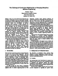
2.2. Methods Scanning electron microscopy (SEM) was applied for visualization, morphological and microstructural investigation using environmental SEM (ESEM Philips XL30), equipped with energy dispersive X-ray (EDX) detector. A set of BSE images were taken at randomly selected locations both at the steel/cement paste interface (the area within a distance of 50 μm away from the steel surface) and in the bulk matrix (the area beyond the distance of 250 μm away from the steel surface) with the magnification of 500×, which is shown in Fig.1 (b). OPTIMAS software was applied for image analysis to derive parameters such as porosity and pore-size distribution. The results were an average value of 35 locations per sample. The image analysis in this study complies with the generally used methodology for pore structure analysis of cement based materials (Hu 2004).
(a)
(b)
Fig.1 Schematic of the investigated reinforced mortar (a) Top, middle and bottom cross sections of the specimen; (b) Areas (interface and bulk) for microstructure analysis
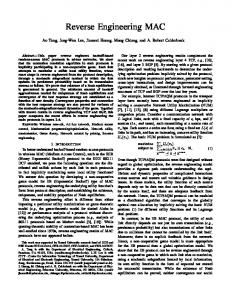
The EDX was used as a qualitative and semi quantitative analysis both on the steel surface and in the cement paste. In addition to general EDX analysis (e.g. EDX mapping), EDX point analysis was conducted (using local area of 5 × 5 μm at magnification of 2000×) in radial direction of the steel bar, starting at 0 μm (the steel surface) up to 3000 μm into the bulk material (direction the edge of the specimen). The image analysis and EDX analysis were conducted at the top, middle and bottom sections of the reinforced mortar cylinders respectively (Fig.1 (a)). 3. Results and Discussion 3.1. Microstructural analysis of the reinforced mortar Fig.2 shows the variation of porosity for all specimens at the top, middle and bottom sections of the reinforced mortar both at the steel/cement paste interface and in the bulk matrix. The porosity results showed the same trend both at the steel/cement paste interface and in the bulk matrix: the admixed micelles can reduce the porosity of the cement paste for specimens immersed in tap water. For example, at the middle section, the micelles-containing specimen NanoW had a denser pore structure, characterized by a porosity of 9.29% for the interface zone and 9.15% for the bulk matrix (Fig.2 (c)). In contrast, the porosity increased to 13.48% for the interface zone and to 11.59% for the bulk matrix for the micelles-
3
347
J. HU et al. / Procedia Engineering 14 (2011) 344–352 4
Author name / Procedia Engineering 00 (2011) 000–000
free specimen RefW (Fig.2 (a)). The porosity reduction in the reinforced mortar caused by the admixed micelles was not very significant, compared to that in plain mortar (Koleva 2009). This result is as expected, considering the curing age of the hereby investigated mortar (114 days), accounting for a more developed cement matrix. Therefore, at this curing age, a 2% difference in the porosity indicates that the microstructure of the cement matrix was altered by the admixed micelles. Further, the steel/cement paste interface was slightly more porous for both specimens, evidenced by higher porosity, compared to that in the bulk matrix (Fig.2 (a) and (c)). However, the micelles-containing specimen NanoW had a more uniform porosity distribution with a smaller porosity gradient in the radial direction (Fig.2 (c)). It confirms the influence of the admixed micelles on refining the pore network i.e. reducing the porosity at the steel/cement paste interface and also causing pore refinement in the bulk matrix. The micelles can act as nucleation sites for the formation of new hydration products, thus resulting in a more uniform distribution of the hydration products and more homogenous microstructure in specimen NanoW (Koleva 2009). It can be also found that there was a slight reduction in porosity from the top (aerated) section to the bottom (immersed) section of the reinforced mortar which is due to the hydration with aging for all sections and the moist gradient from the top section to the bottom section. For specimens immersed in NaCl solution, the porosity was also reduced by the admixed micelles, but the porosity difference was not so evident as for the specimens immersed in tap water (Interface: RefN 10.61%, NanoN 9.53%; Bulk: RefN 9.81%, NanoN 8.82% - middle section). The reason is that the presence of chlorides can promote the cement hydration which can reduce the porosity for both the micelles-free specimen RefN and the micelles-containing specimen NanoN (Suryavanshi et al 1995; Díaz et al 2006). However, for the micelles-containing specimen NanoN, except for the effect of chlorides which can lead to a reduction of the porosity, there is another competitive effect of the admixed micelles: the micelles will collapse and “shrink” in the presence of chlorides (Patel et al 2007) which can increase the porosity. Therefore, the porosity reduction of the micelle-containing specimen NanoN was smaller, compared to the micelles-free specimen RefW. The microstructure alterations derived for the reinforced mortar is consistent with the data derived for the plain mortar as previously reported (Koleva et al 2008; Koleva et al 2009). 16
12
interf_RefW_T
14
interf_RefN_M 10
interf_RefW_B 12
interf_RefN_B bulk_RefN_T
bulk_RefW_T bulk_RefW_M
10
bulk_RefN_M
8
bulk_RefW_B
porosity (%)
porosity (%)
interf_RefN_T
interf_RefW_M
8 6
bulk_RefN_B 6
4
4
2
2
0
0 0.1
1
10
0.1
(a) Specimen RefW
1 pore size (um)
pore size (um)
(b) Specimen RefN
10
348
J. HU et al. / Procedia Engineering 14 (2011) 344–352 5
Author name / Procedia Engineering 00 (2011) 000–000 12
12
interf_NanoW_T
interf_NanoN_T
interf_NanoW_M 10
interf_NanoN_M 10
interf_NanoW_B
interf_NanoN_B
bulk_NanoW_T
bulk_NanoN_M
8
bulk_NanoW_B
porosity (%)
porosity (%)
bulk_NanoN_T
bulk_NanoW_M
8
6
bulk_NanoN_B 6
4
4
2
2
0
0 0.1
1
10
0.1
pore size (um)
(c) Specimen NanoW
1
10
pore size (um)
(d) Specimen NanoN
Fig.2 Pore size distribution of the cement paste at top, middle and bottom section of reinforced mortar
3.2. EDX analysis of the reinforced mortar EDX elemental mapping was performed to evaluate the chemical composition of hydration or corrosion products at the steel/cement paste interface zone of all specimens; Fig.3 shows the data at the middle section of the investigated specimens. No corrosion products were detected for the control specimens RefW and NanoW, evidenced by the lack of Fe in cement paste of the interface zone (Fig.3 (a) and (b)). The results indicate that the steel reinforcement was well passivated in tap water. For the corroding specimens, there was a significant accumulation of corrosion products for the micelles-free specimen RefN, which is shown as “zone A” in Fig.3 (c). In zone A, the elemental mapping result reveals the presence of Fe and O, indicating accumulated corrosion products at the steel/cement paste interface. In contrast, Fig.3 (d) shows that corrosion products were not detected at the steel/cement paste interface for the micelles-containing specimen NanoN. Fig.4 presents the Fe concentration in the cement paste from the steel surface and into the bulk matrix for corroding specimens at the top, middle and bottom sections of the specimens (Fig.1 (b)). For the micellesfree specimen RefN, higher amount of Fe was detected in the vicinity of the steel bar e.g. in the area within a distance of 100-200 μm from the steel surface. The results indicate that corrosion products were accumulated on the steel surface and penetrated into the cement paste (e.g. Fig.3 (c)). In contrast, for the micelles-containing specimen NanoN, except for the top section, very low concentration of Fe was detected in the vicinity of the steel bar. Even at the top sections, corrosion products layer was much thinner for the micelles-containing specimen NanoN (about 40 μm), compared to the micelles-free specimen RefN (about 180 μm). The results indicate that the admixed micelles can cause a delay of corrosion initiation of the steel reinforcement.
J. HU et al. / Procedia Engineering 14 (2011) 344–352 6
Author name / Procedia Engineering 00 (2011) 000–000
(a) RefW
(b) NanoW
(c) RefN
(d) NanoN Fig.3 EDAX elements mapping for specimens immersed in NaCl solution
349
350
J. HU et al. / Procedia Engineering 14 (2011) 344–352 7
Author name / Procedia Engineering 00 (2011) 000–000
Fe (w. %)
70
RefN_T
60
RefN_M
50
RefN_B
40
NanoN_T NanoN_M
30
NanoN_B
20 10 0 0
200
400
600
800
1000
distance from steel surface to bulk matrix (um) Fig.4 Iron concentration from steel surface to bulk matrix for corroding specimens
3.3. Morphological observation of the steel surface Fig.5 (control specimens RefW and NanoW) and Fig.6 (corroding specimens RefN and NanoN) present the morphological observation of the steel surface at the end of the study (curing age of 114d). For the control specimens, there was a more homogeneous and compact layer formed on the steel surface for the micelles-containing specimen NanoW, compared to the micelles-free specimen RefW, as shown in Fig.5. For the corroding specimens, the layer on the steel surface of the micelle-containing specimen NanoN was still more homogenous and compact, compared to the micelles-free specimen RefN (Fig.6 (a) and (b)). Corrosion products can also be observed on the surface of both specimens. For specimen NanoN, corrosion products with smaller dimension were observed at isolated locations on the steel surface (Fig.6 (d)), compared developed corrosion products observed for specimen RefN (Fig.6 (c)). The results confirm that the presence of micelles leads to a corrosion delay for the micelles-containing specimen. The most plausible reasons are as follows: first, the porosity of the cement matrix is reduced by the admixed micelles, which leads to an impeded penetration of chlorides into the cement matrix and then results in the delay of corrosion initiation; second, the micelles present at the steel/cement paste interface can improve the barrier effect of the product layer on the steel surface and contribute to the formation of a more homogenous and compact layer.
J. HU et al. / Procedia Engineering 14 (2011) 344–352 8
Author name / Procedia Engineering 00 (2011) 000–000
(a) RefW
(b) NanoW
Fig.5 Morphological observation of the steel surface for the control specimens
(a) Product layer on specimen RefN
(c) Corrosion products on specimen RefN
(b) Product layer on specimen NanoN
(d) Corrosion products on specimen NanoN
Fig.6 Morphological observation of the steel surface for the corroding specimens
351
352
J. HU et al. / Procedia Engineering 14 (2011) 344–352 Author name / Procedia Engineering 00 (2011) 000–000
4. CONCLUSIONS Based on the experimental results, it can be concluded that the microstructure of the cement matrix was altered by the combined effect of penetrated chlorides, initiated corrosion and the admixed micelles i.e. the admixed micelles can reduce the porosity of the cement paste, evidenced by the microstructural analysis of the reinforced mortar cylinders. Moreover, corrosion initiation of the steel bar was delayed by the admixed micelles, evidenced by a more homogenous layer on the steel surface and none or lower amount of corrosion products accumulated at the steel/cement paste interface for the micelles-containing reinforced mortar. REFERENCES [1]
Berke N.S., Chaker V., Whiting D. (1990), Corrosion of Steel in Concrete, Editors, ASTM STP 1065, ASTM, Philadelphia, PA.
[2]
Bertolini L., Elsener B., Pedeferri P., Polder R. (2004), Corrosion of Steel in Concrete: Prevention, Diagnosis, Repair, Wiley-VCH, Weinheim,.
[3]
Collepardi S., Borsoi A., Ogoumah Olagot JJ., Troli R., Collepardi M., Cursio A.Q., Influence of nano-sized mineral additions on performance of SCC. In: Proceedings of the 6th international congress, global construction, ultimate concrete opportunities, Dundee, UK; 5–7 July 2005.
[4]
Díaz B., Nóvoa X.R., Pérez M.C. (2006), Cem. Concr. Comp., 28, 237-245.
[5]
Hu, J. (2004), Porosity of Concrete, Morphological Study of Model Concrete, PhD thesis, Delft University of Technology, Delft.
[6]
Hui L., Xiao H., Yuan J., Ou J. (2004), Microstructure of cement mortar with nano-particles, Composites B 35 185.
[7]
Koleva, D.A. (2007), Corrosion and Protection in Reinforced Concrete, PhD Thesis, Delft University of Technology, Delft.
[8]
Koleva D.A. (2009), van Breugel K., Ye G., Zhou J., Chamululu G., Koenders E., Porosity and Permeability of Mortar Specimens Incorporating PEO113–b–PS218 Micelles, Special issue of ACI Materials Journal, SP267, 101-110.
[9]
Koleva D.A., Ye G., Zhou J., Petrov P., van Breugel K. (2008), Material properties of mortar specimens at early stage of hydration in the presence of polymeric nano-aggregates, International Conference on "Microstructure related Durability of Cementitious Composites" Nanjing, China, 13th-15th October, 2008, RILEM Publications SARL 2008, pp 161-168.
[10]
Neville A.M. (1996), Properties of concrete. 4th ed. England: ELBS with Addison Wesley Longman.
[11]
Older I. (1998), Lea’s chemistry of cement and concrete. 4th ed. London: Arnold.
[12]
Page C.L., Treadaway K.W.J., Bamforth P.B. (1990), Corrosion of Reinforcement in Concrete, Editors, Elsevier Applied Science, London.
[13]
Parrot L.J. (1987), A Review of Carbonation in Reinforced Concrete, British Cement Association Report C/i-0987.
[14]
Patel K., Bahadur P., Guo C., Ma J.H., Liu H.Z., Yamashita Y., Khanal A., Nakashima K. (2007), European Polymer J. 43: 1699.
[15]
Petrov P., Bozukov M., Tsvetanov C.B. (2005), J. Mater. Chem., 15, 1481.
[16]
Pourbaix M. (1974), Atlas of Electrochemical Equilibria in Aqueous Solutions, NACE-CEBELCOR, Houston, TX.
[17]
Sahoo G., Balasubramaniam R. (2008), Corrosion Science, 50, 131–143.
[18]
Suryavanshi A.K., Scantlebury J.D., Lyon S.B. (1995), Cem. Concr. Res., 25, 980-988.
[19]
Ye, G. (2003), Experimental study and numerical simulation of the development of the microstructure & permeability of
[20]
cementitious materials, PhD thesis, Delft University of Technology, Delft. Zhang M.H., Li H. (2006), Chloride permeability of concrete containing nano-particles for pavement. Structural Health Monitoring and Intelligent Infrastructure, Vols 1 and 2, Proceedings and Monographs in Engineering, Water and Earth Sciences 1469-1474.
9