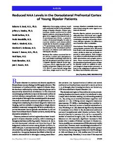
NeuroImage 124 (2016) 214–223
Contents lists available at ScienceDirect
NeuroImage journal homepage: www.elsevier.com/locate/ynimg
Enhanced control of dorsolateral prefrontal cortex neurophysiology with real-time functional magnetic resonance imaging (rt-fMRI) neurofeedback training and working memory practice Matthew S. Sherwood a,b,⁎, Jessica H. Kane a,c, Michael P. Weisend a, Jason G. Parker a,b a b c
Wright State Research Institute, Wright State University, 4035 Colonel Glenn Hwy, Dayton, OH 45431, USA Department of Biomedical, Industrial, and Human Factors Engineering, Wright State University, 3640 Colonel Glenn Hwy, Dayton, OH 45435, USA Department of Neuroscience, Cell Biology, and Physiology, Wright State University, 3640 Colonel Glenn Hwy, Dayton, OH 45435, USA
a r t i c l e
i n f o
Article history: Received 6 May 2015 Accepted 22 August 2015 Available online 5 September 2015 Keywords: Dorsolateral prefrontal cortex Working memory Neurofeedback fMRI
a b s t r a c t Real-time functional magnetic resonance imaging (rt-fMRI) neurofeedback can be used to train localized, conscious regulation of blood oxygen level-dependent (BOLD) signals. As a therapeutic technique, rt-fMRI neurofeedback reduces the symptoms of a variety of neurologic disorders. To date, few studies have investigated the use of self-regulation training using rt-fMRI neurofeedback to enhance cognitive performance. This work investigates the utility of rt-fMRI neurofeedback as a tool to enhance human cognition by training healthy individuals to consciously control activity in the left dorsolateral prefrontal cortex (DLPFC). A cohort of 18 healthy participants in the experimental group underwent rt-fMRI neurofeedback from the left DLPFC in five training sessions across two weeks while 7 participants in the control group underwent similar training outside the MRI and without rt-fMRI neurofeedback. Working memory (WM) performance was evaluated on two testing days separated by the five rt-fMRI neurofeedback sessions using two computerized tests. We investigated the ability to control the BOLD signal across training sessions and WM performance across the two testing days. The group with rt-fMRI neurofeedback demonstrated a significant increase in the ability to self-regulate the BOLD signal in the left DLPFC across sessions. WM performance showed differential improvement between testing days one and two across the groups with the highest increases observed in the rt-fMRI neurofeedback group. These results provide evidence that individuals can quickly gain the ability to consciously control the left DLPFC, and this training results in improvements of WM performance beyond that of training alone. © 2015 The Authors. Published by Elsevier Inc. This is an open access article under the CC BY license (http://creativecommons.org/licenses/by/4.0/).
Introduction New techniques to induce and control neural plasticity hold the promise of enhancing recovery from brain injury (Jenkins and Merzenich, 1987; Wieloch and Nikolich, 2006), combating brain disease (Baroncelli et al., 2011; Sakas et al., 2007), and even improving human performance in healthy subjects (Buschkuehl et al., 2008; Garlick, 2002; Jaeggi et al., 2011; Jausovec and Jausovec, 2012). A number of methods are being explored to induce and control neuroplastic processes, including cognitive training (Kleim et al., 2004; Oleson et al., 2004; Pleger et al., 2003), pharmacotherapy (Delacour et al., 1990; Ehrenreich et al., 2008; Greuel et al., 1988), electrical and magnetic stimulation (Fraser et al.,
Abbreviations: CDT, cyber defense task; EV, explanatory variable; rt-fMRI, real-time functional magnetic resonance imaging; WM, working memory. ⁎ Corresponding author at: Wright State Research Institute, 4035 Colonel Glenn Highway, Dayton, OH 45431, USA. E-mail address:
[email protected] (M.S. Sherwood).
2002; McKinley et al., 2013; Pascual-Leone et al., 1998; Ziemann et al., 2002), and self-modulation of brain regions and networks based on neurofeedback (Birbaumer and Cohen, 2007; Daly and Wolpaw, 2008; Ros et al., 2010). Of these techniques, selfmodulation methods have the advantage of no known side effects, as well as straightforward translation to neurophysiological exercises that could be performed at home without the use of sophisticated equipment and trained professionals (Mak and Wolpaw, 2009; Vaughan et al., 2006). Real-time functional magnetic resonance imaging (rt-fMRI) has been enabled by recent advances in image acquisition, reconstruction, and display of the blood oxygen level-dependent (BOLD) signal. This technique can be used as a tool to deliver region-specific neurofeedback. A number of previous studies have demonstrated the efficacy of rt-fMRI neurofeedback to elicit self-control of localized BOLD signals (Sulzer et al., 2013a) although few show an impact on behavior or cognition. deCharms et al. (2005) applied a controlled rt-fMRI neurofeedback study to influence symptoms of chronic pain. In their study, significant changes in pain perception and chronic pain were produced by learning
http://dx.doi.org/10.1016/j.neuroimage.2015.08.074 1053-8119/© 2015 The Authors. Published by Elsevier Inc. This is an open access article under the CC BY license (http://creativecommons.org/licenses/by/4.0/).
M.S. Sherwood et al. / NeuroImage 124 (2016) 214–223
to control BOLD signals from the rostral anterior cingulate cortex (rACC). Rota et al. (2009) used rt-fMRI neurofeedback of right inferior frontal gyrus (IFG) to improve accuracy on emotional prosodic intonation. Right IFG modulation did not, however, influence syntactic processing. Sham rt-fMRI neurofeedback of the right IFG impeded learning processes in a control group. Ruiz et al. (2013) trained self-regulation of the bilateral anterior insula through rt-fMRI neurofeedback in a group of schizophrenia patients. The success of self-regulation was negatively correlated with negative symptoms and duration of illness. In addition, patients detected significantly more disgust faces in a post-training face emotion recognition evaluation. Young et al. (2014) performed rt-fMRI neurofeedback of the left amygdala on a group of unmedicated patients suffering from major depressive disorder. Significant reductions in self-reported measures of depression, anxiety, anger, restlessness and irritability were measured. A significant increase in the rating of happiness was also observed. These encouraging results demonstrate that direct control over neurophysiology can influence behavior and cognition. We hypothesized that rt-fMRI neurofeedback can be used to learn self-regulation over the BOLD signal in the left DLPFC. Learning selfregulation from rt-fMRI neurofeedback has produced significant behavioral effects (deCharms et al., 2005; Haller et al., 2010; Linden et al., 2012; Rota et al., 2009; Ruiz et al., 2013; Scharnowski et al., 2012; Shibata et al., 2011; Sitaram et al., 2012; Subramanian et al., 2011; Young et al., 2014; Zhang et al., 2013a) resulting from neuroplastic changes of learned self-regulation. Lee et al. (2011) reported that neurofeedback training reinforces pertinent functional networks while extraneous connections are weakened. Haller et al. (2013) exhibited that neurofeedback learning is mediated by widespread alterations in learning networks and the application of learned self-regulation involves more limited and specific network changes. These changes are described by the Hebbian theory which details neuroplasticity during the learning process (Hebb, 1949). Neuroplasticity is engaged through the repetitive stimulation of the postsynaptic cell from the presynaptic cell. In short, the synapses of neurons firing together are reinforced resulting in an increase in synaptic efficiency. The left dorsolateral prefrontal cortex (DLPFC) plays an important role in working memory (WM; D'Esposito et al., 2000) and higher levels of attentional control (Posner and Presti, 1987). Thus, we hypothesize that self-regulation of the left DLPFC would lead to improved synaptic efficiency of the WM and attentional control networks. These changes would lead to improved performance on tasks that rely on WM and attentional control beyond that of training alone. To test these hypotheses, we examined the effect of rt-fMRI neurofeedback on control over the BOLD signal in the left DLPFC as well as pre- and post-training performance of n-back WM, prospective WM, and vigilance tasks. Methods Participants Prior to being enrolled, each participant completed a telephone screening to qualify for the study. All participants gave their written informed consent to participate in the experiment, which was approved by the Institutional Review Board of Wright State University. The study of the experimental group was further approved by the Air Force Surgeon General. All participants were compensated equally for their involvement. Thirty-two individuals participated in the experiment. The participants were separated into two groups which received different training: experimental (those receiving rt-fMRI plus task experience) and control (those receiving task experience alone). Neither the experimental group nor the control group was aware that there was a separate group or condition. The experimental group started with twenty-one individuals. However, one participant did not complete all training sessions, one had incomplete data, and the performance for another was
215
below three standard deviations from the group mean. The remaining eighteen right-handed, healthy volunteers (10 males), ages 19–35 (mean 23.3) with normal or corrected-to-normal vision and not medicated for neurological or psychiatric disorders constituted the experimental group. Eleven individuals started in the control group. However, one participant was unable to complete all training sessions, another had incomplete data, and two had performance scores below two standard deviations from the group mean. The remaining seven right-handed, healthy volunteers (4 males), ages 18–25 (mean 21.9) with normal or corrected-to-normal vision and not medicated for neurological or psychiatric disorders constituted the control group. This group completed n-back training plus attempted self-regulation of working memory by performing similar imagined tasks as the participants in the experimental group but without the additional aid of rt-fMRI neurofeedback. Experimental design An overview of the experimental procedure and subject display is shown in Fig. 1. Prior to training and testing, all participants signed informed consent documents detailing the requirements of participation and were familiarized with the testing apparatus. Training was performed across five sessions, no more than one per day, within 14 days. Training consisted of the n-back task followed by self-regulation training. For the experimental group, self-regulation of DLPFC was visualized with rt-fMRI neurofeedback. Pre- and post-training behavioral assessments were conducted utilizing n-back WM, prospective WM, and vigilance tasks. Practice effects are well documented for n-back WM, prospective WM and vigilance tasks (Jaeggi et al., 2008). Therefore we chose a pure behavioral control group to rule out the possibility that practice effects influenced our behavioral effects. The control group performed the same procedures, including attempted self-regulation over WM, but was not supplied with rt-fMRI neurofeedback. Additionally, MRI scans were not performed on the control group. deCharms et al. (2005) used this type of control group along with four others to evaluate nonspecific effects of rt-fMRI neurofeedback from the rACC. Others have implemented a similar control group within the MRI (Caria et al., 2007; Johnston et al., 2011; Linden et al., 2012; McCaig et al., 2011). Session one began with a behavioral assessment performed outside the scanner for both groups (see Behavioral assessment section) followed by training (see Training section). Sessions two through four involved only training. Session five consisted of training followed by a second behavioral assessment outside the scanner. MRI acquisition MRIs were collected for the experimental group on a 1.5 Tesla (T) MR scanner (Siemens MAGNETOM Avanto, Siemens, Erlangen, Germany) using an 8-channel birdcage head coil. Participants were positioned on the scanner table supine. Their head was stabilized using foam pads attached to the head coil and their arms were placed at their side. Task instructions and the neurofeedback display were delivered using an MR-compatible digital video projection system (BrainLogics MRI Digital Projection System, Psychology Software Tools Inc., Sharpsburg, PA), which the participants viewed via a mirror fixed to the top of the head coil. Noise-canceling headphones (Avotech Audio System, Psychology Software Tools Inc., Sharpsburg, PA) were used for audio communication with the participants. Each training session consisted of task training, self-regulation practice, and self-regulation. BOLD data was acquired using a gradient-recalled-echo (GRE) sequence with a 64 × 64 element matrix, 24 slices parallel to the AC–PC plane, 4 × 4 × 5 mm3 voxel size, 1 mm slice gap, TR/TE = 2000/ 40 ms, and a flip angle of 90°. High-resolution T1-weighted structural images were acquired using a 3D Magnetization-Prepared RapidAcquisition-Gradient-Echo (MPRAGE) sequence with a 256 × 256
216
M.S. Sherwood et al. / NeuroImage 124 (2016) 214–223
Fig. 1. Experimental design overview. The first session began an initial behavioral assessment using the computerized tests followed by training. Only training was completed on the second, third and fourth sessions. The fifth session began with training followed by a final behavioral assessment using the computerized tests.
element matrix, 160 slices oriented in the same plane as in the functional scans, 1 × 1 × 1 mm3 voxel size, TR/TE = 500/15 ms, and a flip angle of 15°. Behavioral assessment Two computer-based tests were used to quantify cognitive performance before and after the training regimen. The first test was a 2back variant of the n-back task, using single letters from the English alphabet as stimuli. Each letter appeared for 500 ms with an interstimulus interval of 2500 ms. The total duration of the n-back task was 6 min. Participants were required to respond to every stimulus signifying whether or not the current letter was the same as 2-back in the list. The probability of the 2-back condition being satisfied on any trial was 40%. The participants were instructed to respond as quickly and accurately as possible with a time-limit of 2500 ms following the presentation of each letter. The second computer test was a dual-task scenario utilizing a dashboard based on cyber defense operations (Cyber Defense Task, CDT). This task was developed in conjunction with the U.S. Air Force and
selected for its applied military relevance. Although this task is not well documented, unpublished pilot experiments indicated bilateral DLPFC, anterior cingulate cortex, and inferior frontal gyrus activation along with other regions. Improving performance on this task may lead to enhanced military effectiveness, a target objective for the funding agency and project scope of work. One of the components was a vigilance task consisting of a plot, scrolling right-to-left, simulating the instantaneous load on a computer network (Fig. 2; referred to as CDT graphical). The plot was updated once per second. Participants were instructed to respond only when a new point was above a warning threshold (indicated by a red line) set at 75% load. The second component was a prospective WM task consisting of a continuously-updated list of destination IP addresses, scrolling top-down, that was representative of computers connected to the network (Fig. 2; referred to as CDT textual). The list was updated once per second. Participants were instructed to respond only when a new addition to the list matched an item from a target list of IP addresses pre-determined to be suspicious. All participants received the same list containing three target destination IP addresses before testing began. Separate target lists were generated for pre- and post-training behavioral assessment sessions.
Fig. 2. Example display of the Cyber Defense Task (CDT). The CDT graphical task contains a threshold line (red) and plotted points (green) which update once per second. The plot scrolls from right-to-left as new points are added. Participants were instructed to respond to each point appearing above the threshold line. The CDT textual task contains a timestamp, source address, source port, destination address, and destinations ports of stimulated network activity. The list scrolls top-to-bottom as the network activity is updated once per second. Participants were instructed to respond when specific target IP addresses appear at the top of the destination address list.
M.S. Sherwood et al. / NeuroImage 124 (2016) 214–223
The entire CDT test was composed of eight two-minute trials. For both components, 15% of the 120 updates in each two-minute trial were target items. Training Training began with a single run of an n-back task executed in a boxcar design with control and n-back blocks repeated four times. The blocks had a duration of 48 s. The n-back condition was identical to that described above. Letters in the n-back task were replaced with a fixation point for the control condition. Participants were instructed to alternate between right and left responses with each presentation of a fixation point. This experimental group's first run was used to identify the left DLPFC and is referred to as the “functional localizer”. Immediately following acquisition, the BOLD data underwent pre-processing implemented in custom MATLAB and C++ software that included standard spatial filtering (2D, 5-point Gaussian low-pass kernel, full-width halfmaximum of 9 mm), motion correction (corrected to the first volume using a rigid-body 3-parameter model), and temporal filtering (5-point Gaussian low-pass kernel, sigma of 3 s) processing functions (Friston et al., 1995). A single explanatory variable (EV) was defined by convolving a boxcar model containing 48 s control and 48 s task conditions with a pre-defined hemodynamic response function (HRF; Ashby, 2011). The BOLD data at each voxel were fit to the model using a general linear model (GLM) by applying a weight of +1 to the EV, representative of activation (positive correlation to the model). The resulting β-parameter maps were converted to t-statistic maps using standard statistical transforms. The activation map from this functional localizer was used to determine the region in the left DLPFC to derive the feedback signal for the neurofeedback training that followed. Voxels were added to the region of interest (ROI) by first locating the axial slice in which the superior surface of the ventricles was visible. Next, activation patterns on the left lateral hemisphere near the anterior side of the ventricles were observed. Voxels within this region responding robustly to the n-back task were added to the ROI to complete the determination of the functional localizer. The placement of the ROI across participants and sessions is represented in Fig. 3. Next, a single run of feedback familiarization was completed. Participants from both groups were instructed to perform the n-back task and not attempt self-regulation. For only the experimental group, the task was overlaid on the feedback signal. A single repetition of the control and n-back blocks, identical to the functional localizer, was applied by presenting the task stimuli on upper-right side of the line plot. For the control group, the task stimuli were presented in the center of the screen. This run familiarized the experimental group with the feedback signal, the expected results of activating the voxels selected from the functional localizer, and the hemodynamic delay associated with this activation. This run was carried out in the control group to mimic the training conducted by the experimental group and ensure the same amount of n-back training was performed.
217
For the experimental group, BOLD data was acquired using the same scan parameters as described for the functional localizer. Eight volumes were acquired prior to the start of the n-back task to determine a baseline BOLD signal value for the selected voxels. During the n-back task, a feedback signal was computed to be displayed to the participants from real-time analysis of BOLD data. This real-time analysis was implemented in custom MATLAB and C++ software that included standard spatial filtering (5-point Gaussian low-pass kernel, full-width half-maximum of 9 mm) and motion-correction (corrected to the first volume of the functional localizer using a rigid-body 3-parameter model) processing functions (Friston et al., 1995). This custom software further compared the average BOLD signal in the voxels selected from the functional localizer at baseline to that of the current volume to derive the percent signal change. The current feedback signal was determined by temporally-filtering (5-point Gaussian low-pass kernel consisting of only past components, sigma of 3 s) the percent BOLD signal change with the feedback signals from previous volumes. This feedback signal was presented to the participants using a continuouslyupdated line plot scrolling from right-to-left. The third run was self-regulation training. Participants performed four repetitions of rest and task blocks in a boxcar-design. During rest, every participant was instructed to relax and clear their mind. During task, every participant was instructed to perform a mindfulness task wherein they could increase brain activity associated with WM by recalling their drive to the experiment site, the walk to the experimental room from the parking lot or a recent phone call, or performing mental math such as square roots. Also, the participants were informed not to use the response devices and to remain as still as possible. Each rest and task block was 48 s. The additional aid of feedback regarding the activity of the left DLPFC during the second and third runs was only supplied to the participants in the experimental group. The baseline and percent BOLD signal change for the voxels selected from the functional localizer were calculated as described above. During this acquisition, the participants viewed the plotted feedback signal and task instructions on the upper right side of the plot. The control group was not supplied with feedback or the feedback line plot but only the task instructions centered on the screen. Both groups were instructed that gaining self-control over the left DLPFC may result in improved WM, and that the supplied training may help them achieve self-control. Moreover, both groups were aware that the goal of the project was to improve cognitive performance. Data processing and analysis Performance on the computer tests was determined by calculating a sensitivity score (d′) for each test (CDT graphical, CDT textual, and nback). Prior to this computation, the data were pre-processed to correct for delayed responses by removing trials where responses occurred within 150 ms of the stimulus onset. For these incidences, the preceding trial was removed only if it did not have a response. On average, 4.075%
Fig. 3. Average rt-fMRI neurofeedback ROI. The proportion of the number of times each voxel was selected from the functional localizer as a part of the left DLPFC for neurofeedback training (blue–light blue). The ROI map is projected on the MNI-152 T1 2 mm standard atlas and displayed in radiological convention at the coordinate z = 26, 32, and 38 mm.
218
M.S. Sherwood et al. / NeuroImage 124 (2016) 214–223
of CDT textual trials, 2.5% of CDT graphical trials, and less than 0.1% of nback trials were removed. Hit and false alarm rates were measured from the pre-processed data. The Z-transform of the hit and false alarm rates were determined from the standard normal distribution. Finally, d′ was calculated for each test by subtracting the Z-transform of the false alarm rate from that of the hit rate. Additionally, the average reaction time was computed from trials with successful acknowledgment of the target. Self-regulation performance was assessed in the experimental group only by defining a single EV using a boxcar model containing 48 s rest and 48 s task conditions convolved with a pre-defined HRF. The feedback signal was fit to the model using a GLM by applying a weight of + 1 to the EV, representative of one's ability to volitionally up-regulate the left DLPFC. The resulting β-parameter was converted to a t-statistic using standard statistical transforms. Additionally, the BOLD data acquired from each functional localizer and self-regulation runs were processed using the FMRIB Software Library (FSL; Smith et al., 2004; Woolrich et al., 2009). Individual (first-level) analyses were first conducted on each of the 4D fMRI data sets. Prior to the individual analysis, the data sets were pre-processed by applying a high-pass temporal filter (Gaussian-weighted leastsquares straight line fitting, cut-off = 48 s) to each voxel. Motion was corrected by registering each volume to the center volume of the data set (rigid-body 12-parameter model; Jenkinson et al., 2002). A brain mask was created from the first volume and applied to each volume (Smith, 2002). Spatial filtering was implemented on each volume using Gaussian convolution (full-width half-maximum of 5 mm). Low-frequency trends were removed using a local fit of a straight line across time at each voxel with Gaussian weighting within the line to create a smooth response. A single EV was defined by convolving a boxcar model containing 48 s rest and 48 s task conditions with a HRF (modeled by a gamma function; phase offset = 0 s, standard deviation = 3 s, mean lag = 6 s). The temporal derivative of the original waveform was added to the result, allowing a small shift in phase which potentially improves the model fit to the measured data. The temporal filter described above was applied to the model, mimicking the pre-processing conducted on the measured data. The data set was fit to the model using a GLM with prewhitening by applying a weight of + 1 to the EV, representative of activation during the task (positive correlation with the model). Z-statistic maps were created using standard statistical transforms to convert the βparameter maps. A clustering method allowed us to account for false positives due to multiple comparisons. The method considered adjacent voxels with a Z-statistic of 2.3 or greater to be a cluster. The significance of each cluster was estimated and compared to a threshold of p b 0.05. Voxels that either did not pass the significance level or did not belong to a cluster were set to zero. A mean image of the data set was registered to the individual's highresolution structural image with motion estimated from a boundarybased registration method including fieldmap-based distortion correction (Greve and Fischl, 2009), then further registered to the MNI-152 T1-weighted 2 mm template provided in FSL (Collins et al, 1995; Mazziotta et al., 2001) using a 12-parameter model (Jenkinson and
Smith, 2001; Jenkinson et al., 2002). The transform responsible for morphing the mean image of each data set to the template was applied to the Z-statistic maps in order to co-register all volumes in standard space. Group (higher level) analyses were performed using a mixedeffects modeling method (Beckmann et al., 2003) and the individual Z-statistic maps from each functional localizer and self-regulation run. Although less sensitive to activation than fixed-effects modeling, this method allows inferences to be made about the populations from which our participants were selected by carrying the variances from the individual analyses to the group. The resulting Z-statistic images were thresholded using the clustering method outlined above. Results Functional localizer The experimental group Z-statistic maps illustrate the consistency of the activation patterns observed in the left DLPFC elicited during the functional localizer across training (Fig. 4). The regions of activity observed across all sessions align with those previously reported in a meta-analysis (Owen et al., 2005). These areas include the lateral premotor cortex, DLPFC, ventrolateral prefrontal cortex, frontal pole, inferior parietal lobule, medial and lateral cerebellum, dorsal cingulate cortex, medial posterior parietal cortex, and thalamus. Self-regulation performance Qualitatively, all participants reported an understanding of the instructions and procedures, and a high level of comfort during the procedures. A wide range of mental/cognitive strategies were used during rtfMRI neurofeedback to attempt up-regulation of the left DLPFC. The strategies of those most successful at voluntarily controlling the left DLPFC included mental math techniques (such as square roots, addition of 3-digit numbers, and computing the Fibonacci sequence), recalling recently learned techniques (such as programming languages), and recalling items from short term memory (events from the past 24 h). 15 of 18 participants reported the perceived ability to successfully self-regulate activity in the left DLPFC during neurofeedback training. The time courses of the neurofeedback signal from a single participant can be used to understand the improvement of self-regulation over the course of training (Fig. 5). For the first session, the feedback signal is not reflective of rest and task instructions given to the participant (t190 = −3.704). In contrast, session five shows a clear differentiation of the task instructions (t190 = 13.509). The differential activity, in both magnitude and extent, of the left DLPFC across training during selfregulation is also illustrated in the group Z-statistic maps (Fig. 6), an effect not observed for the functional localizer (Fig. 4). Across sessions, the dorsal cingulate and right DLPFC were additionally recruited to aid self-regulation of the left DLPFC. Quantitatively, a within-subjects one-way ANOVA was performed using SPSS (IBM SPSS statistics for OSX version 22.0, IBM Corp., Amonk, New York). This analysis revealed
Fig. 4. Mixed-effects analyses of the functional localizer runs. The results illustrate consistent group activation of the left DLPFC across sessions. All activation maps are projected on the MNI-152 T1 2 mm standard atlas and displayed in radiological convention at the coordinate z = 28 mm.
M.S. Sherwood et al. / NeuroImage 124 (2016) 214–223
a significant increase in self-regulation performance across the training sessions (Fig. 7; F (4,68) = 2.216, p = 0.038, sphericity assumed, one-tailed). Behavioral performance Two by two (group by session) mixed-model ANOVAs were performed on each of the task performance measures separately using SPSS. The main effect of session was significant for the n-back (Fig. 8A; F(1,23) = 5.391, p = 0.0145, sphericity assumed, one-tailed) and CDT graphical (Fig. 8C; F(1,23) = 11.689, p = 0.001, sphericity assumed, one-tailed) tasks. The main effect of group was significant for the CDT
219
graphical task (Fig. 8C; F(1,23) = 3.019, p = 0.048, one-tailed) but not for the n-back or CDT textual tasks (p N 0.05). The most interesting behavioral results are the CDT graphical and textual performance improvement for participants who received training plus rt-fMRI neurofeedback from the left DLPFC was greater than those receiving only training. This effect is evidenced by the significant interaction between training and group for the CDT graphical (Fig. 8C; F(1,23) = 4.545, p = 0.022, sphericity assumed, one-tailed) and textual (Fig. 8E; F(1,23) = 5.174, p = 0.0165, sphericity assumed, one-tailed) tasks but not for the n-back task (Fig. 8A; p N 0.05). Post hoc, pairwise, Bonferroni-corrected comparisons revealed that performance did not vary across sessions in the control group for all three tasks (p N 0.05)
Fig. 5. Time-courses of the feedback signal for one participant demonstrate the success of the rt-fMRI neurofeedback training. The feedback signal during the first (A), second (B), third (C), fourth (D), and fifth (E) training sessions is indicated by the light green circles. The dark green solid line in A–E indicates the best fit model created by the GLM. Rest (baseline) and task (up-regulation) periods are marked accordingly.
220
M.S. Sherwood et al. / NeuroImage 124 (2016) 214–223
Fig. 6. Mixed-effects results of the self-regulation runs for the experimental group. The results illustrate an increasing activation, in both magnitude and extent, in the left DLPFC across sessions. All activation maps are projected on the MNI-152 T1 2 mm standard atlas and displayed in radiological convention at the coordinate z = 28 mm.
but increased for the experimental group (p b 0.005, one-tailed). This confirms that the significant interaction effect between training and group observed in the CDT graphical and textual tasks is driven by an increase in performance for the experimental group and not a decrease in performance for the control group. These post hoc comparisons also revealed that performance did not vary at session 1 across groups for all three tasks (p N 0.05). However, a significant difference of CDT graphical (Fig. 8C; p = 0.0185 one-tailed) and textual (Fig. 8E; p = 0.049, onetailed) performance between groups was exposed on session five, with the experimental group showing higher performance. This implies the transfer of training supplemented with rt-fMRI neurofeedback to the CDT graphical and textual tasks. We also applied two by two (group by session) mixed-model ANOVAs to each of the task reaction time measurements separately. No main or interaction effects were detected for the CDT graphical or textual tasks (p N 0.05). The statistical analysis for reaction time on the n-back task revealed a significant main effect of session (Fig. 8B; F(1,23) = 7.261, p = 0.0065, sphericity assumed, one-tailed). Neither the main effect of group nor the interaction effect reached statistical significance for the n-back task (p N 0.05). These results indicate that n-back reaction time decreases similarly in the control and experimental groups across training. Discussion Our study investigated the effect of rt-fMRI neurofeedback on conscious control over the BOLD signal in left DLPFC and associated changes in behavior. Our experimental group attempted self-regulation with the aid of rt-fMRI neurofeedback while the control group attempted selfregulation without the additional aid of neurofeedback. For the experimental group, the left DLPFC was identified both anatomically and functionally using an activation map produced during performance of the n-back task performed at each of the five prior sessions.
Fig. 7. Mean neurofeedback performance for each rt-fMRI neurofeedback training session is indicated by the light green circles. The error bars represent 1 SEM. The result of linear regression is represented by the dark green line (β = 1.078, p b 0.033).
Previous studies have shown that rt-fMRI neurofeedback can be used as a tool in learning to modulate BOLD signals from localized brain regions but few have targeted the DLPFC. One study showed that the DLPFC can be controlled using another form of neurofeedback: low-resolution electromagnetic tomography (Cannon et al., 2007). Although successful, Cannon et al. (2007) did not focus entirely on the DLPFC; but instead on the manipulation of the entire fronto-parietal right hemispheric network in overall attentional processes. In another study, individuals gained the ability to self-regulate the BOLD signal in the rostrolateral prefrontal cortex using rt-fMRI neurofeedback and metacognitive awareness techniques (McCaig et al., 2011). Once again, although the study focused on the prefrontal cortex, it did not specifically identify the DLPFC. Zhang et al. (2013a) specifically targeted the left DLPFC for rt-fMRI neurofeedback. They revealed a progressive increase in mean percent signal changes from a region representative of the left DLFPC defined post hoc. Furthermore, they reveal that behavioral improvements of rt-fMRI neurofeedback outperformed training with sham feedback. Together the literature indicates that individuals may learn volitional control over the BOLD signal, and by inference the activity, of the DLPFC, an ability that may lead to unique improvements in behavior. In the presented study, individuals successfully learned to consciously modify activity in the left DLPFC (Fig. 6), a region active in unconscious processes of WM and cognition. These results add to a growing body of research that demonstrates the success of rt-fMRI neurofeedback in teaching individuals to self-regulate localized brain activity. Self-regulation of the left DLPFC was measured by computing a t-statistic for the feedback signal from each session (Fig. 7). The tstatistic is a measure of the fit to the model which incorporates both change in time course and level of activity with changing task instructions. This metric allowed us to better estimate self-regulation performance than other studies which describe the ability to self-regulate localized BOLD signals using only differences in percent signal change across sessions (deCharms et al., 2005; Rota et al., 2009; Zhang et al., 2013a; 2013b; Zotev et al., 2011). Our results suggest that rt-fMRI neurofeedback can rapidly teach individuals to self-regulate the left DLPFC. In our study, participants underwent 32 min of neurofeedback training divided across five separate sessions. This adds to a wealth of literature suggesting rapid training of self-regulation from rt-fMRI neurofeedback, which has been reportedly achieved within a single day (Berman et al., 2012; 2013; Caria et al., 2007; Chiew et al., 2012; deCharms et al., 2004; Haller et al., 2010; Hamilton et al., 2011; Papageorgiou et al., 2009; Posse et al., 2003; Rota et al., 2011; Sulzer et al., 2013b; Veit et al., 2012; Yoo et al., 2012; Young et al., 2014; Zotev et al., 2011; 2013). Although we did not attempt to train to maximum control, we showed that 32 min of distributed neurofeedback training is adequate to significantly increase control over the left DLPFC. Given the cost of fMRI, rapid training is important if this tool is to be developed as a treatment for disease or performance enhancement in healthy controls. Learning the upper limit of the ability to control BOLD signals is a scientifically-interesting question, but is not necessary in obtaining desired behavioral changes. Future work may examine the boundaries
M.S. Sherwood et al. / NeuroImage 124 (2016) 214–223
221
Fig. 8. Behavioral effect of training. Each bar plots the mean and standard error of n-back performance (A) and reaction time (B), CDT graphical performance (C) and reaction time (D), and CDT textual (E) and reaction time (F) separated by session and group.
of volitional control over brain activity and the resulting behavioral effects to determine the optimal training regimen. The discussion of changes in the BOLD signal that accompany rtfMRI neurofeedback training would not be complete without the inclusion of reasonable alternative hypothesis for the effects described above. In the daily measurements of the BOLD effect, each participant from the experimental group served as their own control. The first day was a baseline measure and changes in the BOLD effect were assessed in the four additional sessions. This effect clearly shows the BOLD signal from the left DLPFC during attempted self-regulation increases throughout training. Further the majority of participants in the experimental group reported successful control over the BOLD signal in their left DLPFC. However the use of a within-subject control does not rule out changes in the BOLD signal due to familiarization to the MR environment or carry-over effects from repeated practice of the n-back task. In addition, the control procedures performed here do not allow us to isolate the contribution of feedback during attempted self-regulation to the measured increase of the BOLD signal across training. Future work will employ additional control groups to assess these potential confounds. We expand upon the extant work by specifically targeting the left DLPFC for neurofeedback, and combine neurofeedback training with behavioral testing in an experimental group to examine the relationship between self-regulation performance and WM. Behavioral testing was performed using two computerized tests and was conducted outside
of the MR environment. These two tests were composed of three tasks (n-back, CDT graphical and CDT textual). Five sessions of training intervened the tests. One of these tasks, the n-back, was additionally performed on each day of training. This was utilized by the experimental group as the functional localizer to identify the left DLPFC. A control group performed the exact same tasks with the same instructions, but was not provided any form of feedback and training was conducted outside of the MR environment. Behavioral performance, measured by d′, was similar on the first day of testing for both groups. Averaged between groups, performance improved across training on the n-back and CDT graphical tasks, but not the CDT textual. Group averages were found to differ significantly for only the CDT graphical task. Furthermore, the improvements in behavioral performance across training varied between the two groups for the CDT graphical and CDT textual tasks but not for the n-back task. These interactions were shown to be driven by the increase across training in the experimental group and not a decrease in the control group. Altogether, this suggests training with and without rt-fMRI neurofeedback transferred similarly to the n-back. Training alone did not transfer to the CDT graphical or textual tasks however training supplemented with rt-fMRI neurofeedback did. Reaction time measurements did not vary between training with and without rt-fMRI neurofeedback for the three tasks. Likewise, reaction time measurements did not change across training for the CDT graphical or textual tasks but was reduced on the n-back task following
222
M.S. Sherwood et al. / NeuroImage 124 (2016) 214–223
training for both groups. These results indicate that reaction time did not offer an advantage to either group. Combined with the lack of an interaction between training and the two groups for the n-back task suggests that the additional practice of the n-back task led to this effect. The use of a pure behavior control group ensures that the reported changes in CDT graphical and textual task performance are not due to repeated practice of the n-back task. However, our choice of a behavior-only control group outside the MRI did limit the set of hypotheses we were able to explore. The experimental group completed training laying supine inside the MRI while the control group was seated in front of a computer monitor. There were differences in the display characteristics such as visual angle, brightness, and contrast. During attempted self-regulation, the experimental group but not the control was supplied with a feedback plot visualizing the BOLD signal from the left DLPFC. The line plot in the feedback and CDT graphical task had similar characteristics that may have led to a practice effect we have not controlled for in this study. Participants who received feedback regarding the BOLD signal in the left DLPFC may have participated in the task more effortfully in the attempt of self-regulation and/or found success to be intrinsically motivating. The same motivators were not available to the control group who did not get the feedback display. Future work will explore these potential confounds. Conclusion This study demonstrated that our technique of identifying the left DLPFC using both functional and anatomical data results in rapid training of conscious control over the BOLD signal with rt-fMRI neurofeedback. Also training self-regulation aided by rt-fMRI neurofeedback can improve task performance more than training alone. Performance on the CDT graphical and CDT textual tasks improved across training in unequal proportions with the group aided by rt-fMRI neurofeedback showing higher improvement. Using the techniques described in this experiment, future work can focus on controlled studies to address the extent of transfer obtained with training supplemented with rt-fMRI neurofeedback. There is a complex and interesting future in research involving the degree to which a human can be aware of and modify functional brain activity for cognitive enhancement and the treatment of brain diseases. Acknowledgments This work was supported by the Air Force Research Laboratory (AFRL) under the Neuroscience and Medical Imaging Program (contract FA8650-11-C-6157). Public release was approved with unlimited distribution (Distribution A; 88ABW-2015-2058). The opinions expressed herein belong solely to the authors. They do not represent and should not be interpreted as being those of or endorsed by the Department of Defense, or any other branch of the federal government. References Ashby, F.G., 2011. Statistical Analysis of fMRI Data. MIT Press, Cambridge, MA. Baroncelli, L., Braschi, C., Spolidoro, M., Begenisic, T., Maffei, L., Sale, A., 2011. Brain plasticity and disease: a matter of inhibition. Neural Plast. 2011, 286073. Beckmann, C., Jenkinson, M., Smith, S.M., 2003. General multi-level linear modelling for group analysis in fMRI. NeuroImage 20 (2), 1052–1063. Berman, B.D., Horovitz, S.G., Venkataraman, G., Hallett, M., 2012. Self-modulation of primary motor cortex activity with motor and motor imagery tasks using real-time fMRI-based neurofeedback. NeuroImage 59 (2), 917–925. Berman, B.D., Horovitz, S.G., Hallett, M., 2013. Modulation of functionally localized right insular cortex activity using real-time fMRI-based neurofeedback. Front. Hum. Neurosci. 7, 638. Birbaumer, N., Cohen, L.G., 2007. Brain–computer interfaces: communication and restoration of movement in paralysis. J. Physiol. 579 (3), 621–636. Buschkuehl, M., Jaeggi, S.M., Hutchison, S., Perriq-Chiello, P., Dӓpp, C., Müller, M., Fabio, B., Hoppeler, H., Perriq, W.J., 2008. Impact of working memory training on memory performance in old-old adults. Psychol. Aging 23 (4), 743–753.
Cannon, R., Lubar, J., Congedo, M., Thornton, K., Towler, K., Hutchens, T., 2007. The effects of neurofeedback training in the cognitive division of the anterior cingulate gyrus. Int. J. Neurosci. 117, 337–357. Caria, A., Veit, R., Sitaram, R., Lotze, M., Weiskopf, N., Grodd, W., Birbaumer, N., 2007. Regulation of anterior insular cortex activity using real-time fMRI. NeuroImage 35 (3), 1238–1246. Chiew, M., LaConte, S.M., Graham, S.J., 2012. Investigation of fMRI neurofeedback of differential primary motor cortex activity using kinesthetic motor imagery. NeuroImage 61 (1), 21–31. Collins, D.L., Holmes, C.H., Peters, T.M., 1995. Automatic 3-D model-based neuroanatomical segmentation. Hum. Brain Mapp. 3 (3), 190–208. Daly, J.J., Wolpaw, J.R., 2008. Brain–computer interfaces in neurological rehabilitation. Lancet Neurol. 7 (11), 1032–1043. deCharms, R.C., Christoff, K., Glover, G.H., Pauly, J.M., Whitfield, S., Gabrieli, J.D.E., 2004. Learned regulation of spatially localized brain activation using real-time fMRI. NeuroImage 21 (1), 436–443. deCharms, R.C., Maeda, F., Glover, G.H., Ludlow, D., Pauly, J.M., Soneji, D., Gabrieli, J.D.E., Mackey, S.C., 2005. Control over brain activation and pain learned by using realtime functional MRI. Proc. Natl. Acad. Sci. U. S. A. 102 (51), 18626–18631. Delacour, J., Houcine, O., Costa, J.C., 1990. Evidence for a cholinergic mechanism of “learned” changes in the responses of barrel field neurons of the awake and undrugged rat. Neuroscience 34 (1), 1–8. D'Esposito, M., Postle, B.R., Rypma, B., 2000. Prefrontal cortical contributions to WM: evidence from event-related fMRI studies. Exp. Brain Res. 133, 3–11. Ehrenreich, H., Bartels, C., Sargin, D., Stawicki, S., Krampe, H., 2008. Recombinant human erythropoietin in the treatment of human brain disease: focus on cognition. J. Ren. Nutr. 18 (1), 146–153. Fraser, C., Power, M., Hamdy, S., Rothwell, J., Hobday, D., Hollaander, I., Tyrell, P., Hobson, A., Williams, S., Thomson, D., 2002. Driving plasticity in human adult motor cortex is associated with improved motor function after brain injury. Neuron 34 (5), 831–840. Friston, K.J., Holmes, A.P., Poline, J.B., Grasby, P.J., Williams, S.C.R., Frackowiak, R.S.J., Turner, R., 1995. Analysis of fMRI time-series revisited. NeuroImage 2 (1), 45–53. Garlick, G., 2002. Understanding the nature of the general factor of intelligence: the role of individual differences in neural plasticity as an explanatory mechanism. Psychol. Rev. 109 (1), 116–136. Greuel, J.M., Luhmann, H.J., Singer, W., 1988. Pharmacological induction of use-dependent receptive field modifications in the visual cortex. Science 242 (4875), 74–77. Greve, D.N., Fischl, B., 2009. Accurate and robust brain image alignment using boundarybased registration. NeuroImage 48 (1), 63–72. Haller, S., Birbaumer, N., Veit, R., 2010. Real-time fMRI feedback training may improve chronic tinnitus. Eur. Radiol. 20, 696–703. Haller, S., Kopel, R., Jhooti, P., Haas, T., Scharnowski, F., Lovblad, K., Scheffler, K., Van De Ville, D., 2013. Dynamic reconfiguration of human brain functional networks through neurofeedback. NeuroImage 81, 243–252. Hamilton, J.P., Glover, G.H., Hsu, J., Johnson, R.F., Gotlib, I.H., 2011. Modulation of subgenual anterior cingulate cortex activity with real-time neurofeedback. Hum. Brain Mapp. 32 (1), 22–31. Hebb, D.O., 1949. The Organization of Behavior: A Neuropsychological Approach. Wiley, New York, NY. Jaeggi, S.M., Buschkuehl, M., Jonides, J., Perrig, W.J., 2008. Improving fluid intelligence with training on working memory. Proc. Natl. Acad. Sci. U. S. A. 105 (19), 6829–6833. Jaeggi, S.M., Buschkeuhl, M., Jonides, J., Shah, P., 2011. Short- and long-term benefits of cognitive training. Proc. Natl. Acad. Sci. U. S. A. 108 (25), 10081–10086. Jausovec, N., Jausovec, K., 2012. Working memory training: improving intelligence — changing brain activity. Brain Cogn. 79 (2), 96–106. Jenkins, W.M., Merzenich, M.M., 1987. Reorganization of neocortical representations after brain injury: a neurophysiological model of the bases of recover from stroke. Prog. Brain Res. 71, 249–266. Jenkinson, M., Smith, S.M., 2001. A global optimisation method for robust affine registration of brain images. Med. Image Anal. 5 (2), 143–156. Jenkinson, M., Bannister, P., Brady, M., Smith, S.M., 2002. Improved optimisation for the robust and accurate linear registration and motion correction of brain images. NeuroImage 17 (2), 825–841. Johnston, S., Linden, D.E., Healy, D., Goebel, R., Habes, I., Boehm, S.G., 2011. Upregulation of emotion areas through neurofeedback with a focus on positive mood. Cogn. Affect. Behav. Neurosci. 11, 44–51. Kleim, J.A., Hogg, T.M., VandenBerg, P.M., Cooper, N.R., Bruneau, R., Remple, M., 2004. Cortical synaptogenesis and motor map reorganization occur during late, but not early, phase of motor skill learning. J. Neurosci. 24 (3), 628–633. Lee, S., Ruiz, S., Caria, A., Veit, R., Birbaumer, N., Sitaram, R., 2011. Detection of cerebral reorganization induced by real-time fMRI feedback training of insula activation: a multivariate investigation. Neurorehabil. Neural Repair 25 (3), 259–267. Linden, D.E., Habes, I., Johnston, S.J., Linden, S., Tatineni, R., Subramanian, L., Sorger, B., Healy, D., Goebel, R., 2012. Real-time self-regulation of emotion networks in patients with depression. PLoS One 7 (6), e38115. Mak, J.N., Wolpaw, J.R., 2009. Clinical applications of brain–computer interfaces: current state and future prospects. IEEE Rev. Biomed. Eng. 2, 187–199. Mazziotta, J., Toga, A., Evans, A., Fox, P., Lancaster, J., Zilles, K., Woods, R., Paus, T., Simpson, G., Pike, B., Holmes, C., Collins, L., Thompson, P., MacDonald, D., Iacoboni, M., Schomann, T., Amunts, K., Palomero-Gallagher, N., Geyer, S., Parsons, L., Narr, K., Kabani, N., Le Goualher, G., Boomsma, D., Cannon, T., Kawashimi, R., Mazoyer, B., 2001. A probabilistic atlas and reference system for the human brain: International Consortium for Brain Mapping (ICBM). Philos. Trans. R. Soc. Lond. Ser. B 356 (1412), 1293–1322.
M.S. Sherwood et al. / NeuroImage 124 (2016) 214–223 McCaig, R.G., Dixon, M., Keramatian, K., Liu, I., Christoff, K., 2011. Improved modulation of rostrolateral prefrontal cortex using real-time fMRI training and meta-cognitive awareness. NeuroImage 55 (3), 1298–1305. McKinley, R.A., McIntire, L., Bridges, N., Goodyear, C., Weisend, M.P., 2013. Acceleration of image analyst training with transcranial direct current stimulation. Behav. Neurosci. 127 (6), 936–946. Oleson, P.J., Westerberg, H., Klingberg, T., 2004. Increased prefrontal and parietal activity after training of working memory. Nat. Neurosci. 7, 75–79. Owen, A.M., McMillan, K.M., Laird, A.R., Bullmore, E., 2005. N-Back working memory paradigm: a meta-analysis of normative functional neuroimaging studies. Hum. Brain Mapp. 25 (1), 46–59. Papageorgiou, T.D., Curtis, W.A., McHenry, M., LaConte, S.M., 2009. Neurofeedback of two motor functions using supervised learning-based real-time functional magnetic resonance imaging. Eng. Med. Biol. Soc., 2009. EMBC 2009. Annu. Int. Conf. IEEE, pp. 5377–5380. Pascual-Leone, A., Tarazona, F., Keenan, J., Tormos, J.M., Hamilton, R., Catala, M.D., 1998. Transcranial magnetic stimulation and neuroplasticity. Neuropsychology 37 (2), 207–217. Pleger, B., Foerster, A.F., Ragert, P., Dinse, H.R., Schwenkreis, P., Malin, J.P., Nicolas, V., Tegenthoff, M., 2003. Functional imaging of perceptual learning in human primary and secondary somatosensory cortex. Neuron 40 (3), 643–653. Posner, M.I., Presti, D.E., 1987. Selective attention and cognitive control. Trends Neurosci. 10 (1), 13–17. Posse, S., Fitzgerald, D., Gao, K., Habel, U., Rosenberg, D., Moore, G.J., Schneider, F., 2003. Real-time fMRI of temporolimbic regions detects amygdala activation during single-trial self-induced sadness. NeuroImage 18 (3), 760–768. Ros, T., Munneke, M.A.M., Ruge, D., Gruzelier, J.H., Rothwell, J.C., 2010. Endogenous control of waking brain rhythms induces neuroplasticity in humans. Eur. J. Neurosci. 31 (4), 770–778. Rota, G., Sitaram, R., Veit, R., Erb, M., Weiskopf, N., Dogil, G., Birbaumer, N., 2009. Selfregulation of regional cortical activity using real-time fMRI: the right inferior frontal gyrus and linguistic processing. Hum. Brain Mapp. 30 (5), 1605–1614. Rota, G., Handjaras, G., Sitaram, R., Birbaumer, N., Dogil, G., 2011. Reorganization of functional and effective connectivity during real-time fMRI-BCI modulation of prosody processing. Brain Lang. 117 (3), 123–132. Ruiz, S., Lee, S., Soekadar, S.R., Caria, A., Veit, R., Kircher, T., Birbaumer, N., Sitaram, R., 2013. Acquired self-control of insula cortex modulates emotion recognition and brain network connectivity in schizophrenia. Hum. Brain Mapp. 34 (1), 200–212. Sakas, D.E., Panourias, I.G., Simpson, B.A., Krames, E.S., 2007. An introduction to operative neuromodulation and functional neuroprosthetics, the new frontiers of clinical neuroscience and biotechnology. In: Sakas, D.E., Simpson, B.A., Krames, E.S. (Eds.), Operative Neuromodulation. Springer, Vienna, pp. 3–10. Scharnowski, F., Hutton, C., Josephs, O., Weiskopf, N., Rees, G., 2012. Improving visual perception through neurofeedback. J. Neurosci. 32 (49), 17830–17841. Shibata, K., Watanabe, T., Sasaki, Y., Kawato, M., 2011. Perceptual learning incepted by decoded fMRI neurofeedback without stimulus presentation. Science 334 (6061), 1413–1415. Sitaram, R., Veit, R., Stevens, B., Caria, A., Gerloff, C., Birbaumer, N., Hummel, F., 2012. Acquired control of ventral premotor cortex activity by feedback training: an exploratory real-time fMRI and TMS study. Neurorehabil. Neural Repair 26 (3), 256–265.
223
Smith, S.M., 2002. Fast robust automated brain extraction. Hum. Brain Mapp. 17 (3), 143–155. Smith, S.M., Jenkinson, M., Woorich, M.W., Beckmann, C.G., Behrens, T.E.J., Johansen-Berg, H., Bannister, P.R., De Luca, M., Drobnjak, I., Flitney, D.E., Niazy, R.K., Saunders, J., Vickers, J., Zhang, Y., De Stefano, N., Brady, J.M., Matthews, P.M., 2004. Advances in functional and structural MR image analysis and implementation as FSL. NeuroImage 23 (S1), S208–S219. Subramanian, L., Hindle, J.V., Johnston, S., Roberts, M.V., Husain, M., Goebel, R., Linden, D., 2011. Real-time functional magnetic resonance imaging neurofeedback for treatment of Parkinson's disease. J. Neurosci. 31 (45), 16309–16317. Sulzer, J., Haller, S., Scharnowski, F., Weiskopf, N., Birbaumer, N., Blefari, M.L., Bruehl, A.B., Cohen, L.G., deCharms, R.C., Gassert, R., Goebel, R., Herwig, U., LaConte, S., Linden, D., Luft, A., Seifritz, E., Sitaram, R., 2013a. Real-time fMRI neurofeedback: progress and challenges. NeuroImage 76, 386–399. Sulzer, J., Sitaram, R., Blefari, M.L., Kollias, S., Birbaumer, N., Stephan, K.E., Luft, A., Gassert, R., 2013b. Neurofeedback-mediated self-regulation of the dopaminergic midbrain. NeuroImage 83, 817–825. Vaughan, T.M., McFarland, D.J., Schalk, G., Sarnacki, W.A., Krusienski, D.J., Sellers, E.W., Wolpaw, J.R., 2006. The Wadsworth BCI research and development program: at home with BCI. IEEE Trans. Neural Syst. Rehabil. Eng. 14 (2), 229–233. Veit, R., Singh, V., Sitaram, R., Caria, A., Rauss, K., Birbaumer, N., 2012. Using real-time fMRI to learn voluntary regulation of the anterior insula in the presence of threat-related stimuli. Soc. Cogn. Affect. Neurosci. 7 (6), 623–634. Wieloch, T., Nikolich, K., 2006. Mechanisms of neural plasticity following brain injury. Curr. Opin. Neurobiol. 16 (3), 258–264. Woolrich, M.W., Jbabdi, S., Patenaude, B., Chappell, M., Makni, S., Behrens, T., Beckmann, C., Jenkinson, M., Smith, S.M., 2009. Bayesian analysis of neuroimaging data in FSL. NeuroImage 45 (1, S1), S173–S186. Yoo, J.J., Hinds, O., Ofen, N., Thompson, T.W., Whitfield-Gabrieli, S., Triantafyllou, C., Gabrieli, J.D.E., 2012. When the brain is prepared to learn: enhancing human learning using real-time fMRI. NeuroImage 59 (1), 846–852. Young, K.D., Zotev, V., Phillips, R., Misaki, M., Yuan, H., Drevets, W.C., Bodurka, J., 2014. Real-time fMRI neurofeedback training of amygdala activity in patients with major depressive disorder. PLoS One 9 (2), e88785. Zhang, G., Li, X., Zhang, H., Long, Z., Zhao, X., 2013a. Improved working memory performance through self-regulation of dorsal lateral prefrontal cortex activation using real-time fMRI. PLoS One 8 (8), e73735. Zhang, G., Zhang, H., Li, X., Zhao, X., Yao, L., Long, Z., 2013b. Functional alteration of the DMN by learned regulation of the PCC using real-time fMRI. IEEE Trans. Neural Syst. Rehabil. Eng. 21 (4), 595–606. Ziemann, U., Wittenberg, G.F., Cohen, L.G., 2002. Stimulation-induced withinrepresentation and across-representation plasticity in human motor cortex. J. Neurosci. 22 (13), 5563–5571. Zotev, V., Krueger, F., Phillips, R., Alvarez, R.P., Simmons, W.K., Bellgowan, P., Drevets, W.C., Bodurka, J., 2011. Self-regulation of amygdala activation using real-time fMRI neurofeedback. PLoS One 6 (9), e24522. Zotev, V., Phillips, R., Young, K.D., Drevets, W.C., Bodurka, J., 2013. Prefrontal control of the amygdala during real-time fMRI neurofeedback training of emotion regulation. PLoS One 8 (11), e79184.