Original Article
J. Clin. Biochem. Nutr., 42, 126–132, March 2008
Journal JCBN the 1880-5086 0912-0009 Kyoto, Original 10.3164/jcbn.2008018 jcnb2008018 Society Japan ofArticle Clinical for Free Biochemistry Radical Research and Nutrition Japan Enhanced Expressions of Endothelin-Converting Enzyme and Endothelin Receptors in Human Colonic Tissues of Crohn’s Disease
Takehisa Suekane1, Yoshihiro Ikura1, Junko Arimoto1, Masashi Nakagawa1, Chizuko Kitabayashi1, Takahiko Naruko2, Toshio Watanabe3, Yasuhiro Fujiwara3, Nobuhide Oshitani3, Kiyoshi Maeda4, Kazuhiko Tanzawa5, Kosei Hirakawa4, Tetsuo Arakawa3, and Makiko Ueda1,* 1
Department of Pathology, Osaka City University Graduate School of Medicine, Osaka 545-8585, Japan Department of Cardiology, Osaka City General Hospital, Osaka 534-0021, Japan 3 Department of Gastroenterology, Osaka City University Graduate School of Medicine, Osaka 545-8585, Japan 4 Department of Surgical Oncology, Osaka City University Graduate School of Medicine, Osaka 545-8585, Japan 5 Biological Research Laboratories, Daiichi Sankyo Co., Ltd., Tokyo 140-8710, Japan 2
3
Received 31 August, 2007; Accepted 17 September, 2007
Copyright © 2008 Endothelin-1, JCBN Summary a powerful vasoconstrictor, forms the endothelin system together
with endothelin-converting enzyme and endothelin type A and type B receptors. These endothelin system components are considered to participate in inflammatory and wound healing responses. Previous reports have suggested a role for the endothelin-1 in the pathology of Crohn’s disease. In the present study, we immunohistochemically investigated the expressions of the endothelin system components in affected human intestinal tissues of Crohn’s disease. Eighteen surgical specimens of colonic tissue obtained from patients with Crohn’s disease and 12 normal colonic tissues as controls were examined. Frozen tissue sections cut from the samples were subjected to the immunohistochemical single and double staining. The endothelin system components were expressed mainly in the muscular layers and blood vessels. In diseased colonic tissues, inflammatory infiltration and fibrotic tissue reactions with marked smooth muscle cell proliferation were frequently seen, and were closely associated with increased expressions of the endothelin system components. These results strongly suggest that endothelin-converting enzyme and endothelin type A and type B receptors collectively play a role in the inflammatory and fibrogenic processes of Crohn’s disease. Especially, submucosal smooth muscle proliferation, a histological hallmark of strictures, may be attributable to the upregulated endothelin system. Key Words: endothelin-1, endothelin-converting enzyme, endothelin type A receptor, endothelin type B receptor, Crohn’s disease flammation with noncaseating granulomas, and inflammationrelated fibrosis with marked smooth muscle cell (SMC) proliferation are histological features of this inflammatory bowel disease, and contribute to the development of its characteristic complications such as fistulas and strictures [1, 2]. Despite recent extensive investigations of patients and through the usage of animal models, the precise pathological mechanisms of the abnormal inflammatory responses remain to be elucidated [1, 3].
Introduction Crohn’s disease (CD) is an idiopathic inflammatory disorder affecting both small and large intestines. Transmural in*To whom correspondence should be addressed. Tel: +81-6-6645-3740 Fax: +81-6-6645-3742 E-mail:
[email protected] 126
Endothelin System Components and Crohn’s Disease
Endothelin-1 (ET-1) is a potent vasoconstrictor, and has a mitogenic effect on SMCs [4, 5]. Because the proliferation of vascular intimal SMCs is an essential process for atherogenesis [6, 7], ET-1 has been considered to play a pathogenic role in the development of atherosclerosis [8, 9]. In addition to atherosclerosis, ET-1 has been considered as a possible pathological factor in various inflammatory disorders, including CD [10–13]. ET-1 can be generated locally by the cleavage of its propeptide, big ET-1, by endothelin-converting enzyme (ECE) [14]. The biological actions of ET-1 are mediated by its specific receptors, endothelin type A receptor (ETA) and endothelin type B receptor (ETB) [15]. Expressions of these ET system components during inflammatory and wound healing processes have been reported and its significance on the disease processes has been investigated [9, 11, 16]. Previous studies demonstrated the increase of ET-1 in sera and colonic tissues of patients with CD [12, 13]. However, the expression of ECE in affected intestinal tissues of CD has not been studied, and the precise roles of ETA and ETB receptors in the inflammatory and fibrogenic processes of CD remain to be elucidated. In the present study, we immunohistochemically investigated the expressions of these ET system components, ECE, ETA and ETB, in affected human intestinal tissues of CD.
Materials and Methods Colonic tissue specimens Eighteen intestinal tissue specimens were sampled from patients with CD [mean age (yr), 31 ± 6 (±SD); 15 men and 3 women] by surgery. Reasons for the surgical resections were intestinal strictures (n = 14), fistulas (n = 3) and abscess (n = 1). Normal colonic tissues as controls were obtained from 12 patients [mean age (yr), 60 ± 7 (±SD); 9 men and 3 women] having surgery for benign or malignant colonic tumors. Informed consent was obtained from every patient, and the Ethics Committee of Osaka City University Hospital approved this study.
Table 1.
127
Each colonic tissue specimen was immediately snapfrozen after sampling and stored at −80°C. The frozen samples were subsequently sectioned serially at 6-µm thickness and fixed in acetone. Every first section was stained with hematoxylin and eosin; the other sections were used for the following immunohistochemical analysis. Immunohistochemistry Single staining—The sources and specificity of all antibodies used in the present immunohistochemical study are listed in Table 1. For the analysis of the expressions of the ET system components in normal and diseased colonic tissues, we used specific anti-ECE antibody (developed by one of us), and anti-ETA and anti-ETB antibodies (kindly provided by Novartis Pharma K.K., Tsukuba Research Institute, Tsukuba, Japan) as primary antibodies [17, 18]. Sections were incubated with one of these primary antibodies at 4°C overnight or for one hour at room temperature, and then subjected to a three-step staining procedure using the streptavidin-biotin complex method. Visualization was performed by horseradish peroxidase-based colorimetric reaction with 3-amino-9-ethylcarbazole (10 minutes, room temperature), and the sections were faintly counterstained with hematoxylin. The specificity and results obtained with the primary antibodies were checked by omission of the antibodies and use of a non-immune mouse IgG antibody (Dako Cytomation, Glostrup, Denmark) as a negative control. Double immunostaining—To identify cell types that express the ET receptors (ETA and ETB), double immunostainings for ET receptors and SMCs or macrophages were performed according to the method previously reported with minor procedural modifications [19]. Alkaline phosphatase was visualized with fast blue BB (blue: SMC and macrophage) and peroxidase with 3-amino-9-ethylcarbazole (red: ET receptors).
Primary antibodies used in the present immunohistochemical study
Designation
Clone or catalog number
Cell identified
Source
Dilution
ECE ETA ETB α-SMC actin CD68 CD31
AEC32-236 — — 1A4 EBM11 JC70A
— — — SMCs Macrophages Endothelial cells
Shimada et al. [17] Sasaki et al. [18] Sasaki et al. [18] Dako Dako Dako
1:200 1:500 1:500 1:200 1:200 1:100
ECE, endothelin-converting enzyme; ETA, endothelin type A receptor; ETB, endothelin type B receptor; SMC, smooth muscle cell; Dako, Dako Cytomation (Glostrup, Denmark)
Vol. 42, No. 2, 2008
128
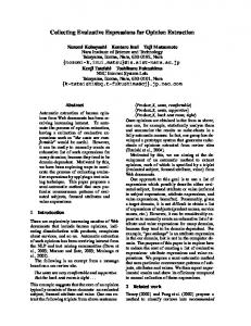
Fig. 1.
T. Suekane et al.
Expression of the ET system in normal colonic tissue. (A) Immunostaining for SMCs (1A4). (B–D) Immunostaining for ECE (B), ETA (C) and ETB (D). Main expression sites of these ET system components are muscular layers and vasculatures. Original magnification: A–D, ×30.
Results Normal colonic tissues Normal colonic tissues histologically demonstrated no fibrotic and no inflammatory changes. Expressions of ECE, ETA and ETB were seen mainly in the muscular layers and vasculatures (Fig. 1). Vascular SMCs were positive for ECE, ETA and ETB (Fig. 2). ETA immunoreactivity in vascular SMCs was stronger than ETB. Of these ET system components, only ECE was detected in endothelial cells (Fig. 2). Colonic tissues of CD In diseased intestinal tissues of CD, marked proliferation and accumulation of SMCs was frequently seen in the submucosa, in association with inflammatory infiltration and fibrotic tissue reaction. In addition to original SMC
tissue components including the muscular layers and vessels, the proliferated SMCs in the submucosa were distinctly positive for the ET system components, ECE, ETA and ETB (Fig. 3). In these proliferated SMCs, ETA immunoreactivity was relatively weak compared to ETB. In contrast, as seen in normal colonic tissues, expression of ETA in the vascular SMCs was stronger than that of ETB (Fig. 3). Macrophages accumulated in the active inflammatory lesions also showed strong expressions of ECE, ETA, and ETB (Fig. 4). Inflammation associated neovascularization was seen in the lesions, and the newly formed vessels were invariably positive for the ET system components (Fig. 4). While ECE expression was confirmed in the endothelial cells, as well as the SMCs, of the newly formed vessels, ETB expression could not be detected in the endothelial cells.
J. Clin. Biochem. Nutr.
Endothelin System Components and Crohn’s Disease
Fig. 2.
129
Expression of the ET system in a vessel in normal colonic tissue. (A) Immunostaining for endothelial cell marker, CD31. (B–D) Immunostaining for the ET system components; ECE (B), ETA (C) and ETB (D). Vascular SMCs are positive for all these ET system components. Of these ET system components, only ECE is detectable in endothelial cells (B; arrow). Original magnification: A–D, ×400.
Discussion This is the first immunohistochemical study based on frozen human colonic sections, that disclosed enhanced expressions of the ET system components, ECE, ETA and ETB, in inflammatory and fibrotic lesions of CD. The finding strongly suggests that these ET system components are collectively involved in inflammatory and fibrogenic processes of CD. ET-1 is a vasoconstrictor peptide that contributes not only to the regulation of vascular tone but also to the repair / fibrogenic processes after tissue injury [11, 16]. In addition to cardiovascular system, ET-1 acts on various normal or diseased organs/tissues [8–13, 16, 20–22]. Inagaki et al. [21] and Egidy et al. [22] reported the immunolocalization of ET system components in human normal colonic tissues and suggested implication of the ET system in physiological functions of human colons. Moreover, the ET system seems to be involved in human colonic diseases, including congenital, neoplastic and inflammatory disorders [12, 13, 23–27]. CD has also been recognized as one of such colonic diseases, in which the ET system potentially plays a pathogenic role. In fact, ET-1 levels in both plasma and colonic tissues were increased in patients with CD compared to normal subjects [12, 13]. However, no study has revealed expression patterns of the ET system components other than Vol. 42, No. 2, 2008
ET-1 in human colonic tissues of CD. ECE is a final key enzyme in the synthesis of ET-1, and regulates the local ET-1 production [14]. The present immunohistochemical investigation revealed increased ECE expression in inflammatory and fibrotic foci in colonic tissues of patients with CD. The finding can provide a likely explanation for the results of prior studies that showed increased ET-1 in CD patients [12, 13]. ECE expression was observed in accumulated macrophages and in proliferated SMCs, both of which are chief effector cells of inflammatory and fibrogenic tissue reactions. Hence, upregulation of ET-1 production by ECE may be an essential process in the development and progression of colonic lesions in CD. The biological effects of ET-1 are expressed via binding to its specific receptors, ETA and ETB [15]. It had been recognized that the vasoconstrictive and mitogenic properties of ET-1 were mediated mainly by ETA [28, 29]. Because ET-1 binding to ETB stimulates the release of nitric oxide from endothelial cells, it has been believed that ETB antagonizes ETA-mediated ET-1 function and therefore induces vasodilatation [30]. However, accumulating evidence has recently shown that ETB also contribute to the vasoconstrictive and mitogenic reactions [11, 16, 31]. We found that both ETA and ETB expressions were enhanced in inflamed and fibrotic colonic lesions of CD. Especially in
130
Fig. 3.
T. Suekane et al.
Expression of the ET system in colonic tissues of CD. (A) Immunostaining for SMCs (1A4). Marked SMC proliferation and accumulation are seen in the submucosa (asterisk). (B–D) Immunostaining for the ET system components; ECE (B), ETA (C) and ETB (D). In addition to SMCs of the muscular layers and the vessels, the proliferated SMCs in the submucosa are positive for all these ET system components. ETA immunoreactivity in these proliferated SMCs is relatively weak. (E, F) Double immunolabeling for ETA (red)/SMC (1A4; blue) (E), and ETB (red)/SMC (1A4; blue) (F). ETA immunoreactivity of the proliferated SMCs is weaker than ETB immunoreactivity (arrows), and conversely, ETA immunoreactivity of the vascular SMCs is stronger than that of ETB immunoreactivity (arrowhead). Original magnification: A–D, ×20; E and F, ×100.
the fibroproliferative foci in the submucosa, ETB was a dominant ET receptor that was expressed in the proliferated SMCs. ETB-mediated nitric oxide production from endothelial cells was thought to be absent in the affected colonic tissues because ETB was not detected in the endothelial cells. The findings could be interpreted that both ETA- and ETB-mediated ET-1 actions might contribute to develop the inflammation-related fibrosis and SMC proliferation, which is a histological hallmark of intestinal strictures in CD [2]. The precise pathway of the development of CD is still obscure. The results of the present study suggest at least that
the ET system plays a pathogenic role in the development of CD and in its characteristic and serious complication, intestinal strictures. Increased ET-1 in the inflamed colonic tissues may induce hypercontraction of the muscular layers and the proliferated SMCs in the submucosa and may worsen the strictures. These mean potential efficacy of ET antagonists in prevention of the disease progression and the complication. Bosentan, a dual ET receptors (ETA and ETB) blockade [31, 32], may be applicable for the treatment of CD patients and may introduce favorable outcomes in the prevention and resolution of intestinal strictures. J. Clin. Biochem. Nutr.
Endothelin System Components and Crohn’s Disease
Fig. 4.
131
Expression of the ET system in an active inflammatory lesion in CD. (A) Immunostaining for macrophage marker, CD68. Abundant macrophages are accumulated in the lesion. (B–D) Immunostaining for the ET system components; ECE (B), ETA (C) and ETB (D). In the lesion with marked macrophage accumulation, enhanced expressions of these ET system components are seen (B–D). (E, F) Double immunolabeling for ETA (red)/macrophage (CD68; blue) (E), and ETB (red)/macrophage (CD68; blue) (F). Many macrophages are positive for ETA and/or ETB. Original magnification: A–D, ×90; E and F, ×150.
In conclusion, the present study demonstrated the enhanced expressions of ECE, ETA and ETB in affected human colonic tissues of CD. These results strongly suggest that the ET system play a pathogenic role in inflammatory and fibrogenic processes of CD. Administration of ET antagonist may improve the prognosis of patients with CD.
Abbreviations CD, Crohn’s disease; ET-1, endothelin-1; ECE, endothelin-converting enzyme; ETA, endothelin type A receptor; ETB, endothelin type B receptor; SMC, smooth muscle cell.
Vol. 42, No. 2, 2008
References [1] Stange, E.F., Travis, S.P., Vermeire, S., Beglinger, C., Kupcinkas, L., Geboes, K., Barakauskiene, A., Villanacci, V., Von Herbay, A., Warren, B.F., Gasche, C., Tilg, H., Schreiber, S.W., Scholmerich, J., and Reinisch, W.: European Crohn’s and Colitis Organisation.: European evidence based consensus on the diagnosis and management of Crohn’s disease: definitions and diagnosis. Gut, 55 Suppl. 1, i1–15, 2006. [2] Van, Assche., Geboes, K., and Rutgeerts, P.: Medical therapy for Crohn’s disease strictures. Inflamm. Bowel Dis., 10, 55– 60, 2004. [3] Dieleman, L.A., Pena, A.S., Meuwissen, S.G., and van Rees, E.P.: Role of animal models for the pathogenesis and treat-
132
[4]
[5]
[6]
[7] [8]
[9]
[10]
[11]
[12]
[13]
[14]
[15]
[16]
[17]
T. Suekane et al. ment of inflammatory bowel disease. Scand. J. Gastroenterol. Suppl., 223, 99–104, 1997. Yanagisawa, M., Kurihara, H., Kimura, S., Tomobe, Y., Kobayashi, M., Mitsui, Y., Yazaki, Y., Goto, K., and Masaki, T.: A novel potent vasoconstrictor peptide produced by vascular endothelial cells. Nature, 332, 411–415, 1988. Hirata, Y., Takagi, Y., Fukuda, Y., and Marumo, F.: Endothelin is a potent mitogen for rat vascular smooth muscle cells. Atherosclerosis, 78, 225–228, 1989. Schwartz, S.M. and Ross, R.: Cellular proliferation in atherosclerosis and hypertension. Prog. Cardiovasc. Dis., 26, 355–372, 1984. Becker, A.: Coronary atherosclerosis revisited. A pathologist’s view. Acta. Med. Scand. Suppl., 694, 69–76, 1985. Jones, G.T., van Rij, A.M., Solomon, C., Thomson, I.A., and Packer, S.G.: Endothelin-1 is increased overlying atherosclerotic plaques in human arteries. Atherosclerosis, 124, 25– 35, 1996. Hai, E., Ikura, Y., Naruko, T., Shirai, N., Yoshimi, N., Kayo, S., Sugama, Y., Fujino, H., Ohsawa, M., Tanzawa, K., Yokota, T., and Ueda, M.: Alterations of endothelin-converting enzyme expression in early and advanced stages of human coronary atherosclerosis. Int. J. Mol. Med., 13, 649–654, 2004. Nakatani, T., Tanabe, S., Han, Y.S., Kayo, S., Yoshimi, N., Hai, E., Shirai, N., Ikura, Y., Ohsawa, M., and Ueda, M.: Enhanced expression of endothelin-A receptor in human transplant renal arteriosclerosis. Int. J. Mol. Med., 11, 153– 156, 2003. Ikura, Y., Ohsawa, M., Naruko, T., Muraguchi, T., Hirayama, M., Suekane, T., Fukushima, H., Sugama, Y., Shirai, N., Kayo, S., Yoshimi, N., Ehara, S., Tanzawa, K., and Ueda, M.: Expression of the hepatic endothelin system in human cirrhotic livers. J. Pathol., 204, 304–310, 2004. Letizia, C., Boirivant, M., De Toma, G., Cerci, S., Subioli, S., Scuro, L., Ferrari, P., and Pallone, F.: Plasma levels of endothelin-1 in patients with Crohn’s disease and ulcerative colitis. Ital. J. Gastroenterol. Hepatol., 30, 266–269, 1998. Murch, S.H., Braegger, C.P., Sessa, W.C., and MacDonald, T.T.: High endothelin-1 immunoreactivity in Crohn’s disease and ulcerative colitis. Lancet, 339, 381–385, 1992. Xu, D., Emoto, N., Giaid, A., Slaughter, C., Kaw, S., deWit, D., and Yanagisawa, M.: ECE-1: a membrane-bound metalloprotease that catalyzes the proteolytic activation of big endothelin-1. Cell, 78, 473–485, 1994. Elshourbagy, N.A., Korman, D.R., Wu, H.L., Sylvester, D.R., Lee, J.A., Nuthalaganti, P., Bergsma, D.J., Kumar, C.S., and Nambi, P.: Molecular characterization and regulation of the human endothelin receptors. J. Biol. Chem., 268, 3873–3879, 1993. Shirai, N., Naruko, T., Ohsawa, M., Ikura, Y., Sugama, Y., Hirayama, M., Kitabayashi, C., Ehara, S., Inoue, T., Itoh, A., Haze, K., Tanzawa, K., Yoshiyama, M., Yoshikawa, J., and Ueda, M.: Expression of endothelin-converting enzyme, endothelin-1 and endothelin receptors at the site of percutaneous coronary intervention in humans. J. Hypertens., 24, 711–721, 2006. Shimada, K., Matsushita, Y., Wakabayashi, K., Takahashi, M., Matsubara, A., Iijima, Y., and Tanzawa, K.: Cloning
[18]
[19]
[20] [21]
[22]
[23]
[24]
[25]
[26]
[27]
[28]
[29]
[30]
[31]
[32]
and functional expression of human endothelin-converting enzyme cDNA. Biochem. Biophys. Res. Commun., 207, 807– 812, 1995. Sasaki, Y., Hori, S., Oda, K., Okada, T., and Takimoto, M.: Both ET(A) and ET(B) receptors are involved in mitogenactivated protein kinase activation and DNA synthesis of astrocytes: study using ET(B) receptor-deficient rats (aganglionosis rats). Eur. J. Neurosci., 10, 2984–2993, 1998. van der Loos, C.M., Becker, A.E., and van den Oord, J.J.: Practical suggestions for successful immunoenzyme doublestaining experiments. Histochem. J., 25, 1–13, 1993. Nambi, P.: Endothelin receptors in normal and diseased kidneys. Clin. Exp. Pharmacol. Physiol., 23, 326–330, 1996. Inagaki, H., Bishop, A.E., Escrig, C., Wharton, J., AllenMersh, T.G., and Polak, J.M.: Localization of endothelinlike immunoreactivity and endothelin binding sites in human colon. Gastroenterology, 101, 47–54, 1991. Egidy, G., Juillerat-Jeanneret, L., Korth, P., Bosman, F.T., and Pinet, F.: The endothelin system in normal human colon. Am. J. Physiol. Gastrointest. Liver. Physiol., 279, G211– G222, 2000. Kusafuka, T., Wang, Y., and Puri, P.: Novel mutations of the endothelin-B receptor gene in isolated patients with Hirschsprung’s disease. Hum. Mol. Genet., 5, 347–349, 1996. Inagaki, H., Bishop, A.E., Eimoto, T., and Polak, J.M.: Autoradiographic localization of endothelin-1 binding sites in human colonic cancer tissue. J. Pathol., 168, 263–267, 1992. Ali, H., Dashwood, M., Dawas, K., Loizidou, M., Savage, F., and Taylor, I.: Endothelin receptor expression in colorectal cancer. J. Cardiovasc. Pharmacol., 36, S69–S71, 2000. Rachmilewitz, D., Eliakim, R., Ackerman, Z., and Karmeli, F.: Colonic endothelin-1 immunoreactivity in active ulcerative colitis. Lancet, 339, 1062, 1992. McCartney, S.A., Ballinger, A.B., Vojnovic, I., Farthing, M.J., and Warner, T.D.: Endothelin in human inflammatory bowel disease: comparison to rat trinitrobenzenesulphonic acid-induced colitis. Life. Sci., 71, 1893–1904, 2002. Maguire, J.J. and Davenport, A.P.: ETA receptor-mediated constrictor responses to endothelin peptides in human blood vessels in vitro. Br. J. Pharmacol., 115, 191–197, 1995. Rodriguez-Vita, J., Ruiz-Ortega, M., Ruperez, M., Esteban, V., Sanchez-Lopez, E., Plaza, J.J., and Egido, J.: Endothelin-1, via ETA receptor and independently of transforming growth factor-beta, increases the connective tissue growth factor in vascular smooth muscle cells. Circ. Res., 97, 125–134, 2005. Hirata, Y., Emori, T., Eguchi, S., Kanno, K., Imai, T., Ohta, K., and Marumo, F.: Endothelin receptor subtype B mediates synthesis of nitric oxide by cultured bovine endothelial cells. J. Clin. Invest., 91, 1367–1373, 1993. Seo, B., Oemar, B.S., Siebenmann, R., von Segesser, L., and Lüscher, T.F.: Both ETA and ETB receptors mediate contraction to endothelin-1 in human blood vessels. Circulation, 89, 1203–1208, 1994. Gardiner, S.M., Kemp, P.A., March, J.E., and Bennett, T.: Effects of bosentan (Ro 47-0203), an ETA-, ETB-receptor antagonist, on regional haemodynamic responses to endothelins in conscious rats. Br. J. Pharmacol., 112, 823–830, 1994.
J. Clin. Biochem. Nutr.