JOURNAL OF CLINICAL MICROBIOLOGY, Oct. 2006, p. 3596–3599 0095-1137/06/$08.00⫹0 doi:10.1128/JCM.02543-05 Copyright © 2006, American Society for Microbiology. All Rights Reserved.
Vol. 44, No. 10
Enumeration of Bacterial Cell Numbers and Detection of Significant Bacteriuria by Use of a New Flow Cytometry-Based Device Hiroshi Okada,1* Toshiro Shirakawa,2 Akinobu Gotoh,2 Yutaka Kamiyama,1 Satoru Muto,1 Hisamitsu Ide,1 Yukio Hamaguchi,3 and Shigeo Horie1 Department of Urology, Teikyo University School of Medicine, Tokyo, Japan,1 and Division of Clinical Genetics and Gene Therapy, Kobe University Graduate School of Medicine,2 and The Sysmex Corp.,3 Kobe, Japan Received 6 December 2005/Returned for modification 22 January 2006/Accepted 15 July 2006
A new, automated flow cytometry-based urine bacterium analyzer (UBA) was developed. We assessed the UBA for linearity of measurement, reproducibility of results, carryover rate, and correlation of measured results with those determined by urine culture. We also evaluated its ability to screen urine samples for significant bacteriuria. The UBA showed excellent linearity and a minor carryover rate. Results from the UBA were highly reproducible, and in between-run precision assays, the coefficients of variation for the UBA results were smaller than those for the urine culture results. Two hundred seventy-three urine specimens from patients attending the outpatient clinics of two university-based hospitals were examined. The results for the UBA were compared with those for urine culture. The UBA detected significant bacteriuria with a sensitivity of 96.6%, a specificity of 79.9%, a positive predictive value of 57.0%, a negative predictive value of 98.8%, a false-positive rate of 15.8%, a false-negative rate of 0.7%, and an accuracy of 83.5%. These results were comparable to or better than those obtained with previously reported screening procedures. The UBA can perform accurate enumeration of bacterial cells automatically in 90 seconds and can be used for the screening of significant bacteriuria.
Although the standard method for diagnosis of urinary tract infection is quantitative urine culture and identification of bacteria, fewer than 30 percent of urine samples sent to the laboratory are proven positive (6). Thus, a rapid screening method is required to reduce these time-consuming and expensive procedures. We have reported that the flow cytometrybased automatic urinalysis device UF-50 can detect significant bacteriuria with a false-negative rate (FN) of 5.4% (7). For the ideal screening method, the false-negative rate should be lower. For this purpose, we developed a new device to perform enumeration of bacterial cells in urine by using flow cytometry technology equipped with a semiconductor laser. In this paper, we evaluated the linearity and reproducibility of the measurement and compared the measured results with those from the conventional urine culture method using clinical urine specimens. We also evaluated the feasibility of using this new device to predict the results of urine culture.
urinalysis, including qualitative measurement of protein and sugar and microscopic examination of centrifuged urinary sediment. The rest of the specimen was processed for assessment of the new urine bacterium analyzer (UBA). All the patients were fully informed of the purpose and study design and consented to provide specimens before starting the study. Approval from the ethical committee and the Institutional Review Board was obtained prior to starting the investigation. Semiquantitative urine culture. Semiquantitative urine cultures were performed by procedures based on the recommendation of Cintron (5), with cystinelactose-electrolyte-deficient (CLED) agar (Nissui Pharmatheutical Co. Ltd., Tokyo, Japan). Most pathogens can grow on CLED agar, and it can inhibit the swarming of Proteus species. For inoculation of urine samples, a 100-l micropipette (Eppendorf Co., Ltd.) was used. Bacterial concentrations were determined by a single trained technician and expressed as numbers of CFU per ml. Samples were considered positive if they contained ⱖ105 or 104 to ⬍105 urinary pathogens/ml of pure culture, if two or more potentially pathological bacterial species were isolated when the individual counts were ⱖ104 CFU, or when the count for one organism was ⱖ104 CFU/ml and it was clearly predominant (i.e., present in numbers at least 10-fold greater than those for the others). The microorganisms isolated were identified by standard biological procedures (6). Urine bacterium analyzer. The UBA, a new device that uses flow cytometry technology and a semiconductor laser as the light source, has been developed for measuring bacterial concentration. The UBA can stain, detect, and analyze cellular components and calculate the number of bacteria in a sample fully automatically. For automated bacterial counts with this device, 200 l of the sample was automatically diluted with 1,360 l of our specially developed diluent, which included citrate buffer solution and 0.1% (wt/wt) cationic surfactant. Then, the mixture was automatically supplemented with 40 l of fluorescent dye which specifically stained nucleic acid in the bacteria (the final concentration of dye was approximately 1 ppm). The mixture was then hydrodynamically focused and passed through a sheath flow cell illuminated with a red semiconductor laser beam ( ⫽ 635 nm). With this method, individual cellular components, including bacteria in urine specimens, fluoresce to various degrees. The forward scatter light intensity and the lateral fluorescent intensity of each cellular component in the urine sample are converted into electric signals by a photomultiplier, and those two parameters are measured simultaneously in each sample. By using these two parameters (forward scatter light intensity and lateral fluorescent intensity), the UBA can distinguish bacteria from other cellular and noncellular components in urine (e.g., erythrocytes, leukocytes, epithelial cells, fungi, and
MATERIALS AND METHODS Bacterial strains and media. Four bacterial strains, Escherichia coli ATCC 11775, Pseudomonas aeruginosa ATCC 27853, Staphylococcus aureus ATCC 29213, and Enterococcus faecalis ATCC 29212, were purchased from ATCC. Clinical specimens. Urine specimens were collected from patients attending the outpatient clinics of two university hospitals. Patients with urinary diversions or orthotopic neobladder with bowel segments were excluded. Also excluded were the patients who had fistulas between the urinary tract and bowels. A sterile plastic container with a wide opening was used to collect midstream urine. Female patients were asked to wipe their external genitalia with a wet tissue before urinating. Ten milliliters of each specimen was used for routine
* Corresponding author. Mailing address: Department of Urology, Teikyo University School of Medicine, 2-11-1 Kaga, Itabashi-ku, Tokyo, Japan 173-8605. Phone: 81 3 3964 2497. Fax: 81 3 3964 8934. E-mail:
[email protected]. 3596
VOL. 44, 2006
AUTOMATED ENUMERATION OF BACTERIAL CELLS
3597
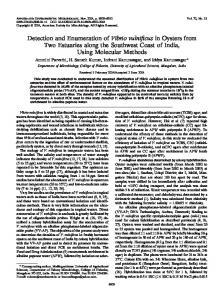
FIG. 2. Linearity of UBA. UBA (y axis) showed perfect linearity with the theoretically estimated concentrations (x axis) of E. coli. y ⫽ 0.9312x (R2 ⫽ 0.9999).
RESULTS
FIG. 1. UBA scattergram pattern of E. coli. The bacterial cluster is shown in the middle of its fluorescent intensity. Basically, other particles, such as erythrocyte, leukocyte, epithelial cells, fungi, and crystals, did not interfere with this bacterial cluster. FSC, forward scatter light intensity; FL, lateral fluorescent intensity.
crystals) (Fig. 1). After data reduction, the number of bacteria in 1 ml of the sample is recorded on the screen, and a hard copy of the results can be obtained. The sample was assessed as containing a significant number of bacteria if it contained ⱖ104 bacterial cells per 1 ml. Between-run imprecision. The quality control sample (0.78-m-diameter latex particle, XPR-652; Duke Scientific Co., CA), which mimicked the size of a bacterium, was used. The concentration of the quality control sample was adjusted to 5.0 ⫻ 105/ml. We measured on 18 separate days, and the coefficients of variation (CVs) were calculated. Within-run imprecision. To test the consistency of our results, repeated measurements were made using four bacterial strains purchased from ATCC: E. coli ATCC 11775, P. aeruginosa ATCC 27853, E. faecalis ATCC 29121, and S. aureus ATCC 29213. The concentrations of the bacteria were measured by the UBA or by semiquantitative urine culture. Measurements were repeated 10 times, and the imprecision of each method was assessed via the CVs obtained. Linearity assessments. Specimens containing E. coli, which were sterilized by the antibiotic minocycline and treated for 1 hour, were prepared by 10-fold dilution with physiological saline. The analytical data were plotted against the theoretical values calculated by the dilution factor from the undiluted sample (2.1 ⫻ 107 particles/ml). Linearity was determined by analyzing specimens in triplicate, and the slope and intercept for the expected value were determined. Analysis of carryover. The urine specimen which included bacteria at a concentration of 4.3 ⫻ 107 CFU/ml was used to assess the between-specimen carryover. Each specimen was analyzed in triplicate, followed by three blank specimens of the diluent. The carryover rate for bacteria was calculated by using the following formula: carryover rate (%) ⫽ [(B1 ⫺ B3)/(S3 ⫺ B3)] ⫻ 100, where B1 is the first measured value for physiological saline, B3 is the third measured value for physiological saline, and S3 is the third measured value for the specimen. Statistical analysis. The Spearman rank correlation coefficient was used to determine the correlation between values obtained by the two methods; P values of less than 0.05 were considered statistically significant. All data were analyzed using Stat View-J5.0 (SAS Institute, Cary, NC). Sensitivity, specificity, positive predictive value (PPV), and negative predictive value (NPV) were calculated by the method developed by Ransohoff and Feinstein (10).
A total of 273 urine specimens were collected and evaluated by the UBA and semiquantitative urine culture with CLED agar. Of these, 151 were from male patients and 122 from female patients. The incidence of positive culture was 21.6%. The microorganisms isolated were of the following species, with the respective numbers of isolates indicated in parentheses: Escherichia coli (21), Pseudomonas aeruginosa (12), Enterococcus spp. (7), coagulase-negative Staphylococcus (5), Staphylococcus aureus (4), Klebsiella pneumoniae (3), Candida albicans (3), Citrobacter freundii (3), Stenotrophomonas maltophilia (2), Morganella morganii (2), Serratia marcescens (2), Proteus vulgaris (1), and Corynebacterium spp. (1). Linearity of results. The values measured by the UBA (Fig. 2, y axis) showed perfect linearity with the theoretically estimated values (Fig. 2, x axis) for samples with concentrations between 107 and 103 particles/ml. The linear correlation was y ⫽ 0.9312x (R2 ⫽ 0.9999, P ⬍ 0.0001) (Fig. 2). Carryover rate. The carryover rate was calculated to be 0.029%, and this result showed that there was no substantial carryover between samples. Between- and within-run imprecisions. The between-run imprecision of the UBA was analyzed by measuring the quality control specimen on 18 different days. The CV was as low as 1.5%. Within-run CVs obtained by measuring bacterial strains of E. coli, P. aeruginosa, E. fecalis, and S. aureus at concentrations of 104 CFU/ml ranged between 9.3% and 18.4% for the UBA and between 21.1% and 63.8% for the conventional semiquantitative culture method. When the bacterial concentration was adjusted to 103 CFU/ml, CVs for the UBA ranged between 27.8% and 35.1%, and those for the semiquantitative method ranged between 38.9% and 83.4%. The UBA showed smaller CVs for measurement of bacterial concentration (Table 1). Comparison with semiquantitative urine culture. Bacterial numbers measured by the UBA correlated well with values measured by the semiquantitative method (P ⬍ 0.001) (Fig. 3). The results for detection of significant bacteriuria by the UBA and semiquantitative urine culture were evaluated in terms of sensitivity (96.6%), specificity (79.7%), PPV (57.0%), NPV
3598
OKADA ET AL.
J. CLIN. MICROBIOL.
TABLE 1. Within-run reproducibilitya CV (%) obtained for indicated concn (CFU/ml) and method Bacterial strain
Semiquantitative urine culture
UBA
E. coli ATCC 11775 P. aeruginosa ATCC 27853 E. faecalis ATCC 29121 S. aureus ATCC 29213
103
104
103
104
25.9 35.1 31.5 27.8
18.4 10.9 11.9 9.3
83.4 68.3 38.9 64.6
45.3 63.8 31.7 21.1
a Measurements were repeated 10 times, and the coefficient of variation for each method was calculated for each bacterial species.
(98.8%), false-positive rate (FP) (15.8%), and false-negative rate (0.7%) (Table 2). DISCUSSION We have developed a series of flow cytometry-based devices for detection of cellular and noncellular components in urine to enable automated urinalysis. The UF-100 and UF-50 have been successfully accepted as devices for performing firstscreening urinalysis without using a microscope (2, 8). When these devices detected abnormal numbers of cellular or noncellular components of urine, these samples were subjected to ordinary urinalysis by observation of urinary sediment with a microscope by trained technicians. The UF-50 was reported to detect significant bacteriuria with a sensitivity of 83.1%, a specificity of 76.4%, a PPV of 62.0%, an NPV of 90.7%, a falsepositive rate of 16.1%, and a false-negative rate of 5.4% (7). However, it still had a rather high false-negative rate. The UF-100 was also reported to have a high false-negative rate in the detection of significant bacteriuria (11). In this study, we developed a new flow cytometry-based automated device which was specific for accurate enumeration of bacterial cells
FIG. 3. Correlation of the bacterial cell numbers measured by the semiquantitative urine culture method and those measured by the UBA. Bacterial numbers measured by the UBA correlated well with values measured by the semiquantitative method (P ⬍ 0.001).
TABLE 2. Correlation between significant bacteriuria detection by the UBA and semiquantitative urine culture No. of urine culture results Results by UBA Positive
Negative
Total
Positive Negative
57 2
43 171
100 173
Total
59
214
273
Samples were considered positive if they contained ⱖ105 or 104 to 105 CFU of urinary pathogens/ml of pure culture or two or more potentially pathological bacterial species when individual cell counts were ⱖ104 CFU/ml or when the count for one predominant organism was ⱖ104 CFU/ml. Sensitivity 关total positive/(total positive ⫹ FN)兴, 57/59 (96.6%); specificity 关total negative/(FP ⫹ total negative)兴, 171/214 (79.9%); PPV 关total positive/(total positive ⫹ FP)兴, 57/100 (57.0%); NPV 关total negative/(FN ⫹ total negative)兴, 171/173 (98.8%); FP (FP/ total), 43/273 (15.8%); FN (FN/total), 2/273 (0.7%); accuracy 关(total positive ⫹ total negative)/total兴, 228/273 (83.5%). a
in urine and tested the feasibility of the device for detecting significant bacteriuria. The new device, named the UBA, basically used the same strategy as the former devices, UF-100 and UF-50, for measurement of cellular components in urine. The UBA uses a semiconductor laser instead of an argon laser to reduce the cost of maintenance. Although the UF-100 and UF-50 were originally designed to perform automated urinalysis, this new device is specially designed to perform detection of bacterial cells. We adopted a cutoff value of 104 particles/ml for significant bacteriuria for the UBA. The UBA showed excellent linearity (R2 ⫽ 1) for concentrations between 102 and 108 particles/ml of target objects (data not shown). Also, the UBA showed good linearity (R2 ⫽ 0.9999) for concentrations between 103 and 107 CFU/ml of bacterial pure culture. The UBA can perform measurement of urine bacterial cells for 90 s per sample without significant carryover. This allows over 30 consecutive measurements of bacterial cells in urine samples in 1 hour. A between-run precision assay found that the CVs for results from the UBA were less than 1.5%. In a within-run precision assay, the UBA showed better precision than urine cultures in measuring bacterial cell numbers at concentrations of 103 and 104 CFU/ml. It is noteworthy that the urine culture method, which is used as the gold standard for measurement of bacterial cell number, showed large CVs (38.9% to 83.4%) in the measurement of lower concentrations of bacteria (Table 1). When we repeated the same experiment with other trained technicians, we got the same results. When clinical specimens were used, the measured results for bacterial cell number obtained by the UBA correlated well with those obtained by the urine culture method. However, in 21 samples, we could detect bacterial cells in urine by the UBA but not by the urine culture method (data not shown). All these samples were from patients who had not received antimicrobial chemotherapy. For these samples, we confirmed the presence of bacteria by microscopic observation after staining. Most bacteria were revealed to be cocci. These bacterial concentrations were estimated to be from 104 to 105 CFU/ml. Since some bacterial cells in urine are very slow to replicate or are dead, not all bacterial cells in urine samples are able to form colonies after incubation. This may explain that phenomenon.
VOL. 44, 2006
AUTOMATED ENUMERATION OF BACTERIAL CELLS
For rapid screening for significant bacteriuria, it is ideal that the method has a low false-negative rate. To date, several rapid screening methods have been reported, including semiautomated microscopy combined with reagent strip chemical determinations (Yellow IRIS) (1), reagent strip chemical determination alone (Clinitek 200) (3), detection of catalase activity (Uriscreen) (9), and microscopic examination of Gram-stained, unspun urine (4). The false-negative rates obtained by Yellow IRIS, Clinitek 200, Uriscreen, and microscopic examination of Gram-stained unspun urine were 9.9%, 0.7%, 8.0%, and 1.0%, respectively (1, 3–4, 9). The false-negative rate obtained by the UBA was 0.7%, the lowest rate, equal to that for the Clinitek 200. With these results combined, the UBA is an ideal device for performing accurate enumeration of bacterial cells in urine and for detecting significant bacteriuria in a short period of time. In the near future, the UBA is expected to detect fungi such as Candida albicans. If we can measure the number of bacterial cells correctly, we can utilize this technique for bacterial susceptibility testing. We are now developing a new device that can perform a rapid, direct antimicrobial susceptibility test to realize an ideal chemotherapeutic strategy in the treatment of urinary tract infections. ACKNOWLEDGMENTS We gratefully acknowledge J. Inoue and Y. Kawashima for their technical assistance.
3599
This study was partly supported by a grant from Sysmex Corp.
REFERENCES 1. Barlett, R. C., D. A. Zern, I. Ratkiewicz, and J. Z. Tetreault. 1992. Screening for urinary tract infection with the Yellow IRIS. Lab. Med. 23:599–602. 2. Ben-Ezra, J., L. Bork, and R. A. McPherson. 1988. Evaluation of the Sysmex UF-100 automated urinalysis analyzer. Clin. Chem. 44:92–95. 3. Bowman, R. A., and T. V. Riley. 1991. Evaluation of Clinitek 200 and RapimatI/T for screening for urinary tract infection. J. Clin. Pathol. 44:58– 60. 4. Cardoso, C. L., C. B. Muraro, V. L. D. Siqueira, and M. Guilhermetti. 1998. Simplified technique for detection of significaqnt bacteriuria by microscopic examination of urine. J. Clin. Microbiol. 36:820–823. 5. Cintron, F. 1992. Initial processing, inoculation, and incubation of aerobic bacteriology specimens. Section 1.4.10–1.4.11. In H. D. Isenberg (ed.), Clinical microbiology procedures handbook. American Society for Microbiology, Washington, D.C. 6. Kellogg, J. A., J. P. Manzella, S. N. Shaffer, and B. B. Schwartz. 1987. Clinical relevance of culture versus screen for the detection of microbial pathogens in urine specimens. Am. J. Med. 83:739–745. 7. Okada, H., Y. Sakai, S. Miyazaki, S. Arakawa, Y. Hamaguchi, and S. Kamidono. 2000. Detection of significant bacteriuria by automated urinalysis using flow cytometry. J. Clin. Microbiol. 38:2870–2872. 8. Okada, H., Y. Sakai, G. Kawabata, M. Fujisawa, S. Arakawa Y. Hamaguchi, and S. Kamidono. 2001. Automated urinalysis. Evaluation of the Sysmex UF-50. Am. J. Clin. Pathol. 115:605–610. 9. Palmer, L. S., I. Richards, and W. Kaplan. 1997. Clinical evaluation of a rapid diagnostic screen (Uriscreen) for bacteria in children. J. Urol. 157: 654–657. 10. Ransohoff, D. F., and A. R. Feinstein. 1978. Problem of spectrum and bias in evaluating the efficacy of diagnostic tests. N. Engl. J. Med. 299:926–930. 11. Zaman, Z., S. Roggeman, and J. Verhagen. 2001. Unsatisfactory performance of flow cytometer UF-100 and urine strip in predicting outcome of urine cultures. J. Clin. Microbiol. 39:4169–4171.